العربية
Part of the Master Guide

General Orthopedics 2026 Practice Questions: Set 13 (Solved)

Orthopedic Sports Medicine 2026 MCQs: Board Review Questions & Answers (Part 3)

23 Apr 2026 85 min read 74 Views
Figure for Sports Medicine 2007 MCQs - Part 3 - Question 51

Key Takeaway

Looking for accurate information on Orthopedic Sports Medicine 2026 MCQs: Board Review Questions & Answers (Part 3)? Top-rated Orthopedic Sports Medicine 2026 MCQs bank. Practice with clinical case questions, orthopedic surgery board review, and evidence-based answers updated for 2026.

Orthopedic Sports Medicine 2026 MCQs: Board Review Questions & Answers (Part 3)

Comprehensive 100-Question Exam


00:00

Start Quiz

Question 1

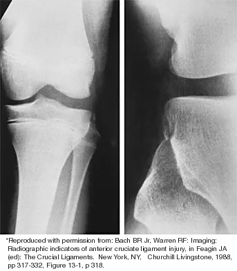
A 22-year-old professional baseball catcher has posterior shoulder pain and severe external rotation weakness with the arm in adduction. Radiographs are normal. MRI scans are shown in Figures 15a through 15c. Management should consist of


Explanation

15b 15c The MRI scans reveal a large posterior paralabral cyst associated with a posterior-superior labral tear. The cyst appears as a well-defined, smoothly marginated mass with low signal intensity on T1-weighted MRI scans and with high signal intensity on T2-weighted MRI scans. MRI also reveals changes in the supraspinatus and infraspinatus muscles secondary to denervation, including decreased muscle bulk and fatty infiltration. MRI has the added advantage, compared with other imaging modalities, of detecting intra-articular lesions, such as labral tears, which are frequently associated with ganglion cysts of the shoulder. In this case of a professional baseball player with a space-occupying lesion causing nerve compression with an associated labral tear, the treatment of choice is arthroscopic decompression of the cyst and repair of the tear. Acromioplasty would not address the primary pathology in this patient. Cummins CA, Messer TM, Nuber GW: Suprascapular nerve entrapment. J Bone Joint Surg Am 2000;82:415-424.

Question 2

A 21-year-old collegiate scholarship football player has an episode of transient quadriplegia. An MRI scan of the cervical spine reveals cord edema and severe congenital spinal stenosis. The athlete has aspirations of playing on a professional level and demands that he be allowed to play. The team physician should give what recommendation to the college?


Explanation

Federal courts have ruled that a student-athlete does not have a constitutional right to participate in athletics against medical advice. As long as the student retains his scholarship, the college is under no legal or ethical obligation to allow the student to participate in sports. A waiver would not hold up in court and would not indemnify the college or the team physician against suit. No equipment has been shown to be effective in preventing transient quadriplegia. Mathias MB: The competing demands of sport and health: An essay on the history of ethics in sports medicine. Clin Sports Med 2004;23:195-214.

Question 3

When performing a posterior cruciate ligament reconstruction with a tibial inlay-type approach, what is the approximate anatomic distance of the popliteal artery from the screws used for fixation of the bone block?


Explanation

Miller and associates reported the results of a cadaveric study of the vascular risk of a posterior approach for posterior cruciate ligament reconstruction using the tibial inlay technique. The average distance from the screw to the popliteal artery was 21.1 mm (range, 18.1 mm to 31.7 mm). Other approaches, such as the transtibial tunnel technique which involves drilling an anterior-posterior tunnel, have also been studied in cadavers. Matava and associates noted that increasing flexion reduces but does not completely eliminate the risk of arterial injury during arthroscopic posterior cruciate ligament reconstruction. However, this study did not use the small, medial utility incision recommended by Fanelli and associates, which creates an interval for the surgeon's finger between the medial gastrocnemius and the posteromedial capsule so that any migration of the guidepin can be palpated and changed prior to any injury to the posterior neurovascular bundle. Matava MJ, Sethi NS, Totty WG: Proximity of the posterior cruciate ligament insertion to the popliteal artery as a function of the knee flexion angle: Implications for posterior cruciate ligament reconstruction. Arthroscopy 2000;16:796-804. Miller MD, Kline AJ, Gonzales J, et al: Vascular risk associated with posterior approach for posterior cruciate ligament reconstruction using the tibial inlay technique. J Knee Surg 2002;15:137-140.

Question 4

Which of the following knee ligament injury patterns is most associated with an increase in external tibial rotation with the knee at 90 degrees of flexion?


Explanation

Cadaveric studies have shown that external rotation of the tibia is most pronounced following transection of the posterior cruciate and lateral collateral ligaments with the knee at 90 degrees of flexion. Isolated release of the lateral collateral ligament results in increased external tibial rotation at 30 degrees. Gollehon DL, Torzilli PA, Warren RF: The role of the posterolateral and cruciate ligaments in the stability of the human knee: A biomechanical study. J Bone Joint Surg Am 1987;69:233-242. Cooper DE: Tests for posterolateral instability of the knee in normal subjects: Results of examination under anesthesia. J Bone Joint Surg Am 1991;73:30-36.

Question 5

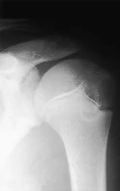
A 28-year-old professional dancer reports a 3-month history of progressive pain in the posterior aspect of the left ankle. Her symptoms are worse when she assumes the en pointe position. Examination reveals tenderness to palpation at the posterolateral aspect of the ankle posterior to the peroneal tendons which is made worse with passive plantar flexion. There is no nodularity, fluctuance, or tenderness of the Achilles tendon. The neurovascular examination is unremarkable. A lateral radiograph and MRI scan are shown in Figures 16a and 16b, respectively. Management should consist of


Explanation

16b The imaging studies reveal findings typical of the os trigonum syndrome. This condition results from inflammation between the os trigonum and the adjacent talus. The symptoms of posterior ankle pain are exacerbated by plantar flexion, which stresses the fibrous union between these two bones. Definitive management of the high-level athlete involves excision of the os trigonum from a medial approach, although arthroscopic excision has also been described. The os trigonum is not an intra-articular structure; therefore, ankle arthroscopy is neither diagnostic nor therapeutic. Abramowitz Y, Wollstein R, Barzilay Y, et al: Outcome of resection of a symptomatic os trigonum. J Bone Joint Surg Am 2003;85:1051-1057. Mouhsine E, Crevoisier X, Leyvraz P, et al: Post-traumatic overload or acute syndrome of the os trigonum: A possible cause of posterior ankle impingement. Knee Surg Sports Traumatol Arthrosc 2004;12:250-253.

Question 6

A professional pitcher reports pain localized to the medial aspect of his throwing elbow. History reveals that he was pitching in a playoff game and heard and felt a pop in his elbow. MRI reveals a complete ulnar-sided avulsion of the medial collateral ligament (MCL). Examination reveals valgus instability and ulnar nerve involvement. What recommendations should be made based on the patient's desire to return to sport?


Explanation

Injuries to the MCL usually result from repetitive high valgus stress on the medial aspect of the elbow joint due to overhead throwing or racquet sports. Excessive stresses during the late cocking and acceleration phase of throwing can injure the anterior band of the MCL. Clinically, the injuries may present as chronic or acute, and a pop may be noted in the latter. Associated ulnar nerve involvement is common. Valgus instability is present in about 25% of patients. Patients typically are athletes who participate in throwing and have localized medial elbow pain and tenderness along the course of a ligament that extends from the medial epicondyle of the distal humerus to the sublime tubercle of the ulna. Surgical reconstruction is the procedure of choice in an athlete desiring a return to a high level of throwing. Miller MD, Cooper DE, Warner JJP (eds): Review of Sports Medicine and Arthroscopy. Philadelphia, PA, WB Saunders, 1995, p 230. Arendt EA (ed): Orthopaedic Knowledge Update: Sports Medicine 2. Rosemont, IL, American Academy of Orthopaedic Surgeons, 1999, p 227.

Question 7

A 20-year-old collegiate football player who sustained blunt head trauma during the first half of a game is emotional and confused. During the halftime intermission, his affect, memory, and disorientation are totally resolved and have returned to preinjury baseline. The only residual finding is a very mild headache. He wants to play the second half. What is the most appropriate course of action?


Explanation

There is almost universal acceptance that an athlete may return to play after blunt head trauma only if he or she is totally asymptomatic. Mild residual symptoms are considered an absolute contraindication for return to play. Returning to play after a cardiovascular challenge or sport-specific activities is permitted on the pretext that the athlete is totally asymptomatic prior to these maneuvers. Neuropsychiatric testing is being used more frequently to monitor residual cognitive effects after head trauma. It has not been used as a return to play criterion. Garrick J (ed): Orthopaedic Knowledge Update: Sports Medicine 3. Rosemont, IL, American Academy of Orthopaedic Surgeons, 2004, pp 29-48.

Question 8

Which of the following actions best enhances performance when an athlete is participating in a 10K race?


Explanation

Proper hydration prior to an athletic event is the most important determinant of performance. It is virtually impossible to keep pace with fluid loss during an athletic competition. When a net loss of fluid occurs and the athlete is properly prehydrated, this fluid loss will not adversely affect performance. It is not necessary to load up on carbohydrates prior to a 10K race, or to replace calories burned during the race. Hyponatremia can develop in ultra-endurance athletes, especially marathoners, if they hydrate without replacing electrolytes lost through sweating; however, this is highly unlikely for a 10K race. Newmark SR, Toppo FR, Adams G: Fluid and electrolyte replacement in the ultramarathon runner. Am J Sports Med 1991;19:389-391.

Question 9

A 25-year-old competitive skier sustains a twisting injury to the right ankle while skiing. She is unable to continue the activity secondary to severe lateral ankle pain. Examination reveals ecchymosis and fullness over the lateral malleolus with pain and weakness on active ankle dorsiflexion and external rotation. There is no medial-sided pain. Neurovascular examination is normal. An AP radiograph and MRI scan are shown in Figures 17a and 17b, respectively. Management should consist of


Explanation

17b The MRI scan shows a dislocated peroneus brevis tendon with disruption of the peroneal retinaculum. This injury is commonly seen in skiers and is the result of peroneal contraction with the ankle everted and dorsiflexed. Nonsurgical management is rarely successful; therefore, repair of the peroneal retinaculum is the treatment of choice. Eckert WR, Davis EA Jr: Acute rupture of the peroneal retinaculum. J Bone Joint Surg Am 1976;58:670-672. Murr S: Dislocation of the peroneal tendons with marginal fracture of the lateral malleolus. J Bone Joint Surg Br 1961;43:563-565.

Question 10

Nonsurgical management of pectoralis major tears is likely to result in weakness of glenohumeral


Explanation

Nonsurgical management is considered for proximal tears as well as partial tears in some individuals. Surgical management is often not appropriate in older or sedentary patients. However, patients treated nonsurgically will have a significant cosmetic defect, as well as weakness in adduction and internal rotation. Schepsis AA, Grafe MW, Jones HP, et al: Rupture of the pectoralis major muscle: Outcome or repair of acute and chronic injuries: Am J Sports Med 2000;28:9-15.

Question 11

A 20-year-old man reports painless snapping about the lateral aspect of the right hip. He denies any history of trauma. Examination reveals no limp or tenderness. Hip range of motion is full, and there is good strength. Radiographs are normal. What anatomic structure is most likely causing these symptoms?


Explanation

Coxa saltans (snapping hip syndrome) can occur in two forms: external/lateral or interior/medial/anterior. This patient has the external/lateral form. The external/lateral form involves the iliotibial band, tensor fascia, or gluteus medius, which snaps over the greater trochanter. The external form usually can be treated with physical therapy alone; however, several recent studies report satisfactory results with surgical treatment. Faraj and associates reported good results from surgical Z-plasty in a series of 10 patients. White and associates reported good results in a series of 16 patients with 17 hips who underwent surgical release of an external snapping hip. The interior/medial/anterior form can involve the iliopsoas tendon, acetabular labrum, subluxation of the hip, and loose bodies. White RA, Hughes MS, Burd T, et al: A new operative approach in the correction of external coxa saltans: The snapping hip. Am J Sports Med 2004;32:1504-1508. Faraj AA, Moulton A, Sirivastava VM: Snapping iliotibial band: Report of ten cases and review of the literature. Acta Orthop Belg 2001;67:19-23.

Question 12

Which of the following statements correctly describes the results of gamma irradiation of musculoskeletal allograft?


Explanation

Low dose gamma irradiation (less than 3.0 megarads) with antibiotic soaks is one of the most common techniques for secondary sterilization. Elimination of HIV with gamma irradiation requires doses estimated to be greater than 3.5 megarads. Gamma irradiation levels of 4 megarads have been shown to alter the mechanical properties of human infrapatellar tendons. Ethylene oxide, also used for allograft sterilization, has been associated with a chronic inflammatory process that resolved after graft removal. Jackson DW, Windler GE, Simon TM: Intraarticular reaction associated with the use of freeze-dried, ethylene oxide-sterilized bone-patella tendon-bone allografts in the reconstruction of the anterior cruciate ligament. Am J Sports Med 1990;18:1-10. Conway B, Tomford W, Mankin HJ, et al: Radiosensitivity of HIV-1: Potential application to sterilization of bone allografts. AIDS 1991;5:608-609.

Question 13

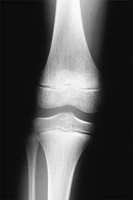
A 35-year-old woman who is a recreational runner reports posterior knee pain and tightness in the knee with flexion during running. She denies any history of trauma. Examination reveals normal patellar glide and tilt and no patellar apprehension. Range of motion is 5 degrees to 120 degrees, and quadriceps function and knee ligamentous examination are normal. Radiographs are normal. An MRI scan is shown in Figure 18. What is the most likely diagnosis?


Explanation

Ganglia involving the cruciate ligaments have been recently reported as a cause of knee pain that interferes with knee flexion and extension. The symptoms are poorly localized in this patient and not along the medial joint line, making the diagnosis of a torn medial meniscus less likely. In addition, the MRI findings do not show a significant medial meniscal lesion. A Baker's cyst is usually posteromedial and extends posterior to the interval between the medial head of the gastrocnemius and semimembranosus. MRI scans show a fluid-filled lesion with an increased signal on T1- and T2-weighted images. A lipoma would be bright on the T1-weighted image only. Deutsch A, Veltri DM, Altchek DW, et al: Symptomatic intraarticular ganglia of the cruciate ligaments of the knee. Arthroscopy 1994;10:219-223.

Question 14

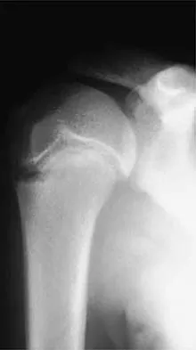
A 12-year-old boy who pitches on two "select" baseball teams has had pain in his dominant right shoulder for the past 6 weeks. The pain is present only with throwing and is associated with decreased throwing velocity and control. He has no radiation of pain or paraesthesias of the upper extremity. An AP radiograph and MRI scan are shown in Figures 19a and 19b, respectively. Management should consist of


Explanation

19b The imaging study demonstrates characteristics of Little Leaguer's shoulder, including physeal widening. This condition is secondary to overuse (typically throwing) and responds well to rest from the inciting activity. There is no evidence from the patient's history or examination that he has an impingement syndrome, nor is there any indication of labral pathology on the MRI scan. The changes in the proximal humerus are classic for this condition and are not suggestive of a neoplastic process requiring biopsy for definitive diagnosis. Lipscomb AB: Baseball pitching injuries in growing athletes. J Sports Med 1975;3:25-34. Cahill BR, Tullos HS, Fain RH: Little league shoulder: Lesions of the proximal humeral epiphyseal plate. J Sports Med 1974;2:150-152.

Question 15

An 18-year-old man underwent open reduction and internal fixation of a tibial spine avulsion and a posterolateral corner repair. Two years later, he underwent lateral collateral ligament (LCL) and posterolateral corner reconstruction because of instability. Examination reveals a pronounced lateral varus knee thrust when ambulating. Varus stress in 30 degrees of flexion produces a 10-mm opening that is eliminated in extension. The Lachman's test is 2 mm with a firm end point, and the posterior drawer test is negative. Standing radiographs show widening of the lateral joint space and a 5-degree mechanical varus alignment. What is the most effective course of treatment?


Explanation

The patient has chronic posterolateral instability with a varus knee alignment; therefore, the most effective treatment is a valgus-producing HTO. A repeat soft-tissue reconstruction without correction of the varus alignment will most likely fail. An ACL reconstruction is not indicated with a normal Lachman's test. Physical therapy and bracing will have little effect. Naudie DD, Amendola A, Fowler PJ: Opening wedge high tibial osteotomy for symptomatic hyperextension-varus thrust. Am J Sports Med 2004;32:60-70.

Question 16

As a baseball player dives to catch a line drive in the outfield, the ball strikes the tip of the player's finger when extended, causing forcible flexion to avulse the extensor tendon from the distal phalanx. Following evaluation and normal radiographic findings, initial management should include


Explanation

Avulsion of the terminal extensor tendon from the distal phalanx (mallet or baseball finger) may or may not be associated with a bony avulsion. The injury is caused by forcible flexion of the DIP joint while catching a ball or hitting an object with the finger extended. Most authorities recommend continuous extension splinting to the DIP joint for 6 weeks, followed by nighttime splinting for an additional 6 weeks. It must be emphasized to the patient that at no time during the initial 6 weeks of treatment should the DIP joint be allowed to fall into flexion or an additional 6 weeks of continuous splinting is required. Miller MD, Cooper DE, Warner JP (eds): Review of Sports Medicine and Arthroscopy. Philadelphia, PA, WB Saunders, 1995, p 255. Rettig AC: Closed tendon injuries of the hand and wrist in the athlete. Clin Sports Med 1992;11:77-99.

Question 17

A favorable outcome following nonsurgical management of a partial tear of the posterior cruciate ligament (PCL) is best associated with


Explanation

Rehabilitation of the quadriceps muscle following a partial tear of the PCL has been associated with a favorable outcome. The quadriceps acts an antagonist to the PCL because its contraction results in anterior tibial translation, which reduces the tensile stress on the injured ligament. Strengthening of the hamstring musculature increases posterior tibial translation and is contraindicated during the early rehabilitative phase following a PCL injury. Brace use has not been found to significantly alter the outcome following nonsurgical management of PCL tears. Parolie JM, Bergfeld JA: Long-term results of nonoperative treatment of isolated posterior cruciate ligament injuries in the athlete. Am J Sports Med 1986;14:35-38.

Question 18

A player on a professional football team sustains a knee injury and is diagnosed with an anterior cruciate ligament rupture. When employed as the team physician, your ethical obligation is to inform


Explanation

When you are employed as a team physician, you are obligated to inform the players and the team organization of all athletically relevant medical issues. This differs significantly from the normal rule of patient confidentiality. If the player came to see you and you were not the team physician, you may not inform the team unless the player so desires. As the team physician, you are not obligated to inform the media. Tucker AM: Ethics and the professional team physician. Clin Sports Med 2004;23:227-241.

Question 19

A 20-year-old basketball player reports a 6-month history of right groin pain that radiates into his testicles with activities of daily living. He denies any history of trauma. Examination reveals tenderness about the groin, and he has full hip range of motion. The abdomen is soft. Radiographs are normal. Nonsurgical management has consisted of rest and physical therapy, but he continues to have pain. What is the next step in management?


Explanation

Sports hernias may be one of the most common causes of groin pain in athletes. Resisted hip adduction is painful in the case of groin disruption. Radiation of pain into the testicles and/or adductor region is often present. Sports hernias are associated with weakening of the posterior inguinal wall. In contrast with sports hernias, traditional or classic hernias can be readily detected on physical examination. Diagnostic imaging studies are not helpful and only serve to help exclude other diagnoses. Systemic high-dose steroids or sacroiliac joint injections have no role in treatment. High success rates have been reported for laparoscopic hernia repair in athletes. Kluin J, den Hoed PT, van Linschoten R, et al: Endoscopic evaluation and treatment of groin pain in the athlete. Am J Sports Med 2004;32:944-949. Genitsaris M, Goulimaris I, Sikas N: Laparoscopic repair of groin pain in athletes. Am J Sports Med 2004;32:1238-1242.

Question 20

A 45-year-old tennis player undergoes surgery for chronic lateral epicondylitis. After returning to play, he notes increasing lateral elbow pain with mechanical catching and locking. Examination shows positive supine posterolateral rotatory instability. What ligament has been injured?


Explanation

The patient has sustained an iatrogenic injury to the lateral ulnar collateral ligament. This injury has been reported after lateral approaches to the elbow. The orbicular, annular, and lateral radial collateral ligaments have a much less important role in lateral elbow stability. The anterior band of the ulnar collateral ligament is on the medial side of the elbow and is important for valgus stability. O'Driscoll SW, Bell DF, Morrey BF: Posterolateral rotatory instability of the elbow. J Bone Joint Surg Am 1991;73:440-446.

Question 21

A female cross-country runner has an insidious onset of right groin pain. Radiographs of the right hip reveal a tension-side stress fracture. History reveals that she was treated for a "foot" fracture 1 year ago. In addition to performing internal fixation of the femoral neck, which of the following should be obtained?


Explanation

Stress fractures in female long distance runners are frequently associated with the Female Athletic Triad. The triad consists of osteoporosis, amenorrhea, and altered eating habits. A thorough menstrual history, including age of menarche, history of amenorrhea, and use of oral contraceptives, is imperative. Amenorrhea leads to osteoporosis and predisposes the athlete to fractures. An MRI of the hip is not necessary because a fracture is evident on the radiograph. Serum calcium levels are normal in osteoporosis, a family history would be noncontributory, and it is highly unlikely that a contralateral hip radiograph will yield useful information. Bennell KL, Malcolm SA, Thomas SA, et al: Risk factors for stress fractures in track and field athletes: A twelve-month prospective study. Am J Sports Med 1996;24:810-818.

Question 22

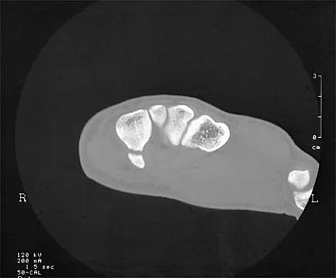
An 18-year-old gymnast has had a 1-year history of foot pain. Examination reveals medial midfoot tenderness without swelling. Non-weight-bearing in a cast for 6 weeks has failed to provide relief. An axial CT scan of the midfoot is shown in Figure 20. What is the optimal treatment for this condition?


Explanation

Stress fractures of the navicular are often seen in running and jumping sports. Whereas most individuals heal with nonsurgical management consisting of 6 weeks of casting, this gymnast has had pain for 1 year and nonsurgical management has failed. Open reduction with bone grafting is the preferred treatment. Quirk RM: Stress fractures of the navicular. Foot Ankle Int 1998;19:494-496.

Question 23

A 20-year-old soccer player who collapsed after a goal kick reports weakness and nausea. He appears slightly confused. Examination reveals that he is not sweating. His skin is warm and dry. The outdoor temperature is 80 degrees F (26.6 degrees C) with a relative humidity of 80%. Management should consist of


Explanation

There is a spectrum of heat-related conditions. Heat cramps are the mildest form of heat illness. In heat exhaustion, cramps are associated with headache and weakness, and the skin is pale and moist. Treatment of heat cramps or heat exhaustion consists of removing and loosening excess clothing, applying ice to the axilla and groin, ingestion of cool water, and cool water sprays. This patient demonstrates symptoms of heat stroke which is a medical emergency. The core body temperature may be as high as 106 to 110 degrees F (41.1 to 43.3 degrees C). In heat stroke, the patient may no longer be sweating, and the skin may be hot and red. The athlete is usually confused, weak, nauseated, and may have seizure activity. Central nervous system depression has been called the most important marker of heat stroke, and progresses from confusion and bizarre behavior to collapse, delirium, and coma. Bizarre behavior is often the first sign of heat stroke. The patient needs to be treated and moved to a medical facility rapidly. During transfer, IV fluids and cooling of the athlete should be initiated. The best treatment of heat-related illness appears to be prevention with adequate hydration and monitoring of conditions (temperature and humidity), with cancellation of competition when conditions do not comply with guidelines. Griffin LY: Emergency preparedness: Things to consider before the game starts. J Bone Joint Surg Am 2005;87:894-902. Barker TA, Motz HA, Gersoff WK: Environmental factors in athletic performance, in Fu FH, Stone DA (eds): Sports Injuries, ed 2. Philadelphia, PA, Lippincott, 2001, pp 67-68.

Question 24

What is the most accurate description of the relationship between gender and knee loading during landing while playing basketball?


Explanation

Ford and associates studied 81 high school basketball players and found that females landed with greater total valgus knee loading and a greater maximum valgus knee angle than male athletes. Hewett and associates reported in a study of 205 female athletes that those with increased dynamic valgus and high abduction loads were at increased risk of anterior cruciate ligament injury. Hewett TE, Myer GD, Ford KR, et al: Biomechanical measures of neuromuscular control and valgus loading of the knee predict anterior cruciate ligament injury risk in female athletes: A prospective study. Am J Sports Med 2005;33:492-501.

Question 25

What is the most common cause of the new onset of amenorrhea in a female endurance athlete who is not sexually active?


Explanation

Insufficient caloric intake caused by either a poor diet or an eating disorder is the most common cause for the loss of menses in a female athlete. In the face of adequate caloric intake, stress is unlikely to cause amenorrhea. Oral contraceptives control menses but do not eliminate it. Diabetes mellitus does not cause the new onset of amenorrhea. Pregnancy can be a cause in a sexually active athlete. Chromosomal abnormalities can result in delayed or absent menarche but not the onset of amenorrhea in a postmenarchal female. Constantini NW: Clinical consequences of amenorrhea. Sports Med 1994;17:213-223.

Question 26

A 45-year-old active female felt a 'pop' in her knee while squatting. MRI reveals a full-thickness tear at the posterior meniscal root.

What biomechanical alteration is most likely present in her knee compared to a normal, uninjured state?


Explanation

Meniscal root tears disrupt the circumferential continuity of the meniscus, leading to a complete loss of hoop stresses. This allows the meniscus to extrude radially under axial loads. Biomechanical studies have demonstrated that a posterior medial meniscus root tear increases peak contact pressures and decreases contact area in the medial compartment, making it biomechanically equivalent to a total meniscectomy.

Question 27

A 24-year-old minor league pitcher presents with posterior shoulder pain during the late cocking phase of throwing. Physical examination reveals a 25-degree loss of internal rotation at 90 degrees of abduction compared to the contralateral side, while external rotation is increased by 10 degrees. What is the most appropriate initial management?


Explanation

This patient exhibits Glenohumeral Internal Rotation Deficit (GIRD), which is commonly seen in overhead throwing athletes. It is characterized by posterior capsular contracture and a loss of internal rotation. A true GIRD is defined as an internal rotation deficit of >20 degrees compared to the contralateral shoulder with a loss of total arc of motion. The initial management for symptomatic GIRD is a targeted physical therapy program focusing on stretching the posterior capsule, utilizing sleeper stretches and cross-body adduction stretches. Operative management is rarely indicated unless prolonged non-operative management fails.

Question 28

A 28-year-old bodybuilder feels a tearing sensation in his anterior chest while performing a heavy bench press. Examination reveals a loss of the anterior axillary fold and significant weakness with internal rotation. Which portion of the pectoralis major is most commonly injured in this mechanism, and what is its anatomic footprint on the humerus?


Explanation

Pectoralis major ruptures most commonly occur at the musculotendinous junction or tendinous insertion of the sternocostal (sternal) head during an eccentric contraction, such as a heavy bench press. The anatomy of the pectoralis major tendon is unique: the tendon twists 180 degrees before inserting on the lateral lip of the bicipital groove. Because of this twist, the lower (sternal) fibers insert superior and deep to the upper (clavicular) fibers.

Question 29

A 21-year-old football player sustains a direct blow to the anteromedial aspect of his knee. Physical examination shows 15 degrees of increased external rotation at 30 degrees of knee flexion, but symmetric external rotation at 90 degrees of knee flexion compared to the uninjured side.

Which structure is most likely injured?


Explanation

The clinical exam describes the dial test. A positive dial test is defined as >10 degrees of increased external rotation compared to the contralateral knee. If the test is positive at 30 degrees of flexion but symmetric (negative) at 90 degrees, it indicates an isolated posterolateral corner (PLC) injury. If it is positive at both 30 and 90 degrees, it indicates a combined PCL and PLC injury.

Question 30

A 22-year-old hockey player complains of insidious onset groin pain exacerbated by hip flexion and internal rotation. Anteroposterior radiographs of the pelvis reveal a lateral center-edge angle (LCEA) of 45 degrees and a positive crossover sign. What is the most likely diagnosis?


Explanation

Pincer-type femoroacetabular impingement (FAI) is caused by focal or global overcoverage of the femoral head by the acetabulum. Radiographic findings diagnostic of pincer FAI include a lateral center-edge angle >39 degrees, acetabular retroversion (indicated by a crossover sign or ischial spine sign), or coxa profunda/protrusio acetabuli. Cam impingement is defined by a lack of femoral head-neck offset, often quantified by an alpha angle >55 degrees.

Question 31

A 25-year-old cyclist falls directly onto his shoulder. Radiographs demonstrate 100% superior displacement of the clavicle relative to the acromion, with the coracoclavicular (CC) distance increased by 50% compared to the contralateral side. A diagnosis of a Type III acromioclavicular (AC) joint separation is made. Which of the following accurately describes the anatomy of the native CC ligaments?


Explanation

The coracoclavicular (CC) ligaments consist of the conoid and trapezoid. The conoid is situated medial and posterior and is the primary restraint to superior displacement of the clavicle. The trapezoid is situated lateral and anterior and is the primary restraint to axial compression (preventing the scapula from moving medially). Understanding this footprint is crucial for anatomic CC ligament reconstruction.

Question 32

A 19-year-old collegiate runner complains of bilateral anterolateral leg pain that reliably begins 15 minutes into her runs and subsides 30 minutes after resting. To confirm the diagnosis of chronic exertional compartment syndrome (CECS), intracompartmental pressures are measured. According to the modified Pedowitz criteria, which of the following post-exercise measurements is diagnostic?


Explanation

The Pedowitz criteria for diagnosing chronic exertional compartment syndrome require one or more of the following intracompartmental pressure criteria: a pre-exercise (resting) pressure > 15 mm Hg, a 1-minute post-exercise pressure > 30 mm Hg, or a 5-minute post-exercise pressure > 20 mm Hg. Therefore, a 1-minute post-exercise pressure > 30 mm Hg meets the diagnostic criteria.

Question 33

During a double-bundle anterior cruciate ligament (ACL) reconstruction, precise knowledge of bundle anatomy and biomechanics is required. Which of the following statements regarding the anteromedial (AM) and posterolateral (PL) bundles of the ACL is correct?


Explanation

The ACL is composed of the anteromedial (AM) and posterolateral (PL) bundles. The AM bundle tightens in flexion and is the primary restraint to anterior tibial translation. The PL bundle tightens in extension and is the primary restraint to rotatory loads (e.g., positive pivot shift). During isolated single-bundle reconstruction, surgeons generally target the center of the footprint or slightly toward the AM bundle position to optimize AP stability.

Question 34

A 21-year-old collegiate baseball pitcher presents with medial elbow pain and decreased pitching velocity. MRI confirms a full-thickness tear of the anterior band of the ulnar collateral ligament (UCL).

During surgical reconstruction of the UCL utilizing the modern docking technique, how is the ulnar nerve typically managed?


Explanation

In the original Jobe technique for UCL reconstruction, the flexor-pronator mass was detached, and a routine ulnar nerve transposition was performed. Modern techniques (such as the docking or modified Jobe technique) utilize a muscle-splitting approach through the flexor carpi ulnaris (FCU). This allows for adequate visualization of the sublime tubercle while leaving the ulnar nerve safely in situ, provided the patient does not have preoperative ulnar nerve symptoms (e.g., cubital tunnel syndrome).

Question 35

A 16-year-old dancer undergoes surgical reconstruction of the medial patellofemoral ligament (MPFL) for recurrent lateral patellar instability. To avoid non-anatomic graft placement, which can result in patellofemoral arthrosis or graft failure, where should the femoral footprint of the MPFL be anatomically positioned?


Explanation

The anatomic femoral origin of the MPFL resides in a saddle-like depression located distal to the adductor tubercle, proximal and posterior to the medial epicondyle, and superficial to the superficial MCL origin. Radiographically, Schottle's point describes this optimal femoral attachment: 1 mm anterior to the posterior cortex line, 2.5 mm distal to the posterior origin of the medial femoral condyle, and proximal to the posterior point of Blumensaat's line.

Question 36

A 24-year-old male presents with loss of knee flexion 6 months after an endoscopic anterior cruciate ligament (ACL) reconstruction using a bone-patellar tendon-bone autograft. Radiographs and an MRI are evaluated.

Which of the following technical errors during graft placement most likely caused this specific complication?


Explanation

An anteriorly placed femoral tunnel creates an ACL graft that is inappropriately tight in flexion and loose in extension, leading to a loss of flexion. Conversely, a tibial tunnel placed too anteriorly leads to graft impingement against the intercondylar notch in extension, leading to a loss of extension.

Question 37

A 19-year-old female collegiate swimmer presents with bilateral shoulder pain and a feeling of joint 'looseness'. Physical examination reveals a positive sulcus sign that does not reduce with external rotation, and apprehension with both anterior and posterior translation. She has failed 6 months of supervised physical therapy. If surgical intervention is planned, what is the most appropriate procedure?


Explanation

The patient has multidirectional instability (MDI) failing conservative management, which is the gold standard initial treatment. Surgical management typically involves reducing capsular volume. Arthroscopic capsular plication (or open inferior capsular shift) is the procedure of choice. Thermal capsulorrhaphy is historical and has high failure and complication rates. Bankart or posterior repairs alone do not address the global capsular redundancy unless a specific labral tear is identified. A sulcus sign that does not reduce with external rotation indicates an incompetent rotator interval.

Question 38

A 50-year-old active male hears a 'pop' in his posterior knee while descending stairs. He presents with posteromedial joint line tenderness. MRI demonstrates a medial meniscal extrusion of 4 mm and a radial tear adjacent to the posterior horn medial meniscus tibial attachment. What is the most likely biomechanical consequence of this injury if left untreated?


Explanation

Posterior root tears of the medial meniscus lead to a complete loss of hoop stresses, effectively mimicking the biomechanical state of a total medial meniscectomy. This drastically increases peak contact pressures in the medial compartment and is highly associated with rapid progression of osteoarthritis and subchondral insufficiency fractures.

Question 39

A 42-year-old weightlifter feels a sudden pop in his right antecubital fossa while performing a deadlift. On examination, he has weakness in forearm supination and elbow flexion. The 'hook test' is positive. During surgical repair through a single anterior incision, which of the following nerves is at greatest risk of injury?


Explanation

The lateral antebrachial cutaneous nerve (LABCN) is the most commonly injured nerve during a single-incision anterior approach for distal biceps tendon repair. The radial nerve or posterior interosseous nerve (PIN) is more at risk during a two-incision approach (specifically the posterolateral incision) or if retractors are placed too vigorously on the radial side in a single incision.

Question 40

A 28-year-old competitive bodybuilder sustains an acute injury to his chest while performing a heavy bench press. Examination reveals ecchymosis over the anterior axillary fold and a palpable defect. Which of the following correctly describes the most common anatomic location and tissue involved in this injury?


Explanation

Pectoralis major ruptures most commonly occur during weightlifting (e.g., bench press). The most common pattern is an avulsion of the tendon of the sternocostal head from its insertion on the humerus. The sternocostal head is under maximal tension when the arm is extended, abducted, and externally rotated (the bottom of a bench press).

Question 41

A 21-year-old collegiate baseball pitcher presents with vague posterior shoulder pain during the late cocking and early acceleration phases of throwing. Physical exam reveals a 25-degree loss of internal rotation compared to the contralateral side, with normal total arc of motion. He has localized tenderness at the posterior joint line. Which of the following is the most appropriate initial management?


Explanation

The patient has Glenohumeral Internal Rotation Deficit (GIRD) symptomatic of a tight posteroinferior capsule. In the throwing athlete, GIRD is defined as a loss of internal rotation >20 degrees compared to the non-throwing shoulder, often with a preserved total arc of motion due to compensatory increased external rotation. The initial treatment is a dedicated physical therapy program utilizing 'sleeper stretches' to stretch the posteroinferior capsule. Surgery is only considered if prolonged nonoperative management fails.

Question 42

A 28-year-old professional volleyball player presents with an insidious onset of right shoulder weakness and vague posterior shoulder pain. Physical examination demonstrates isolated weakness in external rotation. Internal rotation and forward elevation are 5/5. There is noticeable atrophy of the infraspinatus fossa, while the supraspinatus fossa appears normal. MRI reveals a paralabral cyst. Where is the cyst most likely located?


Explanation

The clinical presentation of isolated infraspinatus atrophy and external rotation weakness indicates compression of the suprascapular nerve at the spinoglenoid notch, distal to the innervation of the supraspinatus. Compression at the suprascapular notch would affect both the supraspinatus and infraspinatus muscles, leading to weakness in both abduction and external rotation. Paralabral cysts at the spinoglenoid notch are often associated with posterior superior labral tears.

Question 43

A 14-year-old elite female gymnast presents with lateral elbow pain and catching. Radiographs reveal a radiolucent lesion in the capitellum. MRI demonstrates an osteochondral lesion with a high T2 signal line behind the bone fragment, and an associated loose body in the anterior compartment. What is the most appropriate definitive management?


Explanation

The patient has an unstable osteochondral defect (OCD) of the capitellum, indicated by catching, a high T2 signal line behind the fragment (indicating fluid and instability), and an intra-articular loose body. Conservative management (rest) is indicated for stable lesions with an open capitellar physis. For unstable lesions or those with loose bodies, surgical intervention is required. Arthroscopic loose body removal and debridement/microfracture of the base is the standard of care for fragments that are completely detached or unsuitable for fixation.

Question 44

A 17-year-old female undergoes medial patellofemoral ligament (MPFL) reconstruction for recurrent lateral patellar instability. To ensure isometry of the graft, the femoral attachment must be placed precisely. In terms of anatomic landmarks on the medial femur, where is the origin of the MPFL located?


Explanation

The anatomic origin of the MPFL is located in a saddle-shaped groove between the medial epicondyle and the adductor tubercle. On a true lateral radiograph, Schottle's point defines this radiographic location: 1 mm anterior to the posterior cortical line, 2.5 mm distal to the posterior origin of the medial femoral condyle, and proximal to the level of the posterior portion of Blumensaat's line.

Question 45

A 25-year-old professional hockey player sustains a direct blow to the point of his shoulder. Radiographs demonstrate an acromioclavicular (AC) joint injury with the clavicle displaced 150% superiorly relative to the acromion. There is palpable trapezius and deltoid fascia stripping. Which of the following is the classification and recommended management for this injury?


Explanation

The injury described is a Type V acromioclavicular (AC) joint separation, characterized by 100% to 300% superior displacement of the clavicle relative to the acromion and extensive stripping of the deltotrapezial fascia. Type III injuries have 25% to 100% displacement and are typically treated non-operatively initially, except in certain high-demand overhead athletes. Type V injuries are highly symptomatic, alter shoulder biomechanics significantly, and generally require early surgical reconstruction of the coracoclavicular ligaments.

Question 46

A 19-year-old collegiate soccer player undergoes anterior cruciate ligament (ACL) reconstruction using an anteromedial portal technique for femoral tunnel drilling. To avoid a critically short femoral tunnel and prevent posterior cortical blowout, at what approximate knee flexion angle should the femoral tunnel be drilled?


Explanation

When drilling the femoral tunnel through an anteromedial (AM) portal during ACL reconstruction, the knee must be hyperflexed (typically 120 degrees or more). This maneuver changes the trajectory of the drill in relation to the femur, ensuring a longer femoral tunnel and minimizing the risk of posterior cortical blowout. Drilling at 90 degrees or less via the AM portal typically results in a short tunnel and a high risk of violating the posterior femoral cortex.

Question 47

A 25-year-old professional baseball pitcher complains of posterior shoulder pain during the late cocking phase of throwing. Physical examination reveals a loss of internal rotation of 25 degrees compared to the contralateral side, with normal total arc of motion. What is the most appropriate initial management for this patient's condition?


Explanation

The patient is presenting with Glenohumeral Internal Rotation Deficit (GIRD), common in overhead throwing athletes due to contracture of the posteroinferior capsule. The hallmark is a loss of internal rotation with a corresponding gain in external rotation, maintaining a normal total arc of motion. The initial and most effective treatment is a conservative physical therapy regimen focusing on stretching the posterior capsule, specifically utilizing the 'sleeper stretch'.

Question 48

A 20-year-old female presents with recurrent lateral patellar instability and has failed conservative management. A medial patellofemoral ligament (MPFL) reconstruction is planned. Which of the following best describes the anatomical origin of the MPFL on the femur?


Explanation

The femoral footprint of the MPFL is situated in a 'saddle' area that is proximal and posterior to the medial epicondyle, and distal to the adductor tubercle. Radiographically, this is described by Schöttle's point: 1 mm anterior to the posterior cortical line, 2.5 mm distal to the posterior intersecting line of the femoral condyle, and proximal to Blumensaat's line.

Question 49

A 28-year-old hockey player undergoes hip arthroscopy for a symptomatic CAM lesion (femoroacetabular impingement). Following the osteochondroplasty of the femoral head-neck junction, what complication is significantly increased if the resection depth exceeds 30% of the femoral neck diameter?


Explanation

During osteochondroplasty for a CAM lesion, resection of the anterolateral femoral head-neck junction is performed. Biomechanical studies have demonstrated that resecting greater than 30% of the femoral neck diameter significantly alters the load-bearing capacity of the proximal femur, drastically increasing the risk of a post-operative femoral neck fracture.

Question 50

A 32-year-old recreational athlete sustains an acute Achilles tendon rupture. Based on recent Level I evidence comparing operative repair to nonoperative management with an early functional rehabilitation protocol, what is the expected outcome?


Explanation

Recent high-level evidence (such as the Willits et al. trial and subsequent meta-analyses) has demonstrated that when nonoperative management is paired with an early functional rehabilitation protocol (early weight-bearing and ROM in a functional brace), the re-rupture rates are equivalent to operative repair. However, operative management carries a higher risk of complications, particularly superficial and deep infections, and sural nerve injury.

Question 51

A 24-year-old cyclist falls directly onto his shoulder. Clinical examination demonstrates a prominence of the distal clavicle. Radiographs confirm a Type III acromioclavicular (AC) joint separation. Which structure is the primary restraint to anterior-posterior translation of the distal clavicle?


Explanation

The primary stabilizer against anterior-posterior translation of the clavicle relative to the acromion is the acromioclavicular (AC) joint capsule and its intrinsic ligaments (specifically the superior and posterior AC ligaments). The coracoclavicular (CC) ligaments (conoid and trapezoid) are the primary restraints to superior-inferior translation.

Question 52

A 45-year-old woman experiences a 'pop' in the back of her knee while squatting. MRI reveals a complete radial tear of the posterior horn of the medial meniscus at its root attachment. If left untreated, the alteration in knee joint biomechanics most closely mimics which of the following conditions?


Explanation

A posterior horn medial meniscal root tear disrupts the circumferential hoop stresses of the meniscus. Biomechanical studies have shown that a root tear leads to meniscal extrusion and alters contact areas and peak contact pressures in the medial compartment to a degree that is functionally equivalent to a total medial meniscectomy. This leads to rapid progression of osteoarthritis if not repaired.

Question 53

A 14-year-old male presents with vague medial knee pain. Radiographs and MRI demonstrate an osteochondritis dissecans (OCD) lesion on the lateral aspect of the medial femoral condyle. The physis is wide open, and the MRI shows intact overlying cartilage with no fluid behind the lesion.

What is the most appropriate initial treatment?


Explanation

Juvenile osteochondritis dissecans (OCD) in a patient with open physes and a stable lesion (intact cartilage, no fluid behind the fragment on MRI) has a high healing potential with conservative management. The initial treatment of choice is nonoperative, focusing on activity modification, restricted weight-bearing, and immobilization if symptomatic.

Question 54

A 65-year-old man presents with chronic, profound shoulder weakness. MRI demonstrates a massive, retracted tear of the supraspinatus and infraspinatus tendons with Goutallier stage 4 fatty infiltration.

During attempted arthroscopic mobilization and lateral traction of these chronically retracted tendons, which neurologic structure is at greatest risk of stretch injury?


Explanation

The suprascapular nerve innervates the supraspinatus and infraspinatus muscles. It is relatively fixed at the suprascapular notch and the spinoglenoid notch. In the setting of a massive, chronically retracted rotator cuff tear, the muscle belly shortens. Aggressive lateral traction during mobilization or repair places significant tension on the suprascapular nerve, increasing the risk of a traction neuropraxia.

Question 55

A 21-year-old collegiate baseball pitcher is undergoing an ulnar collateral ligament (UCL) reconstruction utilizing an autograft.

Which native anatomical structure is the primary restraint to valgus stress at the elbow during the late cocking and early acceleration phases of throwing?


Explanation

The anterior bundle of the ulnar collateral ligament (UCL) is the primary restraint to valgus stress at the elbow, particularly between 30 and 120 degrees of flexion, which corresponds to the late cocking and early acceleration phases of throwing. The posterior bundle is a secondary restraint, and the transverse ligament provides no significant stability.

Question 56

A 24-year-old male is 3 months post-operative from an anterior cruciate ligament (ACL) reconstruction using a bone-patellar tendon-bone autograft. He complains of a painful clunk and inability to fully extend the knee. An MRI shows a nodular mass anterior to the ACL graft. What is the most likely diagnosis and appropriate next step in management?


Explanation

A cyclops lesion is a localized form of anterior arthrofibrosis that occurs after ACL reconstruction. It typically presents with a loss of terminal extension and a painful clunk at terminal extension as the fibrotic nodule gets trapped between the femur and tibia. MRI classically demonstrates a soft-tissue nodule anterior to the tibial insertion of the ACL graft. The definitive treatment is arthroscopic excision, which generally restores full extension and resolves symptoms.

Question 57

A 28-year-old soccer player sustains a twisting knee injury. On physical examination, the Dial test reveals 15 degrees of increased external rotation at 30 degrees of knee flexion compared to the contralateral side. However, at 90 degrees of knee flexion, the external rotation is symmetric bilaterally. Which of the following structures is most likely injured?


Explanation

The Dial test evaluates external rotation of the tibia relative to the femur and is used to assess the integrity of the posterolateral corner (PLC) and the posterior cruciate ligament (PCL). An increase of more than 10 degrees of external rotation compared to the normal side at 30 degrees of flexion, but not at 90 degrees, indicates an isolated injury to the PLC (which includes the lateral collateral ligament, popliteus tendon, and popliteofibular ligament). If the test is positive at both 30 and 90 degrees, it suggests a combined PCL and PLC injury.

Question 58

A 22-year-old rugby player undergoes an open Latarjet procedure for recurrent anterior shoulder instability with 25% glenoid bone loss. Postoperatively, he exhibits weakness in elbow flexion and supination, along with decreased sensation over the lateral aspect of the forearm. Which nerve was most likely injured during the procedure?


Explanation

The musculocutaneous nerve is at significant risk during the Latarjet procedure (coracoid transfer) due to its proximity to the conjoined tendon (coracobrachialis and short head of biceps). It typically penetrates the coracobrachialis 5 to 8 cm distal to the coracoid process. Injury to this nerve leads to weakness in elbow flexion (biceps, brachialis) and supination (biceps), as well as sensory loss in the distribution of the lateral antebrachial cutaneous nerve (lateral forearm).

Question 59

A 26-year-old male hockey player presents with chronic groin pain exacerbated by hip flexion and internal rotation. Radiographs show an alpha angle of 65 degrees and a positive crossover sign.

The patient's radiographic findings are most consistent with which of the following?


Explanation

An increased alpha angle (>50-55 degrees) is indicative of a Cam-type morphology (aspherical femoral head-neck junction). The crossover sign indicates focal cranial retroversion of the acetabulum, which is a classic radiographic marker of Pincer-type impingement. The combination of both an elevated alpha angle and a crossover sign indicates mixed femoroacetabular impingement (FAI), which is the most common clinical presentation of FAI.

Question 60

A 32-year-old professional basketball player presents with a symptomatic full-thickness focal chondral defect on the weight-bearing surface of the medial femoral condyle. The lesion measures 3.5 cm in diameter. He has failed conservative management and desires to return to high-impact sports. What is the most appropriate surgical intervention for this specific lesion?


Explanation

For large (>2 to 3 cm^2) full-thickness chondral or osteochondral defects in young, high-demand patients, fresh osteochondral allograft transplantation or Matrix-induced Autologous Chondrocyte Implantation (MACI) are the preferred treatments. Microfracture and OATS (autograft) are generally reserved for smaller lesions (<2 cm^2) due to the poorer biomechanical properties of fibrocartilage (microfracture) and donor site morbidity (OATS). Given the 3.5 cm diameter, fresh osteochondral allograft is highly indicated.

Question 61

A 21-year-old collegiate baseball pitcher presents with medial elbow pain and decreased pitching velocity. MRI confirms a full-thickness tear of the anterior bundle of the ulnar collateral ligament (UCL). He undergoes a UCL reconstruction utilizing a palmaris longus autograft via the modified Jobe technique. During the exposure and preparation of the medial epicondyle for the humeral tunnels, what structure is at greatest risk of iatrogenic injury and must be meticulously protected?


Explanation

The ulnar nerve runs directly posterior to the medial epicondyle in the cubital tunnel and is at significant risk during UCL reconstruction. Whether the surgeon performs a routine ulnar nerve transposition or leaves it in situ, the nerve must be meticulously identified and protected, particularly during the creation of the humeral tunnels in the medial epicondyle.

Question 62

A 45-year-old male sustains an acute posterior root tear of the medial meniscus while performing a deep squat.

Biomechanical studies have demonstrated that if this lesion is left untreated, the resultant changes in knee contact pressures are most equivalent to which of the following?


Explanation

The posterior root of the medial meniscus anchors the meniscus to the tibial plateau, allowing it to convert axial loads into hoop stresses. A complete radial tear at or near the root disrupts these hoop stresses entirely, leading to meniscal extrusion. Biomechanical studies have shown that the peak contact pressures and contact areas in a knee with a medial meniscus root tear are biomechanically equivalent to those in a knee that has undergone a total medial meniscectomy.

Question 63

A 28-year-old recreational volleyball player presents with deep shoulder pain and clicking. An MR arthrogram demonstrates a SLAP tear characterized by a bucket-handle tear of the superior labrum that extends into the long head of the biceps tendon. According to the Snyder classification, what type of SLAP tear is this?


Explanation

According to the Snyder classification of SLAP (Superior Labrum Anterior and Posterior) tears: Type I is superior labral fraying with an intact biceps anchor. Type II is detachment of the superior labrum and biceps anchor from the superior glenoid. Type III is a bucket-handle tear of the superior labrum with an intact biceps anchor. Type IV is a bucket-handle tear of the superior labrum that extends into the biceps tendon.

Question 64

A 17-year-old female is undergoing a medial patellofemoral ligament (MPFL) reconstruction for recurrent lateral patellar instability.

Intraoperative fluoroscopy is used to identify the anatomic femoral attachment of the MPFL (Schöttle's point). Which of the following radiographic descriptions best defines this exact location on a true lateral radiograph?


Explanation

Schöttle's point is the radiographic landmark for the femoral origin of the MPFL on a true lateral radiograph. It is defined geometrically as: 1 mm anterior to a line extending the posterior cortex of the femoral shaft, 2.5 mm distal to a perpendicular line intersecting the posterior origin of the medial femoral condyle articular surface, and proximal to a perpendicular line intersecting the posterior extent of Blumensaat's line.

Question 65

A 16-year-old male sprinter feels a sudden 'pop' and experiences severe pain in his buttock during a 100-meter dash. Radiographs demonstrate an avulsion fracture of the ischial tuberosity with 3.5 cm of displacement. Which of the following muscles or muscle groups is primarily responsible for the displacement of this fracture?


Explanation

The ischial tuberosity is the anatomic origin of the hamstring muscle complex (long head of the biceps femoris, semitendinosus, and semimembranosus). An avulsion fracture of the ischial tuberosity in a skeletally immature athlete is typically caused by a sudden, forceful eccentric contraction of the hamstrings. The rectus femoris originates at the AIIS, the sartorius at the ASIS, and the iliopsoas inserts at the lesser trochanter.

Question 66

A 24-year-old male presents with stiffness and loss of terminal knee flexion 6 months after an anterior cruciate ligament (ACL) reconstruction using a bone-patellar tendon-bone autograft. Radiographs show the femoral tunnel positioned too anteriorly in the intercondylar notch. What is the primary clinical consequence of this specific tunnel malposition?


Explanation

Non-anatomic graft placement is a leading cause of ACL reconstruction failure and stiffness. A femoral tunnel placed too anteriorly (high in the notch) results in the graft being overtensioned as the knee goes into flexion, functionally capturing the joint and causing a loss of terminal knee flexion. Conversely, a tibial tunnel placed too anteriorly leads to graft impingement against the intercondylar roof during extension, resulting in a loss of terminal knee extension.

Question 67

A 28-year-old soccer player sustains a twisting knee injury. Physical examination reveals a positive dial test at 30 degrees of knee flexion, but symmetrical external rotation at 90 degrees of knee flexion compared to the uninjured contralateral knee. Which of the following injury patterns is most consistent with these clinical findings?


Explanation

The dial test is used to evaluate injury to the posterolateral corner (PLC) and the posterior cruciate ligament (PCL). An increase in external rotation of more than 10 degrees compared with the normal knee at 30 degrees of flexion, but not at 90 degrees, is indicative of an isolated PLC injury. If external rotation is increased at both 30 and 90 degrees of flexion, it indicates a combined injury to both the PLC and the PCL.

Question 68

A 20-year-old rugby player undergoes an open Latarjet procedure for recurrent anterior shoulder instability with 25% glenoid bone loss. Postoperatively, the patient exhibits weakness with elbow flexion and forearm supination, accompanied by numbness over the lateral aspect of his forearm. Which nerve is most likely to have been injured during the retraction of the conjoint tendon?


Explanation

The musculocutaneous nerve is highly vulnerable during the Latarjet procedure, particularly during the mobilization and medial retraction of the conjoint tendon. It typically penetrates the coracobrachialis muscle 3 to 8 cm distal to the coracoid process. Injury to this nerve leads to denervation of the biceps brachii and brachialis (causing weakness in elbow flexion and supination) and sensory deficits in the lateral antebrachial cutaneous nerve distribution.

Question 69

When performing a medial patellofemoral ligament (MPFL) reconstruction for recurrent patellofemoral instability, identifying the exact isometric femoral attachment point is critical to avoid overtensioning the graft during flexion. Radiographically, where is the anatomic femoral origin of the MPFL (Schöttle's point) located?


Explanation

The anatomic femoral insertion of the MPFL is located in a distinct saddle region that lies strictly between the adductor tubercle (proximal) and the medial epicondyle (distal). Schöttle et al. described this radiographically on a true lateral x-ray as 1 mm anterior to a line extending the posterior femoral cortex, 2.5 mm distal to the posterior origin of the medial femoral condyle, and proximal to the level of the posterior point of the Blumensaat line. Positioning the femoral tunnel non-anatomically, particularly too far proximally, will inappropriately overtension the graft during knee flexion.

Question 70

A 22-year-old elite collegiate baseball pitcher presents with vague posterior shoulder pain, a 'dead arm' sensation, and a decrease in pitching velocity.

He is diagnosed with a Type II superior labrum anterior and posterior (SLAP) tear. What biomechanical mechanism is primarily responsible for the propagation of this specific lesion during the throwing cycle?


Explanation

Type II SLAP tears in overhead throwing athletes are primarily driven by the 'peel-back' mechanism, originally described by Burkhart and Morgan. During the late cocking phase, the arm is in a position of maximal abduction and external rotation. This shifts the vector of the biceps tendon posteriorly, generating significant torsional 'peel-back' forces at the superior labrum, causing it to detach from the superior glenoid rim.

Question 71

A 24-year-old professional baseball pitcher undergoes ulnar collateral ligament (UCL) reconstruction utilizing the docking technique. Concurrently, the surgeon addresses concomitant valgus extension overload (VEO) syndrome. To avoid catastrophic failure of the newly reconstructed UCL, the surgeon must exercise extreme caution to prevent which of the following errors?


Explanation

Valgus extension overload (VEO) leads to osteophyte formation at the posteromedial olecranon due to abutment in the olecranon fossa. However, the olecranon acts as a critical bony restraint to valgus stress. Resection of more than 3 mm of the posteromedial olecranon drastically alters this bony constraint, significantly increasing the strain transferred to the anterior bundle of the UCL (or the newly placed graft), predisposing the patient to early graft failure and recurrent valgus instability.

Question 72

A 21-year-old collegiate hockey player complains of deep anterior groin pain exacerbated by hip flexion, adduction, and internal rotation (FADIR test).

An AP pelvis radiograph demonstrates a prominent 'crossover sign.' What specific morphological abnormality is most closely associated with this radiographic finding?


Explanation

The 'crossover sign' on a properly aligned anteroposterior (AP) pelvis radiograph represents the anterior wall of the acetabulum crossing over the posterior wall before reaching the lateral sourcil. It is the hallmark radiographic indicator of acetabular retroversion, which results in focal anterior overcoverage and predisposes the patient to pincer-type femoroacetabular impingement (FAI).

Question 73

A 45-year-old recreational runner sustains a sudden pop in the posterior aspect of his knee while descending stairs. MRI confirms a complete radial tear immediately adjacent to the medial meniscus posterior root attachment. If managed conservatively, the knee biomechanics will be altered. The resulting tibiofemoral contact mechanics are most equivalent to which of the following conditions?


Explanation

The posterior root anchors the medial meniscus, allowing it to convert axial loads into circumferential hoop stresses. A complete tear of the meniscal root disrupts this structural continuity, resulting in meniscal extrusion. Biomechanical studies have demonstrated that a medial meniscus posterior root tear leads to a complete loss of meniscal load-sharing ability, causing peak contact pressures in the medial compartment to increase to levels functionally equivalent to those seen after a total medial meniscectomy, accelerating the onset of osteoarthritis.

Question 74

A 26-year-old professional mountain biker falls directly onto his right shoulder. Clinical examination reveals an irreducible, posterior displacement of the distal clavicle.

Radiographs confirm a posterior dislocation of the clavicle relative to the acromion on the axillary lateral view. Which Rockwood classification and optimal treatment paradigm applies to this injury?


Explanation

This describes a Rockwood Type IV acromioclavicular (AC) joint injury. Type IV injuries are characterized by posterior displacement of the distal clavicle into or through the trapezius muscle fascia. Unlike Types I, II, and many Type III injuries which can be managed nonoperatively, Type IV injuries generally require surgical reduction and stabilization due to the severe soft tissue interposition and static non-reducibility that prevents adequate ligamentous healing.

Question 75

A 13-year-old male gymnast complains of intermittent right knee swelling, pain, and mechanical catching.

Radiographs demonstrate a classic presentation of osteochondritis dissecans (OCD) in the knee. What is the most common anatomic location for this pathology?


Explanation

Osteochondritis dissecans (OCD) of the knee predominantly affects the femoral condyles. By far the most common location, accounting for roughly 70-80% of all cases, is the lateral aspect of the medial femoral condyle (often remembered by the acronym LAME - Lateral Aspect Medial Epicondyle/Condyle). This is thought to be related to repetitive microtrauma from the tibial spine impinging upon the condyle during internal tibial rotation.

Question 76

During an anatomic single-bundle anterior cruciate ligament (ACL) reconstruction, identifying the native footprint is critical. With the knee in 90 degrees of flexion, the native ACL femoral footprint is located immediately posterior to which of the following arthroscopic bony landmarks?


Explanation

The lateral intercondylar ridge, also known as resident's ridge, is the most consistent and reliable anatomic landmark for identifying the anterior border of the ACL on the lateral femoral condyle. The entire ACL femoral footprint is located posterior to this ridge. The lateral bifurcate ridge separates the anteromedial (AM) and posterolateral (PL) bundles of the ACL, but it is less consistently identified than the lateral intercondylar ridge.

Question 77

A 50-year-old female presents with acute medial knee pain and a popping sensation after squatting. MRI reveals a posterior medial meniscus root tear. Biomechanical studies have shown that a complete medial meniscus posterior root tear alters knee joint kinematics most similarly to which of the following?


Explanation

A complete tear of the posterior root of the medial meniscus leads to the complete loss of circumferential hoop stresses within the meniscus, resulting in meniscal extrusion under load. Biomechanical cadaveric studies have demonstrated that this increases peak contact pressures and decreases contact area in the medial compartment to levels equivalent to those seen after a total medial meniscectomy. This accelerates the progression of osteoarthritis if left untreated.

Question 78

A 19-year-old collegiate baseball pitcher presents with medial elbow pain during the late cocking and early acceleration phases of throwing.

Based on the history, physical exam, and imaging, a decision is made to perform a Ulnar Collateral Ligament (UCL) reconstruction using the docking technique. What is the primary biomechanical and surgical advantage of the docking technique compared to the classic figure-of-eight (Jobe) technique?


Explanation

The docking technique for UCL reconstruction involves securing the two ends of the graft into a single bony tunnel in the medial epicondyle. This minimizes the amount of bone removed and reduces the number of drill holes required compared to the classic figure-of-eight (Jobe) technique, which requires multiple intersecting tunnels. This substantially reduces the risk of iatrogenic medial epicondyle fracture while providing excellent biomechanical fixation.

Question 79

A 28-year-old weightlifter feels a sharp "pop" and tearing sensation in his anterior axilla while performing a heavy bench press. Physical examination reveals loss of the anterior axillary fold and weakness in internal rotation. Operative exploration is planned. Which portion of the pectoralis major is most commonly injured in this scenario, and what is its correct anatomic insertion on the humerus relative to the other head?


Explanation

The pectoralis major tendon undergoes a 180-degree twist before inserting onto the lateral lip of the bicipital groove. Because of this twist, the lower (sternal) fibers insert most proximally and posteriorly, while the upper (clavicular) fibers insert distally and anteriorly. The sternal head is under maximal tension when the arm is extended and abducted (such as at the bottom of a bench press) and is therefore the most commonly ruptured segment in weightlifting injuries.

Question 80

A 24-year-old hockey player underwent right hip arthroscopy for femoroacetabular impingement (cam and pincer resection with labral repair) 3 weeks ago.

He now complains of numbness over the dorsum of his right foot and difficulty extending his toes. Which of the following intraoperative factors most likely contributed to this specific complication?


Explanation

The patient's symptoms (dorsal foot numbness, weak toe extension) are classic for a common peroneal nerve neuropraxia, which is a branch of the sciatic nerve. During hip arthroscopy, prolonged traction places the pudendal and sciatic nerves at significant risk for neuropraxia. It is generally recommended to limit traction time to less than 2 hours and use a traction force of less than 50 pounds. Damage to the lateral femoral cutaneous nerve (often during anterolateral portal placement) would cause anterolateral thigh numbness, not foot symptoms.

Question 81

A 17-year-old female experiences recurrent lateral patellar instability, and a medial patellofemoral ligament (MPFL) reconstruction is planned.

To maintain proper graft isometry, the femoral tunnel must be placed accurately at the anatomic footprint. Radiographically, Schöttle's point is best described on a true lateral view as being located:


Explanation

Schöttle's point is a radiographic landmark for the anatomic femoral origin of the MPFL. On a strict true lateral radiograph, it is identified as 1 mm anterior to a line extending the posterior femoral cortex, 2.5 mm distal to the posterior border of the medial femoral condyle articular surface, and proximal to the posterior extent of Blumensaat's line. Placing the graft at this isometric point prevents over-constraining the patellofemoral joint during flexion.

Question 82

A 19-year-old female collegiate swimmer presents with bilateral shoulder pain and a sensation of "looseness" during her butterfly stroke. Physical examination reveals a positive Beighton score, positive sulcus signs bilaterally that do not reduce with external rotation, and apprehension with both anterior and posterior translation. What is the most appropriate initial management?


Explanation

This patient presents with multidirectional instability (MDI) of the shoulder, characterized by generalized ligamentous laxity and a positive sulcus sign. The hallmark of initial management for MDI is a comprehensive, prolonged physical therapy program (typically 3 to 6 months) focusing on strengthening the dynamic stabilizers of the shoulder, particularly the periscapular muscles and rotator cuff. Surgical intervention (such as an open or arthroscopic capsular shift/plication) is reserved for patients who fail an extended course of rigorous nonoperative management.

Question 83

A 45-year-old manual laborer undergoes shoulder arthroscopy for a massive, irreparable rotator cuff tear with significant long head of the biceps (LHB) tenosynovitis.

Which of the following is an established advantage of performing a biceps tenotomy instead of a biceps tenodesis in this patient population?


Explanation

Biceps tenotomy is technically simpler, faster, and avoids the use of implants, thereby eliminating the risk of implant-related failure and pain at the tenodesis site. It also allows for immediate, unrestricted rehabilitation and is associated with a lower incidence of postoperative stiffness. Conversely, tenodesis is associated with a lower rate of cosmetic deformity (Popeye muscle) and cramping. Biomechanical studies have generally shown no clinically significant difference in final elbow flexion or supination strength between tenodesis and tenotomy in non-elite athletes.

Question 84

A 14-year-old male gymnast complains of chronic lateral elbow pain and mechanical catching for the past 6 months. Radiographs demonstrate a radiolucent defect in the capitellum. MRI reveals a fragmented, unstable 1.2 cm osteochondral lesion with fluid tracking behind the fragment. What is the most appropriate definitive management?


Explanation

The patient has osteochondritis dissecans (OCD) of the capitellum. While nonoperative management (rest, cessation of throwing/weight-bearing) is indicated for stable lesions in patients with open physes, this patient has mechanical symptoms and an MRI showing fluid behind a fragmented lesion, indicating instability. For unstable, non-reconstructable fragments smaller than 1.5 cm, arthroscopic excision, loose body removal, and marrow stimulation (microfracture) of the base is the standard of care to stimulate fibrocartilage repair.

Question 85

The posterior cruciate ligament (PCL) consists of two functional bundles: the larger anterolateral (AL) bundle and the smaller posteromedial (PM) bundle. Which of the following statements accurately describes their respective biomechanical tensioning patterns during knee range of motion?


Explanation

The PCL is the primary restraint to posterior tibial translation. The anterolateral (AL) bundle is the larger and stiffer of the two; it is relatively lax in extension and becomes tight in flexion (maximally taut around 80-90 degrees). The smaller posteromedial (PM) bundle exhibits the opposite pattern: it is tight in full extension and becomes lax as the knee flexes. This reciprocal tensioning allows the PCL to function effectively throughout the entire arc of motion.

Question 86

A 16-year-old female soccer player undergoes primary ACL reconstruction with a bone-patellar tendon-bone autograft. Which of the following radiographic anatomical factors is most highly associated with an increased risk of primary ACL tear and subsequent graft failure?


Explanation

Increased posterior tibial slope is a well-documented independent risk factor for both primary anterior cruciate ligament (ACL) tears and subsequent ACL reconstruction graft failure. A steeper slope increases the anterior translational force on the tibia during axial loading, placing higher strain on the native ACL or the graft. Decreased intercondylar notch width (stenosis), not increased width, is also a recognized risk factor.

Question 87

A 19-year-old female gymnast presents with bilateral shoulder pain and a sensation of 'slipping.' Clinical examination demonstrates a positive sulcus sign and apprehension in multiple positions. Initial management has included 6 months of supervised physical therapy focusing on periscapular strengthening, with no improvement.

What is the most appropriate next step in management?


Explanation

For multidirectional instability (MDI) that has failed an extensive (typically >6 months) course of physical therapy, capsular shift or plication is indicated. Arthroscopic capsular plication has become the modern gold standard, replacing open capsular shifts, yielding equivalent outcomes with less morbidity. The Latarjet procedure is reserved for recurrent anterior instability with significant anterior glenoid bone loss. Thermal capsulorrhaphy is no longer recommended due to high failure rates and capsular necrosis.

Question 88

A 45-year-old male recreational tennis player presents with acute posterior knee pain after a deep lunge. MRI reveals a complete radial tear of the posterior horn of the medial meniscus at its root attachment.

Biomechanically, this injury is equivalent to which of the following?


Explanation

A complete radial tear at the meniscal root completely disrupts the circumferential hoop stresses of the meniscus. Biomechanically, it leads to extrusion of the meniscus and an increase in peak tibiofemoral contact pressures that is equivalent to a total meniscectomy. This severely predisposes the knee to rapid articular cartilage degeneration and early-onset osteoarthritis if left untreated.

Question 89

A 14-year-old male baseball pitcher complains of lateral elbow pain. MRI reveals an osteochondritis dissecans (OCD) lesion of the capitellum with fluid tracking behind the subchondral bone, but the articular cartilage cap remains intact. What is the most appropriate surgical management?


Explanation

The presence of fluid tracking behind the OCD fragment on MRI indicates an unstable lesion. Because the articular cartilage cap is intact and the patient is young, the fragment is salvageable. Unstable, salvageable OCD lesions are best treated with internal fixation (e.g., bioabsorbable pins or screws) to promote healing while preserving the native hyaline cartilage. Retrograde drilling is indicated for stable lesions without fluid tracking. Excision/microfracture or OATS are reserved for unsalvageable fragments or frank osteochondral defects.

Question 90

A 22-year-old female dancer complains of a painful, audible 'snap' in her lateral right hip when extending her hip from a flexed position. Clinical examination demonstrates a reproducible snap over the greater trochanter. An ultrasound-guided corticosteroid injection provided transient relief. What anatomical structure is most commonly implicated in this specific condition?


Explanation

This clinical scenario describes external snapping hip syndrome (coxa saltans externa), which is caused by the iliotibial band (ITB) or the anterior border of the gluteus maximus snapping over the greater trochanter during hip flexion and extension. Internal snapping hip is caused by the iliopsoas tendon snapping over the iliopectineal eminence or the femoral head.

Question 91

A 17-year-old female suffers an acute lateral patellar dislocation. MRI shows a tear of the medial patellofemoral ligament (MPFL). During an MPFL reconstruction, identifying the isometric point on the femur is critical. According to Schöttle's radiographic landmarks, where is the anatomic femoral attachment of the MPFL on a true lateral radiograph?


Explanation

Schöttle's point reliably identifies the femoral footprint of the MPFL on a true lateral radiograph. It is located 1.3 mm anterior to the posterior cortical line of the femur, 2.5 mm distal to the posterior origin of the medial femoral condyle, and proximal to the level of the posterior aspect of Blumensaat's line. Misplacement of the femoral tunnel, particularly too proximal or anterior, leads to abnormal graft tensioning and high failure rates.

Question 92

A 24-year-old professional hockey player sustains an external rotation injury to his right ankle.

Examination reveals tenderness over the anterior inferior tibiofibular ligament (AITFL) and a positive squeeze test. Gravity stress radiographs show an increased medial clear space. According to the Lauge-Hansen classification for a typical pronation-external rotation (PER) injury, which of the following describes the correct order of ligamentous/bony failure?


Explanation

In a Lauge-Hansen Pronation-External Rotation (PER) injury, the foot is pronated (tensioning medial structures) and an external rotation force is applied. The sequence of injury is: 1) Deltoid ligament rupture or medial malleolus avulsion, 2) Anterior inferior tibiofibular ligament (AITFL) tear, 3) High fibular fracture (or interosseous membrane tear up to the level of the fracture), and 4) Posterior inferior tibiofibular ligament (PITFL) tear or posterior malleolus fracture.

Question 93

A 38-year-old male construction worker presents with deep anterior shoulder pain, particularly when lifting heavy objects. An MRI reveals a type II SLAP (Superior Labrum Anterior and Posterior) tear. After failing 4 months of conservative management, he undergoes arthroscopic evaluation. Given his age and occupation, what is the most appropriate surgical management for an isolated type II SLAP tear?


Explanation

In patients over the age of 35-40, particularly those with heavy manual labor occupations, primary biceps tenodesis is preferred over SLAP repair for type II SLAP tears. SLAP repairs in this older demographic have significantly higher rates of postoperative stiffness, residual pain, and subsequent revision surgery. Biceps tenodesis provides reliable pain relief while maintaining strength and avoiding the 'Popeye' deformity associated with tenotomy.

Question 94

A 26-year-old male sustains an isolated grade III posterior cruciate ligament (PCL) injury during a motorcycle collision. Biomechanically, the anterolateral (AL) bundle of the PCL is tightest in which position, and what is its primary role?


Explanation

The PCL consists of two main functional bundles: the anterolateral (AL) bundle and the posteromedial (PM) bundle. The AL bundle is larger and stronger, and it is tightest in knee flexion (typically between 80 to 90 degrees). Its primary role is to act as the primary restraint to posterior tibial translation when the knee is flexed. The PM bundle is tightest in knee extension.

Question 95

A 20-year-old collegiate baseball pitcher is undergoing an ulnar collateral ligament (UCL) reconstruction using a palmaris longus autograft (Tommy John surgery).

During the preparation of the tunnels, where is the optimal location for the femoral (humeral) tunnel to best recreate the native anatomy and isometry of the anterior bundle of the UCL?


Explanation

The anterior bundle of the UCL is the primary restraint to valgus stress at the elbow. Its native humeral origin is located on the anterior-inferior aspect of the medial epicondyle. Recreating this anatomic origin is critical for maintaining graft isometry throughout the elbow's range of motion during UCL reconstruction. The sublime tubercle is the anatomical insertion site on the ulna, not the humerus.

Question 96

A 24-year-old professional rugby player undergoes a multiligament knee reconstruction, including an anatomic posterolateral corner (PLC) reconstruction using a fibular-based technique. During the creation of the fibular tunnel, the drill is passed from anterolateral to posteromedial.

Which of the following structures is at greatest risk of iatrogenic injury during this specific step, and what is its primary clinical manifestation if injured?


Explanation

The common peroneal nerve is intimately associated with the fibular head and neck. During anatomic posterolateral corner (PLC) reconstruction, creating the fibular tunnel (especially when dissecting distally on the fibular head or drilling from anterolateral to posteromedial) places the common peroneal nerve at significant risk. Injury to the common peroneal nerve leads to weakness in ankle dorsiflexion and eversion (foot drop) and decreased sensation over the anterolateral leg and the dorsum of the foot. The deep peroneal nerve branches further distally and isolated injury here during a fibular tunnel drill is less likely than a main trunk injury.

Question 97

A 19-year-old female collegiate gymnast presents with chronic, bilateral shoulder pain and a sensation of her shoulders 'sliding out of place' during routines. She denies any specific traumatic event. Physical examination reveals a 2+ sulcus sign bilaterally, positive apprehension, and positive relocation tests. If this patient fails a comprehensive 6-month physical therapy program emphasizing periscapular stabilization and proceeds to surgical intervention, what is the primary pathoanatomic target that must be addressed?


Explanation

This patient's presentation is classic for multidirectional instability (MDI), which is primarily characterized by generalized capsular redundancy rather than a specific traumatic labral detachment. The essential pathoanatomy in MDI is a patulous inferior capsule and a widened rotator interval. Surgical management, if conservative measures fail, typically involves an arthroscopic or open inferior capsular shift to reduce capsular volume, combined with closure or plication of the rotator interval.

Question 98

A 22-year-old collegiate hockey player undergoes hip arthroscopy for symptomatic CAM-type femoroacetabular impingement (FAI). Intraoperatively, extensive osteochondroplasty of the femoral head-neck junction is performed to restore the femoral head-neck offset.

Three weeks postoperatively, the patient reports a sudden onset of severe groin pain and an inability to bear weight. What is the most likely catastrophic complication, and what is the generally accepted biomechanical threshold for the maximum recommended depth of the femoral neck resection to prevent it?


Explanation

Extensive osteochondroplasty of the femoral head-neck junction can weaken the proximal femur, predisposing the patient to an iatrogenic femoral neck fracture. Biomechanical studies have demonstrated that resecting more than 30% of the femoral neck diameter significantly reduces the load to failure and dramatically increases the risk of postoperative fracture. Therefore, a 30% depth limit is strictly advised during CAM resection.

Question 99

A 23-year-old elite collegiate baseball pitcher is undergoing ulnar collateral ligament (UCL) reconstruction using a palmaris longus autograft. To accurately reproduce the kinematics of the native UCL and restore valgus stability, the surgeon must precisely locate the anatomic footprints. Which of the following accurately describes the anatomic insertion of the anterior bundle of the UCL on the ulna?


Explanation

The anterior bundle of the UCL is the primary restraint to valgus stress at the elbow. Anatomic studies (such as those by Dugas et al.) have demonstrated that the ulnar footprint of the anterior bundle is not merely a single point at the sublime tubercle but rather a broad insertion that begins near the sublime tubercle and tapers distally along the ulnar ridge for an average length of approximately 18 mm. Recognizing this broad, tapering footprint is critical for proper tunnel placement and graft tensioning during reconstruction.

Question 100

A 35-year-old recreational basketball player suffers an acute, closed mid-substance Achilles tendon rupture. He is treated nonoperatively utilizing a modern functional rehabilitation protocol that incorporates early weight-bearing in a functional brace. Based on current high-level evidence, how do his long-term clinical outcomes compare to a similar patient treated with acute surgical repair?


Explanation

Recent high-level evidence, including large randomized controlled trials and meta-analyses, has demonstrated that when acute Achilles tendon ruptures are treated with a modern functional rehabilitation protocol (involving early functional bracing and early weight-bearing), the re-rupture rates and long-term functional outcomes are equivalent to those of operative repair. However, surgical repair carries a significantly higher risk of complications, such as surgical site infections, delayed wound healing, and sural nerve injury.

None

Clinic OS
Medically Verified Content by
Prof. Clinic OS
Consultant Orthopedic & Spine Surgeon
Chapter Index