العربية
Part of the Master Guide

General Orthopedics 2026 Practice Questions: Set 13 (Solved)

Orthopedic Upper Extremity 2026 MCQs: Board Review Questions & Answers (Part 3)

23 Apr 2026 85 min read 60 Views
Figure for Upper Extremity 2005 MCQs - Part 3 - Question 51

Key Takeaway

Your ultimate guide to Orthopedic Upper Extremity 2026 MCQs: Board Review Questions & Answers (Part 3) starts here. Top-rated Orthopedic Upper Extremity 2026 MCQs bank. Practice with clinical case questions, orthopedic surgery board review, and evidence-based answers updated for 2026.

Orthopedic Upper Extremity 2026 MCQs: Board Review Questions & Answers (Part 3)

Comprehensive 100-Question Exam


00:00

Start Quiz

Question 1

A 42-year-old woman with a long-standing history of rheumatoid arthritis undergoes total shoulder arthroplasty for persistent pain that has failed to respond to nonsurgical management. Intraoperative radiographs reveal an oblique, minimally displaced fracture of the greater tuberosity. Based on these findings, what is the best course of action?


Explanation

The risk of intraoperative fracture in osteoporotic bone in patients with rheumatoid arthritis is significant. Fractures most often occur during humeral head dislocation and positioning for canal reaming. If the fracture occurs at the greater tuberosity, cerclage suture fixation of the tuberosity fracture with autogenous cancellous bone graft from the resected humeral head is the treatment of choice. Wright TW, Cofield RH: Humeral fractures after shoulder arthroplasty. J Bone Joint Surg Am 1995;77:1340-1346. Norris TR (ed): Orthopaedic Knowledge Update: Shoulder and Elbow. Rosemont, IL, American Academy of Orthopaedic Surgeons, 1997, pp 215-225.

Question 2

A 13-year-old gymnast has had recurrent right elbow pain for the past year. She denies any history of trauma. Rest and anti-inflammatory drugs have failed to provide relief. Examination reveals no localized tenderness and only slight loss of both flexion and extension (10 degrees). What is the most likely diagnosis?


Explanation

Osteochondritis of the capitellum is characterized by pain, swelling, and limited motion. Catching, clicking, and giving way also can occur. It commonly affects athletes who participate in competitive sports with high stresses, such as pitching or gymnastics. Krijnen MR, Lim L, Willems WJ: Arthoscopic treatment of osteochondritis dissecans of the capitellum: Report of 5 female athletes. Arthroscopy 2003;19:210-214.

Question 3

The incidence of ipsilateral phrenic nerve blockade after an interscalene block approaches


Explanation

The most common side effect of an interscalene block is ipsilateral phrenic nerve blockade. The phrenic nerve arises chiefly from the fourth cervical ramus (with contributions from the third and fifth) and is the sole motor supply to the diaphragm. Phrenic nerve palsy usually is well tolerated in healthy patients but should be avoided in patients with limited pulmonary function (severe restrictive or obstructive lung disease, myasthenia gravis, or contralateral hemidiaphragmatic dysfunction). The incidence of ipsilateral phrenic nerve blockade afer interscalene block approaches 100%. Long T, Wass C, Burkle C: Perioperative interscalene blockade: An overview of its history and current clinical use. J Clin Anesthesia 2002;14;546-556.

Question 4

What is the most consistent finding regarding glenohumeral kinematics in patients with symptomatic tears of the rotator cuff?


Explanation

Normal glenohumeral kinematics are represented by ball-and-socket modeling when the rotator cuff is intact. This is true for motion that involves more than 30 degrees of abduction. In patients with shoulder pain and symptomatic rotator cuff tears, superior translation occurs with abduction beyond 30 degrees. This is quite evident in massive tears but is seen consistently to a lesser degree with smaller tears. Yamaguchi K, Sher JS, Anderson WK, et al: Glenohumeral motion in patients with rotator cuff tears: A comparison of asymptomatic and symptomatic shoulders. J Shoulder Elbow Surg 2000;9:6-11.

Question 5

A 28-year-old man sustained numerous injuries in an accident including a dislocation of the elbow and a severe closed head injury that resulted in unconsciousness. The elbow was reduced in the emergency department. After 1 month of rehabilitation, the patient reports pain and stiffness. A radiograph is shown in Figure 23. Management should now consist of


Explanation

In a young individual with a chronic dislocation of the elbow and heterotopic bone formation, the treatment of choice is open reduction, heterotopic bone excision, anterior and posterior capsular releases, and a dynamic hinged fixator to begin protected early postoperative range of motion. It is important to understand that the fixator protects the reconstruction and allows early range of motion, but it does not maintain the reduction and should not be expected to do so. Pin fixation across the elbow delays early motion and is not recommended. Total elbow arthroplasty is not indicated, and ulnohumeral arthroplasty is for a primary arthritic condition. Garland DE, Hanscom DA, Keenan MA, et al: Resection of heterotopic ossification in the adult with head trauma. J Bone Joint Surg Am 1985;67:1261-1269.

Question 6

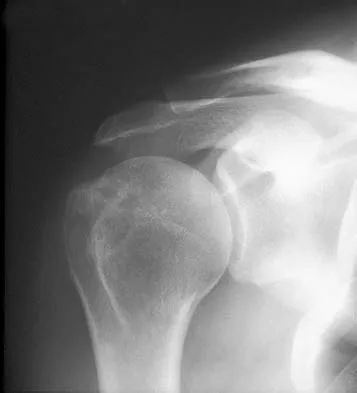
A 52-year-old man has had right shoulder pain in the deltoid region that increases at night for the past 2 months. He denies any history of trauma. Examination reveals mild tenderness over the greater tuberosity, and the Neer and Hawkins impingement signs are positive. AP and outlet lateral radiographs are shown in Figures 24a and 24b. Initial management should consist of


Explanation

24b The patient has the findings of classic subacromial impingement. Initial management should consist of stretching exercises directed at the posterior capsule and a program of rotator cuff and deltoid strengthening exercises performed below the horizontal in a "safe" plane. The judicious use of subacromial cortisone injections (one or two) may be helpful. Anterior acromioplasty is reserved for patients who have failed to respond to nonsurgical management. Morrison DS, Frogameni AD, Woodworth P: Non-operative treatment of subacromial impingement syndrome. J Bone Joint Surg Am 1997;79:732-737. Neer CS: Impingement lesions. Clin Orthop 1983;173:70-77.

Question 7

A 70-year-old woman is brought to the emergency department with a two-part greater tuberosity fracture with an anterior subcoracoid dislocation. One day after successful closed reduction, examination reveals marked swelling of the involved arm, forearm, and hand, as well as large amounts of "weeping" serous fluid but no obvious lacerations. The fingers are warm and pink, and the pulses are normal distally with good refill. Edema is present. There is no pain with passive and active motion of the elbow, wrist, and fingers. What is the next most appropriate step in management?


Explanation

Although not as common as arterial injury, venous thrombosis secondary to trauma of the subclavian or axillary vein can be problematic; therefore, venous duplex ultrasound scanning is the diagnostic study of choice. Arteriography may not show venous thrombosis in the venous run-off phase. The clinical history does not fit the usual presentation of a compartment syndrome or complex regional pain syndrome.

Question 8

A baseball pitcher has intractable posterior and superior shoulder pain. The arthroscopic view seen in Figure 25 shows no Bankart or Hill-Sachs lesion and a negative drive-through sign. There are no signs of ligamentous laxity, but active compression and anterior slide tests are positive. Treatment should consist of


Explanation

According to Morgan and associates, a type II SLAP lesion can create or is associated with a superior instability pattern. They suggest that this can exist without a co-existing anteroinferior instability pattern. They reported that repair of the SLAP lesion alone resulted in satisfactory outcomes in 90% of patients and a return to throwing in more than 90% of pitchers. The arthroscopic findings in this patient do not support a diagnosis of anteroinferior laxity or instability; therefore, thermal capsular shift or capsular placation is not necessary. Morgan CD, Burkhart SS, Palmeri M, et al: Type II SLAP lesions: Three subtypes and their relationships to superior instability and rotator cuff tears. Arthroscopy 1998;14:553-565. Mileski RA, Snyder RJ: Superior labral lesions in the shoulder: Pathoanatomy and surgical management. J Am Acad Orthop Surg 1998;6:121-131.

Question 9

With increasing abduction in the scapular plane, maintaining neutral rotation, contact area, and contact pressure per unit area between the humeral head and glenoid follows what pattern if the total load across the joint is held constant?


Explanation

The glenohumeral joint becomes more congruent at higher levels of abduction. As a consequence, contact area increases. As the load is spread more evenly across the joint, contact pressure per unit area decreases as long as the total load across the joint is held constant. Warner JJP, Bowen MK, Deng XH, et al: Articular contact patterns of the normal glenohumeral joint. J Shoulder Elbow Surg 1998;7:381-388.

Question 10

A 21-year-old patient has had pain and a marked decrease in active and passive shoulder motion after having had a seizure 2 months ago as the result of alcohol abuse. Current AP and axillary radiographs and a CT scan are shown in Figures 26a through 26c. Management should consist of


Explanation

26b 26c Open reduction and subscapularis and lesser tuberosity transfer into the defect is the treatment of choice in young individuals who have defects that involve between 20% to 45% of the head. Disimpaction and bone grafting is an option in injuries that are less than 3 weeks old. Closed reduction 2 to 3 months after injury usually is unsuccessful and increases the risk of fracture or neurovascular injury. Total shoulder arthroplasty is reserved for defects of greater than 50% or with associated glenoid surface damage. Hemiarthroplasty should be avoided in young individuals unless 50% or more of the head is involved. Gerber C: Chronic locked anterior and posterior dislocations, in Warner JJ, Iannotti JP, Gerber C (eds): Complex and Revision Problems in Shoulder Surgery. Philadelphia, PA, Lippincott-Raven, 1997, pp 99-113.

Question 11

Which of the following ligaments are the primary static restraints to inferior translation of the arm when the shoulder is in 0 degrees of abduction and neutral rotation?


Explanation

Biomechanical ligament sectioning studies have implicated both the superior glenohumeral and coracohumeral ligaments as restraints to inferior translation when the shoulder is in 0 degrees of abduction and neutral rotation. Although there is controversy over the significance of each ligament, both are involved to some degree. The middle glenohumeral ligament is more important in the midranges of abduction, and the inferior ligament is more important at 90 degrees of abduction. The coracoacromial and coracoclavicular ligaments play no role in glenohumeral restraint. Warner JJ, Deng XH, Warren RF, et al: Static capsuloligamentous restraints to superior-inferior translation of the glenohumeral joint. Am J Sports Med 1992;20:675-685.

Question 12

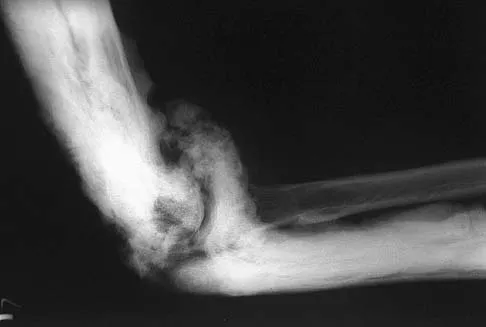
A 44-year-old man who sustained an elbow dislocation 3 months ago now reports pain and restricted elbow motion. Radiographs are shown in Figures 27a and 27b. Management should consist of


Explanation

27b The treatment of choice for an ankylosed chronically dislocated elbow is surgical reduction. Open reduction with application of an external fixator provides excellent results for this complex problem. Radial head arthroplasty is indicated for a radial head fracture that cannot be reconstructed. Attempts at closed reduction will be unsuccessful and should not be attempted in a stiff elbow. In chronic dislocations, direct reinsertion of injured ligaments is not feasible because of soft-tissue contracture. Jupiter J, Ring D: Treatment of unreduced elbow dislocation with hinged external fixation. J Bone Joint Surg Am 2002;84:1630-1635.

Question 13

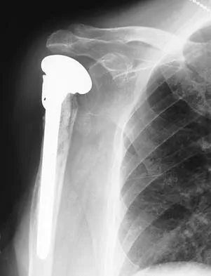
A 67-year-old man who underwent humeral head arthroplasty for a four-part fracture 6 months ago reports that he is still unable to actively elevate his arm. Rehabilitation after surgery consisted of a sling with passive range-of-motion exercises for 2 weeks and then progressed to active-assisted and strengthening exercises at 3 weeks. Radiographs are shown in Figures 28a and 28b. What is the primary cause of his inability to elevate the arm?


Explanation

28b The radiographs show nonunion of both the greater and lesser tuberosities. Tuberosity pull-off and nonunion remain among the most common causes of failed humeral head arthroplasty for fracture. Strict attention to securing the tuberosities to each other and to the shaft, and autogenous bone grafting from the excised humeral head will decrease the incidence of pull-off and improve healing rates. Active-assisted range-of-motion and strengthening exercises should be delayed until tuberosity healing is noted radiographically, usually at 6 to 8 weeks postoperatively. Hartsock LA, Estes WJ, Murray CA, et al: Shoulder hemiarthroplasty for proximal humeral fractures. Orthop Clin North Am 1998;29:467-475. Hughes M, Neer CS: Glenohumeral joint replacement and postoperative rehabilitation. Phys Ther 1975;55:850-858.

Question 14

Initial postoperative management after repair of an acute rotator cuff tear includes


Explanation

In the immediate postoperative period following repair of an acute rotator cuff tear, passive forward elevation and external rotation should be performed within the safe zone determined at surgery. Early active range of motion (prior to tendon healing), internal rotation behind the back, and resistive exercises increase the risk of rupture of the repair. Iannotti JP: Full-thickness rotator cuff tear: Factors affecting surgical outcome. J Am Acad Orthop Surg 1994;2:87-95.

Question 15

A 34-year-old woman reports constant midlateral arm pain after sustaining minimal trauma to the shoulder. Radiographs and a biopsy specimen are shown in Figures 29a and 29b. What is the most likely diagnosis?


Explanation

29b Eighty percent of giant cell tumors occur in patients older than age 20 years, with the peak incidence in the third decade of life. Most of these tumors are eccentrically located and epiphyseal in location. They are lytic in nature as in this patient. Although named for the hallmarked multinucleated giant cells seen in the lesion, the basic cell type is the spindle-shaped stromal cell. Chondroblastoma is highly cellular and contains large multinucleated giant cells with intercellular chondroid material, some of which is calcified. Chondromyxoid fibroma has chondroid tissue separated by strands of more cellular tissue with occasional multinucleated giant cells. Desmoplastic fibroma is characterized by poorly cellular fibrous tissue, and lymphoma is highly cellular with characteristic round cells. Campanacci M, Baldini N, Boriani S, et al: Giant cell tumor of bone. J Bone Joint Surg Am 1987;69:106-114.

Question 16

A 25-year-old professional baseball pitcher reports a 4-month history of gradually increasing medial elbow pain that occurs during the late cocking and acceleration phases of throwing. The pain occasionally refers distally along the ulnar aspect of the forearm. He denies any weakness; however, he notes occasional paresthesias. A nerve conduction velocity study demonstrates increased latency across the cubital tunnel. Management consisting of 6 weeks of rest and rehabilitation fails to provide relief as the symptoms returned when he resumed throwing. What is the best course of action?


Explanation

In the thrower's elbow, ulnar neuritis is felt to result from both chronic compression and traction on the nerve that occurs during the throwing motion. Occasionally, subluxation of the nerve also can lead to symptoms. If nonsurgical management fails to provide relief, transposition of the nerve to an anterior subcutaneous location is the surgical procedure of choice. The nerve is held in its new position by one or two fascial slings created from the fascia of the common flexor origin. Schickendantz MS: Diagnosis and treatment of elbow disorders in the overhead athlete. Hand Clin 2002;18:65-75.

Question 17

What artery provides the only direct vascularizaton to both the intraneural and extraneural blood supply of the ulnar nerve just proximal to the cubital tunnel?


Explanation

The superior ulnar collateral, inferior ulnar collateral, and posterior ulnar recurrent arteries provide consistent vascular supply to the ulnar nerve. This supply is segmental in nature. No identifiable direct anastomosis is seen between the superior ulnar collateral and the posterior ulnar recurrent arteries. The inferior ulnar collateral artery provides the only direct vascularization to the nerve and is located in the region just proximal to the cubital tunnel. The segmental nature of the blood supply to the ulnar nerve underscores the importance of its preservation during transposition.

Question 18

A 56-year-old man underwent right total shoulder arthroplasty 2 months ago. Recently while reaching with his shoulder in a flexed and adducted position, he noted shoulder pain and afterwards he could not externally rotate his arm. An axillary radiograph is shown in Figure 30. What is the most likely cause of this problem?


Explanation

Anteversion of the humeral component may result in anterior instability of the component. Posterior instability after total shoulder arthroplasty is usually the result of some combination of the following factors: untreated anterior soft-tissue contractures, excessive posterior capsular laxity, and excessive retroversion of the humeral and/or glenoid components. Cofield RH, Edgerton BC: Total shoulder arthroplasty: Complications and revision surgery. Instr Course Lect 1990;39:449-462.

Question 19

A 70-year-old man seen in the emergency department has had left shoulder pain and a fever of 101.5 degrees F (38.6 degrees C) for the past 3 days. He denies any history of trauma. Examination reveals tenderness anterosuperiorly and at the posterior glenohumeral joint line. He has very limited range of motion (passive and active). Laboratory studies show a WBC count of 12,000/mm3 and an erythrocyte sedimentation rate of 48 mm/h. Initial management should consist of


Explanation

It appears that the patient has septic arthritis of the glenohumeral joint; therefore, initial management should consist of aspiration of the glenohumeral joint and subacromial space separately, followed by Gram stain and culture of the fluid. Based on the findings, broad-spectrum IV antibiotics should be started. If the diagnosis of septic arthritis is confirmed, then arthroscopic or open surgical drainage usually is indicated. Sawyer JR, Esterhai JL Jr: Shoulder infections, in Warner JJ, Iannotti JP, Gerber C (eds): Complex and Revision Problems in Shoulder Surgery. Philadelphia, PA, Lippincott-Raven, 1997.

Question 20

A 40-year-old man who is an avid weight lifter has had chronic pain in the proximal anterior shoulder for the past year. He denies any history of trauma. Examination reveals tenderness at the intertubercular groove, a positive speed test, and a positive Neer impingement sign. Nonsurgical management has failed to provide relief, and he is now considering surgery. Arthroscopic findings in the glenohumeral joint are shown in Figure 31. Based on these findings, treatment should consist of


Explanation

The arthroscopic image shows a tear through more than 50% of the biceps tendon; therefore, treatment should consist of tenodesis or tenotomy of the tendon. However, because this patient is relatively young and active, the treatment of choice is tenodesis of the biceps tendon. Sethi N, Wright R, Yamaguchi K: Disorders of the long head of the biceps tendon. J Shoulder Elbow Surg 1999;8:644-654. Eakin CL, Faber KJ, Hawkins RJ, et al: Biceps tendon disorders in athletes. J Am Acad Orthop Surg 1999;7:300-310.

Question 21

A 59-year-old man reports moderate shoulder pain and very restricted range of motion after undergoing humeral arthroplasty for osteoarthritis 1 year ago. An AP radiograph is shown in Figure 32. Management should now consist of


Explanation

The radiograph reveals that an insufficient amount of the proximal humerus was excised in the index procedure, resulting in malalignment of the humeral component, overstuffing of the glenohumeral joint, and glenoid arthritis. It is unlikely that physical therapy or soft-tissue releases alone will be adequate. Revision of the humeral component, recutting of the proximal humerus to allow a more anatomic alignment of the humeral component, appropriate soft-tissue releases, and glenoid arthroplasty will offer the best chance of improvement in this difficult situation. Neer CS II, Kirby RM: Revision of humeral head and total shoulder arthroplasties. Clin Orthop 1982;170:189-195.

Question 22

A 70-year-old woman has a preoperative anterior interscalene block prior to undergoing a total shoulder arthroplasty. After seating her in the beach chair position, she becomes acutely hypotensive. What is the most likely cause for the hypotension?


Explanation

The beach chair position may cause sudden hypotension and bradycardia as a result of the Bezold-Jarisch reflex. This reflex occurs when venous pooling and increased sympathetic tone induce a low-volume, hypercontractile ventricle, resulting in activation of the parasympathetic nervous system and sympathetic withdrawal. The reported incidence of this phenomenon associated with the sitting position is between 13% to 24%. Left untreated, the result may be cardiac arrest. Pneumothorax or central nervous system toxicity after interscalene block is rare and has an incidence of less than 0.2%. Laryngeal nerve block associated with interscalene nerve block can occur but usually results in hoarseness secondary to ipsilateral vocal cord palsy. Long T, Wass C, Burkle C: Perioperative interscalene blockade: An overview of its history and current clinical use. J Clin Anesthesia 2002;14:546-556.

Question 23

What structure is considered the single most important soft-tissue restraint to anterior-posterior stability of the sternoclavicular joint?


Explanation

In a cadaver ligament sectioning study, the posterior capsular ligament was considered the most important structure for anterior-posterior stability of the sternoclavicular joint. The anterior capsular ligament also helps prevent anterior displacement but not to the same degree as the posterior ligament. The interclavicular ligament provides little support for anteroposterior translation. Spencer EE, Kuhn JE, Huston LJ, et al: Ligamentous restraints to anterior and posterior translation of the sternoclavicular joint. J Shoulder Elbow Surg 2002;11:43-47.

Question 24

A 35-year-old man has atraumatic painless limited elbow motion. Radiographs are shown in Figures 33a and 33b. What is the most likely diagnosis?


Explanation

33b Based on the radiographic findings, the patient has melorheostosis, a rare, benign connective tissue disorder that is characterized by a cortical thickening of bone. It produces a "dripping candle wax" appearance with dense hyperostosis that flows along the cortex. Ectopic bone formation is a consideration but is associated with injuries or burns. Bone infarcts produce intraosseous sclerosis typically affecting the distal femur with the "smoke up chimney" appearance. Infection is always a consideration but typically does not have the linear osteitis seen in melorheostosis. Juxacortical chondroma is a benign cartilage growth that arises from the capsule and may involve the underlying cortical bone but rarely the medullary canal. Campbell CJ, Papademetriou T, Bonfiglio M: Melorheostosis: A report of the clinical, roentgenographic, and pathological findings in fourteen cases. J Bone Joint Surg Am 1968;50:1281-1304.

Question 25

A 64-year-old man who underwent total shoulder arthroplasty 4 weeks ago is making satisfactory progress in physical therapy, but his therapist notes limitations in external rotation to neutral. A stretching program is started, and the patient suddenly gains 90 degrees of external rotation but now reports increased pain and weakness. What is the best course of action?


Explanation

Nearly all approaches to shoulder arthroplasty require detachment of the subscapularis tendon from the humerus and subsequent repair. Healing of this tenotomy is one of the limiting factors in postoperative recovery. Failure of the tenotomy repair must be recognized and treated early with repeat repair or pectoralis muscle transfer for optimal results. Failure of the subscapularis is diagnosed clinically as excessive external rotation and weakness, especially in the lift-off or belly press position. Muscle testing can be difficult in the postoperative period and may not be possible to assess in those positions. Although MRI might be useful to confirm the diagnosis, studies may be limited by artifact. CT or electromyography would not be diagnostic. Wirth MA, Rockwood CA Jr: Complications of total shoulder-replacement arthroplasty. J Bone Joint Surg Am 1996;78:603-616.

Question 26

A 28-year-old competitive weightlifter complains of painful snapping on the medial aspect of his dominant right elbow when performing triceps extensions. Physical examination reveals a palpable snap over the medial epicondyle during active elbow flexion and extension. Dynamic ultrasound confirms the diagnosis of snapping triceps syndrome. What is the most common anatomical variant associated with this condition?


Explanation

Snapping triceps syndrome is characterized by the concurrent subluxation of the ulnar nerve and the medial head of the triceps over the medial epicondyle during elbow flexion. It is most commonly associated with hypertrophy of the medial head of the triceps, often seen in weightlifters or manual laborers. It must be differentiated from isolated ulnar nerve subluxation to ensure both pathologies are addressed surgically if conservative measures fail.

Question 27

In the standard surgical management of a terrible triad injury of the elbow (elbow dislocation, radial head fracture, coronoid fracture), which of the following represents the correct sequence of reconstruction to restore stability?


Explanation

The standard "inside-out" sequence for repairing a terrible triad injury of the elbow is: 1) Coronoid fracture fixation or anterior capsular repair (addressing the deepest structure first), 2) Radial head fixation or replacement, and 3) Lateral ulnar collateral ligament (LUCL) repair. If the elbow remains unstable in extension after these steps, the medial collateral ligament (MCL) may be repaired or a hinged external fixator applied.

Question 28

A 65-year-old man presents with persistent shoulder pain, weakness, and increased passive external rotation 6 months following an anatomic total shoulder arthroplasty (TSA). Physical examination reveals a positive belly-press test and increased passive external rotation compared to the contralateral side. Radiographs show no evidence of hardware loosening. What is the most likely cause of his symptoms?


Explanation

Subscapularis failure is a known complication following anatomic total shoulder arthroplasty, especially since the tendon is typically taken down (via tenotomy, peel, or lesser tuberosity osteotomy) to gain joint access. Clinical signs include weakness in internal rotation (positive belly-press, bear-hug, or lift-off tests) and increased passive external rotation. Early recognition is crucial, as delayed repair results in poorer functional outcomes.

Question 29

A 78-year-old female with severe osteoporosis sustains a 3-part proximal humerus fracture. Due to the high risk of avascular necrosis and poor bone quality, a reverse total shoulder arthroplasty (RTSA) is performed. In this setting, healing of which of the following structures is most critical to ensure adequate functional external rotation and optimal clinical outcomes?


Explanation

In the setting of reverse total shoulder arthroplasty (RTSA) for proximal humerus fractures, anatomic healing of the greater tuberosity is strongly associated with improved active forward elevation and external rotation. Failure of greater tuberosity healing often leads to decreased functional outcomes due to the loss of the posterior rotator cuff (infraspinatus and teres minor) acting as a force couple and external rotator.

Question 30

A 55-year-old construction worker presents with a symptomatic type II SLAP tear and biceps tendinopathy. He undergoes arthroscopic biceps tenodesis. Compared to biceps tenotomy, which of the following is a recognized advantage of biceps tenodesis?


Explanation

Biceps tenodesis and tenotomy are both accepted treatments for long head of the biceps pathology. Tenodesis maintains the length-tension relationship of the muscle, which theoretically preserves supination strength and significantly decreases the incidence of a cosmetic "Popeye" deformity compared to tenotomy. However, tenotomy typically requires a shorter operative time, allows a faster return to unrestricted activities, and carries a lower risk of postoperative stiffness.

Question 31

A 24-year-old male presents with chronic radial-sided wrist pain. He sustained a hyperextension injury to his wrist 2 years ago but did not seek medical treatment. Radiographs reveal a scaphoid waist nonunion with a "humpback" deformity and a dorsal intercalated segment instability (DISI) pattern. There is no radiographic evidence of radiocarpal arthritis. Which of the following is the most appropriate surgical treatment?


Explanation

A scaphoid nonunion with a "humpback" deformity (volar angulation of the scaphoid) and a resulting DISI posture indicates a collapse of the normal carpal architecture. To correct the deformity and restore carpal kinematics, a volar approach with a structural wedge bone graft (usually from the iliac crest) and internal fixation is required. Salvage procedures (PRC or 4-corner fusion) are reserved for cases with established radiocarpal arthritis (SNAC wrist).

Question 32

A 62-year-old female who was treated nonoperatively for a nondisplaced distal radius fracture presents 6 weeks post-injury with a sudden inability to actively extend her right thumb. Examination reveals a lack of active retropulsion of the thumb, but she is able to extend the interphalangeal joint when the thumb is held in adduction. Which of the following is the most appropriate surgical intervention?


Explanation

Extensor pollicis longus (EPL) rupture is a classic complication following distal radius fractures, particularly those treated nonoperatively. The rupture is typically due to attrition over the bony callus or ischemia within the third dorsal compartment. Because the tendon ends are usually retracted and degenerated, primary end-to-end repair is rarely feasible. The standard of care is a tendon transfer, with the extensor indicis proprius (EIP) to EPL transfer being the most common and reliable procedure.

Question 33

A 25-year-old male is involved in a high-speed motorcycle accident and sustains a severe traction injury to his right brachial plexus. On physical examination, he has flaccid paralysis of the right upper extremity, associated with right-sided ptosis, miosis, and anhidrosis. The presence of Horner's syndrome suggests an injury at which of the following anatomic levels?


Explanation

Horner's syndrome (ptosis, miosis, anhidrosis) in the setting of a brachial plexus injury strongly suggests avulsion of the T1 nerve root. The preganglionic sympathetic fibers to the head and neck exit the spinal cord at the T1 level and travel with the T1 nerve root before joining the sympathetic chain. Injury to the lower trunk or avulsion of the T1 root disrupts these sympathetic fibers, indicating a preganglionic injury with a poor prognosis for spontaneous recovery.

Question 34

A 7-year-old boy presents for evaluation of a left elbow deformity. He sustained a displaced supracondylar humerus fracture 2 years ago, which was treated with closed reduction and percutaneous pinning. Physical examination reveals a significant cubitus varus deformity. Which of the following statements regarding this condition is most accurate?


Explanation

Cubitus varus ("gunstock deformity") is the most common long-term complication of supracondylar humerus fractures, occurring primarily due to malunion rather than physeal growth arrest. The malunion is typically a combination of extension, medial tilt, and internal rotation. Long-term studies have shown it can lead to functional issues, including an increased risk of lateral condyle fractures, posterolateral rotatory instability (PLRI), and tardy ulnar nerve palsy. Coronal plane deformities do not remodel with growth.

Question 35

A 58-year-old woman with a history of poorly controlled type 2 diabetes mellitus presents with a locked trigger finger of the right ring finger. She has had no prior treatments. Which of the following statements regarding the management of trigger finger in diabetic patients is most accurate?


Explanation

Diabetic patients with trigger finger (stenosing tenosynovitis) tend to have a more recalcitrant disease course than non-diabetics. Extensive literature demonstrates that diabetic patients have a significantly higher rate of treatment failure with corticosteroid injections and are more likely to eventually require surgical release of the A1 pulley. They also have a higher incidence of multiple digit involvement. Despite the higher failure rate, an initial injection is still often utilized, but patients should be counseled on the increased likelihood of needing surgery.

Question 36

A 65-year-old woman presents with the sudden inability to extend her thumb. She reports that 5 weeks ago she sustained a nondisplaced distal radius fracture, which was treated nonoperatively in a short arm cast. Physical examination reveals an inability to retro-extend the thumb at the interphalangeal joint, but tenodesis effect is intact. What is the most appropriate surgical management?


Explanation

The patient has experienced an extensor pollicis longus (EPL) tendon rupture, a known complication of nondisplaced distal radius fractures. The rupture typically occurs at Lister's tubercle due to a combination of mechanical attrition and vascular ischemia within the intact third extensor compartment. Because of the delay in presentation and degeneration of the tendon ends, primary end-to-end repair is usually impossible. The gold standard treatment is a tendon transfer utilizing the extensor indicis proprius (EIP).

Question 37

A 35-year-old man sustains a 'terrible triad' injury to his elbow. During surgical reconstruction, standard protocol is followed: the coronoid fracture is fixed and the comminuted radial head is replaced with a prosthesis. Intraoperative fluoroscopy reveals that the elbow remains unstable in extension. What is the next most appropriate step in the surgical algorithm?


Explanation

The 'terrible triad' of the elbow includes an elbow dislocation, a radial head fracture, and a coronoid process fracture. The standard surgical algorithm progresses from deep to superficial and typically involves: (1) fixation or replacement of the radial head, (2) fixation of the coronoid, and (3) repair of the lateral collateral ligament (LCL) complex, specifically the lateral ulnar collateral ligament (LUCL), to the lateral epicondyle. If the elbow remains unstable after LUCL repair, the MCL is addressed or a hinged external fixator is applied.

Question 38

Reverse total shoulder arthroplasty (RTSA) is designed to alter the biomechanics of the glenohumeral joint to compensate for a massive, irreparable rotator cuff tear. Which of the following best describes the biomechanical alterations achieved with a classic Grammont-style prosthesis?


Explanation

The classic Grammont-style reverse total shoulder arthroplasty works by medializing and distalizing (inferiorly translating) the center of rotation. Medialization limits torque at the glenoid bone-implant interface (reducing shear forces) and recruits more deltoid fibers. Inferior translation tensions the deltoid muscle, increasing its moment arm and allowing it to act as the primary elevator of the shoulder in the absence of a functional rotator cuff.

Question 39

A 28-year-old professional volleyball player complains of insidious, deep posterior shoulder pain and weakness with external rotation. Clinical examination shows isolated atrophy of the infraspinatus fossa. MRI demonstrates a paralabral cyst causing nerve compression. Based on the examination findings, where is the cyst most likely located and what is the typical associated labral pathology?


Explanation

The patient has isolated infraspinatus weakness and atrophy, which localizes the suprascapular nerve compression to the spinoglenoid notch (after the nerve has already given off its motor branches to the supraspinatus muscle at the suprascapular notch). Paralabral cysts at the spinoglenoid notch are strongly associated with posterosuperior labral tears or posterior SLAP lesions. A cyst at the suprascapular notch would typically cause weakness of both the supraspinatus and infraspinatus.

Question 40

A 40-year-old bodybuilder undergoes a single-incision anterior approach for the repair of a distal biceps tendon rupture. During his first postoperative visit, he complains of numbness and tingling along the radial aspect of his forearm. Which nerve was most likely injured during the procedure, and what is the most common mechanism?


Explanation

The lateral antebrachial cutaneous nerve (LABCN), the terminal sensory branch of the musculocutaneous nerve, is the most commonly injured nerve during a single-incision anterior distal biceps repair. Injury is typically due to traction or stretch from retractors during the superficial approach, as the nerve runs closely alongside the cephalic vein in the lateral subcutaneous tissue. Posterior interosseous nerve (PIN) injury is more classically associated with the two-incision technique or deep retractor placement.

Question 41

A 32-year-old carpenter presents with a 6-month history of dorsal wrist pain and decreased grip strength. Radiographs reveal sclerosis, cystic changes, and early fragmentation of the lunate, without carpal collapse. Ulnar variance is noted to be negative 2 mm. Which of the following is the most appropriate surgical intervention?


Explanation

The patient has Kienböck's disease (avascular necrosis of the lunate) stage IIIa (sclerosis and fragmentation without carpal collapse or scaphoid rotation). In a patient with negative ulnar variance and an intact carpal architecture (stages I, II, and IIIa), a joint-leveling procedure such as a radial shortening osteotomy or ulnar lengthening is indicated to biomechanically unload the lunate. Proximal row carpectomy or intercarpal fusions are reserved for more advanced stages (IIIb or IV) with carpal collapse or arthritis.

Question 42

A 24-year-old man presents with chronic radial-sided wrist pain following a fall onto an outstretched hand one year ago. Imaging confirms a nonunion of the proximal pole of the scaphoid with avascular necrosis, but no radiocarpal arthritis is present. The surgeon elects to perform a pedicled vascularized bone graft based on the 1,2 intercompartmental supraretinacular artery (1,2 ICSRA). From which anatomic location is this graft harvested?


Explanation

The 1,2 intercompartmental supraretinacular artery (1,2 ICSRA) vascularized bone graft is harvested from the dorsal aspect of the distal radius. It utilizes a pedicle located between the first and second extensor compartments. It is a well-described option for the treatment of scaphoid proximal pole nonunions complicated by avascular necrosis. The medial femoral condyle is used for free vascularized bone grafting, not a local pedicled flap.

Question 43

A 19-year-old rugby player presents to the emergency department after a direct blow to the anteromedial shoulder. He reports shortness of breath, mild dysphagia, and right-sided neck fullness. Physical exam reveals a palpable defect over the medial clavicle. Standard radiographs are equivocal. What is the most appropriate next step in diagnostic imaging, and which surgical specialty should ideally be available on standby if closed reduction is attempted?


Explanation

The clinical presentation is highly suspicious for a posterior sternoclavicular dislocation, which can compress posterior mediastinal structures including the trachea, esophagus, and great vessels (subclavian or innominate artery/vein). A CT scan is the imaging modality of choice to accurately assess the direction of dislocation and proximity to vascular structures. If closed reduction is attempted, it should be done in the operating room with cardiothoracic surgery available, due to the life-threatening risk of lacerating a great vessel upon reduction.

Question 44

During a routine in situ ulnar nerve decompression for cubital tunnel syndrome, the surgeon must systematically evaluate and release potential sites of nerve compression. Which of the following structures represents the most proximal potential site of ulnar nerve entrapment?


Explanation

The ulnar nerve can be compressed at several sites around the elbow. From proximal to distal, these sites are: the arcade of Struthers (a fascial band extending from the medial intermuscular septum to the medial head of the triceps, located about 8 cm proximal to the medial epicondyle), the medial intermuscular septum, the medial epicondyle, Osborne's ligament (the retinaculum forming the roof of the cubital tunnel), and the aponeurosis of the two heads of the flexor carpi ulnaris (FCU). Therefore, the arcade of Struthers is the most proximal site.

Question 45

A 45-year-old man sustains a closed, spiral fracture of the distal third of the humeral shaft (Holstein-Lewis fracture) following a fall. On initial examination in the emergency department, he exhibits a dense radial nerve palsy. According to current orthopaedic literature, what is the most appropriate initial management for this injury?


Explanation

A primary radial nerve palsy associated with a closed humeral shaft fracture (including Holstein-Lewis types) is predominantly a neurapraxia or axonotmesis. The overall rate of spontaneous recovery is high (greater than 85%). Therefore, the standard of care is initial nonoperative management of the fracture with a coaptation splint or functional brace, and clinical observation. Surgical exploration of the nerve is indicated if the palsy occurs after a closed reduction attempt (secondary palsy), in open fractures, or if there is no clinical or electromyographic evidence of recovery by 3 to 4 months.

Question 46

A 65-year-old woman undergoes volar locked plating for a displaced intra-articular distal radius fracture. Postoperative lateral radiographs demonstrate that the plate is positioned distally, bridging the watershed line. Three months postoperatively, she presents with an inability to actively flex the interphalangeal joint of her thumb. Which of the following is the most likely cause of her current presentation?


Explanation

The watershed line is a critical anatomic landmark for volar plating of distal radius fractures. Plates placed distal to this line become prominent and impinge on the flexor tendons, particularly the flexor pollicis longus (FPL), leading to attrition and secondary rupture. Soong et al. classified volar plate position relative to the watershed line, with grade 2 (plate completely distal to the watershed line) having the highest risk of FPL rupture. Dorsal screw penetration is a well-known risk for EPL rupture, not FPL.

Question 47

A 45-year-old man falls from a height and sustains a 'terrible triad' injury of the elbow. Which of the following describes the most appropriate sequence of surgical reconstruction to restore elbow stability?


Explanation

The classic 'terrible triad' of the elbow includes an elbow dislocation, radial head fracture, and coronoid fracture. The standard surgical sequence to restore elbow stability is typically performed from deep to superficial: 1) fixation of the coronoid fracture (or anterior capsule repair if the fragment is too small), 2) repair or replacement of the radial head, 3) repair of the lateral ulnar collateral ligament (LUCL/LCL complex), and 4) if the elbow remains unstable in extension after these steps, repair of the medial collateral ligament (MCL) or application of a hinged external fixator.

Question 48

The Grammont design of a reverse total shoulder arthroplasty (RTSA) alters the biomechanics of the shoulder to compensate for a massive, irreparable rotator cuff tear. Which of the following accurately describes the primary biomechanical advantage of this design?


Explanation

The Grammont design principles for reverse total shoulder arthroplasty include medializing and inferiorly translating the center of rotation. Medialization decreases the torque on the glenoid component, reducing the risk of loosening, while inferior translation tensions the deltoid muscle. Together, these biomechanical changes significantly increase the deltoid's moment arm and recruit more of its anterior and posterior fibers, allowing the deltoid to compensate for the deficient rotator cuff and elevate the arm effectively.

Question 49

A 24-year-old man falls on an outstretched hand and sustains a fracture of the proximal pole of the scaphoid. He delays seeking treatment for 3 months. What is the primary anatomical reason for the high risk of nonunion and avascular necrosis in this specific fracture pattern?


Explanation

The major blood supply to the scaphoid arises from branches of the radial artery (specifically the dorsal carpal branch), which enter the scaphoid at the distal pole and waist. From there, the blood flows in a retrograde fashion to supply the proximal pole. Because of this tenuous retrograde blood supply, fractures at the proximal pole carry a particularly high risk of disrupting the vascularity to the proximal fragment, strongly predisposing the patient to nonunion and avascular necrosis.

Question 50

A 30-year-old elite volleyball player presents with insidious onset of right shoulder pain and weakness. Physical examination reveals isolated atrophy of the infraspinatus muscle with normal bulk of the supraspinatus. Weakness is noted exclusively with external rotation. Where is the most likely location of nerve entrapment?


Explanation

The suprascapular nerve innervates both the supraspinatus and infraspinatus muscles. Entrapment at the suprascapular notch affects both muscles, leading to weakness in abduction and external rotation. However, entrapment at the spinoglenoid notch occurs after the nerve has already given off its motor branches to the supraspinatus. This results in isolated denervation and atrophy of the infraspinatus muscle, causing weakness exclusively in external rotation. This condition is frequently seen in overhead athletes and is often associated with paralabral cysts arising from posterior labral tears.

Question 51

A 19-year-old male is brought to the emergency department after a severe tackle in a rugby match. He complains of chest pain, difficulty swallowing, and a feeling of fullness in his neck. Physical examination reveals a palpable depression over the medial aspect of the right clavicle. Radiographs and a subsequent CT scan confirm a posterior sternoclavicular joint dislocation. Which of the following anatomical structures is at the highest risk of injury in this setting?


Explanation

Posterior sternoclavicular dislocations are considered orthopedic emergencies due to the close proximity of the joint to critical mediastinal structures. The most commonly injured or compressed structures in this scenario are the great vessels, specifically the brachiocephalic (innominate) vein, which lies directly posterior to the sternoclavicular joint. Other structures at risk include the trachea, esophagus, and subclavian vessels. Closed reduction should be performed urgently in the operating room with cardiothoracic surgery backup.

Question 52

In the surgical management of recalcitrant trigger finger (stenosing tenosynovitis) in the middle digit, the primary structure targeted for release is the A1 pulley. During this procedure, great care must be taken to avoid violating the adjacent A2 pulley. What is the primary biomechanical consequence of a complete iatrogenic transection of the A2 pulley?


Explanation

The flexor tendon pulley system of the hand consists of annular and cruciate pulleys that keep the flexor tendons closely applied to the phalanges. The A2 (located over the proximal phalanx) and A4 (over the middle phalanx) pulleys are the most critical mechanically. Complete release or rupture of the A2 pulley results in 'bowstringing' of the flexor tendons away from the axis of rotation during active flexion. While this increases the tendon's moment arm, it significantly limits the active range of motion and decreases overall grip strength, as a large amount of tendon excursion is wasted in pulling the tendon away from the bone rather than flexing the joint.

Question 53

A 55-year-old carpenter presents with progressive numbness and tingling in his small and ring fingers of his right hand, along with clumsiness when handling small tools. Examination reveals intrinsic muscle wasting and a positive Froment's sign. Which of the following findings would differentiate a compressive neuropathy at the cubital tunnel from one at Guyon's canal?


Explanation

The dorsal ulnar cutaneous nerve branches off the main ulnar nerve approximately 5 to 8 cm proximal to the wrist joint and provides sensation to the dorsoulnar aspect of the hand. A compressive ulnar neuropathy at the elbow (cubital tunnel syndrome) will affect this branch, resulting in sensory loss over the dorsal ulnar hand. In contrast, compression at Guyon's canal (at the wrist) occurs distal to the takeoff of the dorsal ulnar cutaneous nerve, thereby sparing dorsal ulnar sensation. Both compression sites can cause intrinsic muscle wasting, a positive Froment's sign, Wartenberg's sign, and volar sensory loss in the ulnar digits.

Question 54

During open reduction and internal fixation of a displaced 3-part proximal humerus fracture using a deltopectoral approach, the surgeon needs to carefully retract the deltoid and protect the axillary nerve. At what approximate distance from the lateral edge of the acromion does the axillary nerve typically traverse the deep surface of the deltoid?


Explanation

The axillary nerve travels through the quadrilateral space and courses anteriorly around the surgical neck of the humerus along the deep surface of the deltoid muscle. It typically lies approximately 5 to 7 cm distal to the lateral edge of the acromion. When performing surgery on the proximal humerus, especially via lateral approaches (such as the deltoid-splitting approach) or when placing retractor blades deep to the deltoid, it is critical to respect this anatomical 'safe zone' (less than 5 cm from the acromion) to avoid iatrogenic injury to the axillary nerve, which would result in catastrophic denervation of the anterior and middle deltoid.

Question 55

A 32-year-old basketball player presents with a 'dropped' distal phalanx of his long finger after the ball struck the tip of his extended finger. Radiographs show a small dorsal avulsion fracture of the distal phalanx involving 15% of the articular surface with no volar subluxation. Nonoperative management is chosen. What is the most appropriate splinting protocol for this Zone I extensor tendon injury?


Explanation

Mallet finger injuries (Zone I extensor tendon injuries) with or without a small avulsion fracture (less than 30% to 50% of the articular surface and without volar subluxation) are best treated nonoperatively. The standard protocol involves continuous, uninterrupted splinting of the distal interphalangeal (DIP) joint in extension (or slight hyperextension) for 6 to 8 weeks. The proximal interphalangeal (PIP) joint must be left free to allow active motion and prevent stiffness. If the splint is removed and the distal phalanx is allowed to flex at any point during this period, the healing tissue is disrupted, and the 6-to-8-week clock must be restarted.

Question 56

A 45-year-old woman presents with vague shoulder pain and inability to elevate her arm above 90 degrees. She underwent an excisional biopsy of a posterior triangle cervical lymph node 3 months ago. On examination, the affected shoulder droops, and the scapula is translated laterally and rotated downward. Winging is exacerbated by arm abduction. Which of the following is the most appropriate surgical treatment if conservative management fails?


Explanation

The clinical scenario describes a spinal accessory nerve (CN XI) palsy, which denervates the trapezius muscle, leading to lateral winging of the scapula (downward rotation and lateral translation). The Eden-Lange procedure involves transferring the levator scapulae to the acromion, and the rhomboid major and minor to the infraspinatus fossa, substituting for the paralyzed trapezius to stabilize the scapula. Pectoralis major transfer is typically used for serratus anterior palsy (medial winging).

Question 57

A 40-year-old man sustains a 'terrible triad' injury to his elbow. Intraoperatively, the radial head is replaced, the coronoid fracture is fixed securely, and the lateral ulnar collateral ligament (LUCL) is repaired. On fluoroscopic examination, the elbow is noted to subluxate posteriorly when extended beyond 30 degrees of flexion. What is the most appropriate next step in management?


Explanation

The treatment algorithm for terrible triad injuries involves systematic restoration of the anterior and lateral stabilizers (coronoid, radial head, and LUCL). If the elbow remains unstable or subluxates during terminal extension after adequately addressing these structures, the medial collateral ligament (MCL) should be repaired. An articulated external fixator is a salvage option if instability persists despite MCL repair.

Question 58

A 58-year-old woman undergoes volar locked plating for a displaced distal radius fracture. Six months postoperatively, she presents with a sudden inability to actively flex the interphalangeal joint of her thumb. Radiographs demonstrate that the volar plate is positioned distal to the watershed line. Which of the following is the most likely mechanism for her current deficit?


Explanation

Placement of a volar plate distal to the watershed line of the distal radius increases the risk of flexor tendon irritation and subsequent attritional rupture. The flexor pollicis longus (FPL) tendon is the most commonly affected due to its close anatomical proximity to the prominent distal edge of the plate. Treatment typically involves plate removal and tendon transfer (e.g., brachioradialis or FDS to FPL) or grafting.

Question 59

A 72-year-old man presents with chronic right shoulder pain and pseudoparalysis. Radiographs reveal superior migration of the humeral head and acetabularization of the coracoacromial arch. The patient undergoes a reverse total shoulder arthroplasty (RTSA). During the procedure, the glenoid baseplate is deliberately positioned with an inferior tilt. What is the primary biomechanical rationale for this baseplate positioning?


Explanation

In reverse total shoulder arthroplasty (RTSA), inferior positioning and inferior tilt of the glenosphere/baseplate are utilized primarily to reduce the incidence of inferior scapular notching. Scapular notching occurs when the medial aspect of the humeral tray impinges on the inferior neck of the scapula during adduction. Inferior tilt clears the scapular neck and decreases this mechanical impingement.

Question 60

A 48-year-old typist presents with numbness in his small and ring fingers. Examination reveals a positive Tinel's sign at the cubital tunnel and weakness in finger abduction. EMG confirms severe ulnar neuropathy at the elbow. During a submuscular ulnar nerve transposition, which of the following fascial structures represents a potential site of nerve compression that MUST be released to prevent postoperative failure?


Explanation

During an ulnar nerve transposition, it is critical to release all potential sites of compression. These sites include the Arcade of Struthers (a fascial band extending from the medial intermuscular septum to the medial head of the triceps), the medial intermuscular septum, Osborne's ligament, and the deep flexor-pronator aponeurosis. The Ligament of Struthers and Lacertus fibrosus compress the median nerve. The Arcade of Frohse compresses the posterior interosseous nerve.

Question 61

A 25-year-old man presents with chronic wrist pain. Radiographs reveal a scaphoid waist nonunion with a 'humpback' deformity (volar angulation) and a dorsal intercalated segment instability (DISI) pattern. There is no midcarpal arthritis. Which of the following is the most appropriate surgical management to restore carpal alignment and heal the nonunion?


Explanation

A scaphoid nonunion with a 'humpback' deformity requires correction of the scaphoid alignment to restore normal carpal kinematics and treat the DISI posture. This is best achieved using a volar approach, inserting an anterior (volar) wedge graft combined with rigid internal fixation. A dorsal approach with 1,2 ICSRA graft cannot easily correct significant volar angulation. Proximal row carpectomy or four-corner fusions are salvage operations for SNAC arthritis.

Question 62

A 62-year-old woman presents with debilitating pain at the base of her thumb, unresponsive to conservative care. Radiographs demonstrate Eaton-Littler Stage III advanced trapeziometacarpal joint arthritis with complete loss of joint space and subluxation. Which of the following surgical procedures provides the most reliable long-term pain relief and functional restoration for this specific presentation?


Explanation

Trapeziectomy, with or without ligament reconstruction and tendon interposition (LRTI), is the most commonly performed and reliable procedure for advanced (Eaton-Littler Stage III or IV) thumb carpometacarpal (CMC) arthritis. Metacarpal osteotomy is indicated for early-stage disease (Stage I or II). MCP arthrodesis does not address CMC joint pathology, though it may be an adjunct if hyperextension instability is present.

Question 63

A 32-year-old carpenter presents with dorsal wrist pain and decreased grip strength. Radiographs reveal sclerosis and fragmentation of the lunate (Lichtman stage IIIA). Ulna variance is measured at -3 mm. Which of the following is the most appropriate initial surgical treatment?


Explanation

This patient has Lichtman stage IIIA Kienböck's disease and ulnar minus variance. Joint-leveling procedures are indicated to decrease the compressive forces across the radiolunate joint. Radial shortening osteotomy is the preferred and most reliable joint-leveling procedure due to a high union rate. Ulnar lengthening is associated with a higher risk of nonunion. PRC or wrist fusion are reserved for stage IV disease with extensive carpal arthritis.

Question 64

A 24-year-old motorcyclist sustains a severe closed traction injury to his right brachial plexus. Clinical examination at 4 months reveals complete pan-plexus paralysis. Horner's syndrome is present. MRI demonstrates pseudomeningoceles at C8 and T1, and EMG shows denervation of the cervical paraspinal muscles. What is the most appropriate reconstructive strategy for restoring elbow flexion?


Explanation

The presence of Horner's syndrome, pseudomeningoceles, and denervated paraspinals indicates a pre-ganglionic pan-plexus root avulsion. Primary nerve grafting is impossible because there are no viable proximal nerve roots. Extra-plexal nerve transfers are required. Intercostal nerves (usually 3 or 4) are frequently transferred to the musculocutaneous nerve to restore elbow flexion. The spinal accessory nerve is typically prioritized for suprascapular nerve transfer for shoulder stability. Tendon transfers are not viable as all native muscles are paralyzed.

Question 65

A 28-year-old man sustains a laceration to the volar aspect of his index finger in Zone II, transecting both the flexor digitorum superficialis and profundus tendons. Primary repair of both tendons is performed using a 4-strand core suture and an epitendinous suture. To optimize functional outcome and minimize adhesion formation, which postoperative rehabilitation protocol is most appropriate?


Explanation

In Zone II flexor tendon repairs, the risk of adhesion formation within the fibro-osseous sheath is exceptionally high. Modern robust multi-strand repairs (4-strand or greater) allow for early active motion protocols (using a dorsal blocking splint to prevent excessive extension while permitting controlled active flexion). This significantly reduces adhesions, stimulates intrinsic tendon healing, and improves functional range of motion compared to prolonged immobilization.

Question 66

A 45-year-old carpenter presents with numbness and tingling in his small and ring fingers, which is exacerbated by prolonged elbow flexion. Electrodiagnostic studies confirm isolated ulnar neuropathy at the elbow. Which of the following is the most common site of ulnar nerve compression in this condition?


Explanation

The most common site of ulnar nerve compression at the elbow is between the two heads of the flexor carpi ulnaris (FCU), specifically under Osborne's ligament (the cubital tunnel retinaculum). Compression can also occur at the arcade of Struthers, medial intermuscular septum, or deep flexor pronator aponeurosis, but Osborne's ligament is statistically the most frequent location.

Question 67

A 62-year-old woman sustained a minimally displaced distal radius fracture treated nonoperatively in a short arm cast for 6 weeks. Three weeks after cast removal, she suddenly loses the ability to actively extend her thumb interphalangeal joint. She denies any new trauma. What is the most appropriate management?


Explanation

Extensor pollicis longus (EPL) tendon ruptures typically occur 3 to 8 weeks after minimally displaced distal radius fractures. This is due to ischemia or mechanical attrition at the Lister tubercle. Because the tendon ends retract and undergo necrosis, primary repair is usually not possible. An EIP to EPL tendon transfer is the gold standard surgical treatment, providing an expendable donor with a matching vector and excursion.

Question 68

A 24-year-old man falls onto an outstretched hand and sustains a fracture of the scaphoid proximal pole. He is at high risk for avascular necrosis (AVN) and nonunion. Which of the following best describes the predominant arterial supply to the scaphoid that makes this fracture pattern vulnerable?


Explanation

The scaphoid receives 70-80% of its blood supply from the dorsal carpal branch of the radial artery, which enters along the dorsal ridge in the distal half of the bone. This retrograde blood supply puts proximal pole fractures at a disproportionately high risk for AVN, as the fracture disrupts the blood flow from distal to proximal.

Question 69

A 28-year-old elite volleyball player presents with isolated weakness in shoulder external rotation. She denies any shoulder pain. Physical examination reveals obvious atrophy of the infraspinatus, but supraspinatus strength and muscle bulk are normal. Where is the most likely site of nerve compression?


Explanation

The suprascapular nerve supplies both the supraspinatus and infraspinatus muscles. It passes first through the suprascapular notch (innervating the supraspinatus) and then continues through the spinoglenoid notch to innervate the infraspinatus. Isolated infraspinatus weakness and atrophy indicate compression at the spinoglenoid notch, frequently due to a paralabral cyst associated with superior labral tears in overhead athletes.

Question 70

A 40-year-old man sustains a 'terrible triad' injury of the elbow consisting of a posterior elbow dislocation, a radial head fracture, and a coronoid fracture. During open surgical reconstruction, what is the generally recommended sequence of fixation to reliably restore elbow stability?


Explanation

The standard surgical algorithm for terrible triad injuries builds from deep to superficial and anterior to posterior. First, the coronoid fracture is fixed or its anterior capsule repaired (restoring the anterior buttress). Second, the radial head is fixed or replaced (restoring the anterior column). Finally, the lateral collateral ligament (LCL) complex is repaired to the lateral epicondyle to restore posterolateral rotatory stability.

Question 71

A 72-year-old man undergoes a reverse total shoulder arthroplasty (RTSA) for severe rotator cuff tear arthropathy. How does the biomechanical design of the Grammont reverse prosthesis improve active forward elevation compared to the native shoulder?


Explanation

The Grammont design of reverse total shoulder arthroplasty medializes and distalizes the center of rotation of the glenohumeral joint. Medialization recruits more deltoid fibers (anterior and posterior) for elevation, while distalization tensions the deltoid and increases its moment arm, allowing the deltoid to effectively compensate for the deficient rotator cuff.

Question 72

A 45-year-old male weightlifter feels a 'pop' in his anterior elbow while performing a heavy deadlift. Clinical examination and MRI confirm a complete rupture of the distal biceps tendon. If the patient elects for nonoperative management, which of the following best describes the expected persistent functional deficit?


Explanation

Nonoperative management of distal biceps tendon ruptures results in a significant functional deficit, primarily in supination. Patients typically experience an approximately 40% loss of supination strength and a 30% loss of elbow flexion strength, along with decreased endurance.

Question 73

A 32-year-old carpenter presents with dorsal wrist pain and decreased grip strength. Radiographs reveal sclerosis and early fragmentation of the lunate, but the carpal height is maintained. An MRI confirms avascular necrosis of the lunate. Radiographs confirm an ulnar variance of negative 2 mm. What is the most appropriate surgical intervention?


Explanation

The patient has Lichtman Stage IIIa Kienböck disease (fragmentation of the lunate but maintained carpal height) and ulnar negative variance. A joint-leveling procedure, specifically a radial shortening osteotomy, is indicated to offload the lunate and shift loads to the radioulnar joint. Ulnar lengthening is rarely performed due to higher nonunion rates.

Question 74

During an open carpal tunnel release, the surgeon must carefully identify and protect the recurrent motor branch of the median nerve. In the most common anatomical variation (Lanz Group 1), how does the recurrent motor branch exit the median nerve in relation to the transverse carpal ligament (TCL)?


Explanation

According to the Lanz classification of the median nerve motor branch, the extraligamentous (post-ligamentous) pattern is the most common (approximately 46-90%). In this variation, the nerve branches from the radial aspect of the median nerve distal to the transverse carpal ligament and courses recurrently to innervate the thenar musculature.

Question 75

A 28-year-old man sustains a spiral fracture of the distal third of the humeral shaft (Holstein-Lewis fracture) following a fall. On presentation to the emergency department, he is unable to extend his wrist or fingers, but triceps function is intact. The fracture is closed. Which of the following scenarios is considered an absolute indication for early surgical exploration of the radial nerve?


Explanation

Primary radial nerve palsies associated with closed humeral shaft fractures (including Holstein-Lewis fractures) are generally observed, as up to 90% resolve spontaneously (representing a neurapraxia or axonotmesis). However, a secondary radial nerve palsy that occurs after a closed reduction attempt is an absolute indication for surgical exploration, because the nerve may have been drawn into the fracture site and entrapped during the reduction maneuver.

Question 76

A 72-year-old woman undergoes reverse total shoulder arthroplasty for cuff tear arthropathy. Postoperatively, she does well but at 2-year follow-up, radiographs show grade 2 scapular notching. Which of the following surgical modifications would have most likely decreased the risk of this complication?


Explanation

Scapular notching is a well-recognized complication of reverse total shoulder arthroplasty (RTSA), occurring when the medial aspect of the humeral tray impinges on the inferior scapular neck during adduction. Placing the glenosphere inferiorly with overhang, using a larger glenosphere, lateralizing the center of rotation, and applying an inferior tilt to the baseplate are technical modifications that reduce the risk of scapular notching. Superior placement, superior tilt, and medialization of the center of rotation increase the risk.

Question 77

A 55-year-old woman presents with the inability to actively flex the interphalangeal joint of her right thumb 8 months after undergoing volar locked plating for a distal radius fracture.

Lateral radiographs demonstrate a prominent plate edge over the volar cortex. What is the most likely cause of her current symptom?


Explanation

The patient has suffered a rupture of the flexor pollicis longus (FPL) tendon, which is a known complication of volar plating of distal radius fractures. The FPL tendon is at particular risk for attrition and rupture when the volar plate is placed distal to the watershed line of the distal radius. This leads to prominence of the hardware that rubs against the tendon during excursion. Dorsal penetration of screws causes extensor tendon irritation or rupture (e.g., EPL), not flexor tendon injury.

Question 78

A 22-year-old collegiate baseball pitcher reports medial elbow pain during the late cocking and early acceleration phases of throwing. MRI confirms a partial-thickness tear of the medial ulnar collateral ligament (MUCL). Which bundle of the MUCL is the primary restraint to valgus stress at the elbow during these specific phases of the throwing motion?


Explanation

The medial ulnar collateral ligament (MUCL) complex is composed of the anterior, posterior, and transverse bundles. The anterior bundle is the primary restraint to valgus stress from 30 to 120 degrees of elbow flexion. The anterior bundle itself is functionally divided into anterior and posterior bands. The anterior band is the most important restraint to valgus stress and is taut in extension and early flexion, which corresponds to the extreme valgus stress encountered during the late cocking and early acceleration phases of throwing.

Question 79

A 45-year-old manual laborer sustains a distal biceps tendon rupture after a sudden eccentric load to his flexed elbow. He wishes to pursue nonoperative management. He should be counseled that he will experience the greatest percentage loss of which of the following functional strengths?


Explanation

Nonoperative management of distal biceps tendon ruptures results in permanent weakness. The greatest functional deficit is a loss of forearm supination strength, which decreases by approximately 40% to 50% compared to the contralateral side. Elbow flexion strength is also affected but to a lesser degree, typically decreasing by about 30%. Pronation and elbow extension strengths remain relatively unaffected.

Question 80

A 35-year-old man fell from a ladder 6 months ago, sustaining a radial head fracture that was treated nonoperatively. He now presents with chronic, progressive wrist pain and ulnar-sided swelling. Examination reveals tenderness at the distal radioulnar joint (DRUJ) and a positive ulnar variance on radiographs.

What is the most appropriate management for this chronic condition?


Explanation

The patient has a missed Essex-Lopresti injury (radial head fracture, interosseous membrane tear, and DRUJ disruption). In the chronic setting, longitudinal radioulnar dissociation leads to proximal migration of the radius, causing ulnar impaction and DRUJ arthritis. Treatment requires restoring the radiocapitellar joint and leveling the radioulnar joint. Radial head arthroplasty alone cannot pull the radius back out to length once soft tissues have contracted. Therefore, an ulnar shortening osteotomy combined with radial head arthroplasty is indicated to restore DRUJ congruity and unload the ulnar carpus.

Question 81

A 50-year-old man presents with chronic, worsening radial-sided wrist pain. He recalls a severe wrist sprain 15 years ago. Radiographs reveal scaphoid nonunion advanced collapse (SNAC) with arthritis involving the radioscaphoid and capitolunate joints, while the radiolunate joint is perfectly spared. What is the most appropriate surgical treatment?


Explanation

The patient has Stage III SNAC wrist (radioscaphoid and midcarpal/capitolunate arthritis). The radiolunate joint is typically spared because the lunate facet of the radius and the proximal lunate have congruent spherical shapes that preserve cartilage. Proximal row carpectomy (PRC) is contraindicated in Stage III SNAC because it relies on a healthy articulation between the capitate head and the lunate fossa; in Stage III, the capitate head is already arthritic. Therefore, scaphoid excision and four-corner fusion (capitate, hamate, lunate, triquetrum) is the procedure of choice.

Question 82

A 28-year-old man presents to the emergency department after a motor vehicle collision with a closed, significantly displaced spiral fracture of the middle third of the humerus. On initial physical exam, he has 5/5 wrist and finger extension. Following closed reduction and placement of a coaptation splint, he is completely unable to extend his wrist or fingers, and lacks sensation over the dorsal first web space. What is the most appropriate next step in management?


Explanation

Primary radial nerve palsy associated with a closed humeral shaft fracture is typically observed, as up to 90% of cases spontaneously recover. However, a secondary (iatrogenic) radial nerve palsy that develops strictly after closed reduction is a strong indication for surgical exploration. This is because the nerve may be physically entrapped between the fracture fragments. Immediate surgical exploration of the radial nerve and internal fixation of the fracture is the most appropriate management.

Question 83

A 62-year-old woman presents with base of thumb pain that is exacerbated by gripping and pinching. On examination, she has tenderness directly over the flexor carpi radialis (FCR) tendon and pain with resisted wrist flexion and radial deviation.

Radiographs reveal isolated scaphotrapezialtrapezoid (STT) arthritis. The first carpometacarpal (CMC) joint is radiographically normal. If conservative management fails, which of the following surgical procedures allows for preservation of carpal kinematics and avoids the risk of nonunion?


Explanation

The patient has isolated STT arthritis. While STT arthrodesis is a traditional option, it carries risks of nonunion and significantly alters carpal kinematics, specifically decreasing wrist radial deviation and flexion. Distal pole scaphoid excision is an effective alternative for isolated STT arthritis that preserves carpal kinematics, has a shorter recovery time, and completely avoids the complication of nonunion associated with STT fusion. Trapeziectomy is reserved for CMC arthritis, which is not present here.

Question 84

During a standard deltopectoral approach to the shoulder for open reduction internal fixation of a proximal humerus fracture, the surgeon attempts to identify the axillary nerve to protect it. At the inferior border of the subscapularis muscle, the axillary nerve passes posteriorly through the quadrangular space. Which of the following structures forms the superior border of this anatomic space?


Explanation

The axillary nerve passes through the quadrangular space alongside the posterior humeral circumflex artery to innervate the deltoid and teres minor. The borders of the quadrangular space are: superiorly, the subscapularis (when viewed anteriorly) and the teres minor (when viewed posteriorly); inferiorly, the teres major; medially, the long head of the triceps; and laterally, the surgical neck of the humerus. Understanding these borders is critical during anterior shoulder approaches to avoid iatrogenic nerve injury.

Question 85

A 40-year-old mechanic complains of recurrent numbness and tingling in his small and ring fingers, 6 months after an in situ open ulnar nerve decompression at the elbow. On examination, the ulnar nerve is palpated subluxating over the medial epicondyle during elbow flexion.

What is the best surgical option for this patient?


Explanation

The patient has recurrent cubital tunnel syndrome with a subluxating ulnar nerve following a previous in situ release. Instability and subluxation of the ulnar nerve over the medial epicondyle is a primary indication for anterior transposition (subcutaneous, intramuscular, or submuscular). A revision in situ decompression would not address the dynamic instability causing neuritis. Anterior transposition relocates the nerve anterior to the axis of elbow motion, preventing subluxation and decreasing tension on the nerve during active flexion.

Question 86

A 45-year-old woman sustains a terrible triad injury of the elbow after a fall from a height. She is taken to the operating room for surgical stabilization. After standard surgical approaches are made and the joint is debrided of loose bodies, what is the most widely accepted sequence of reconstruction to restore elbow stability?


Explanation

The standard and most widely accepted surgical sequence for a terrible triad injury of the elbow proceeds from deep to superficial and anterior to posterior: first addressing the coronoid process, followed by the radial head, and finally the lateral ulnar collateral ligament (LUCL). If the elbow remains unstable after these structures are addressed, MCL repair or a hinged external fixator may be considered.

Question 87

Scapular notching is a well-documented complication of reverse total shoulder arthroplasty (rTSA). Which of the following baseplate and glenosphere positioning strategies is most strongly associated with a decreased incidence of inferior scapular notching?


Explanation

Inferior translation and inferior tilt of the glenosphere, along with lateralization, help reduce the incidence of scapular notching. This positioning minimizes mechanical impingement of the medial aspect of the humeral component against the inferior scapular neck during arm adduction.

Question 88

A 52-year-old man presents with a highly comminuted intra-articular distal radius fracture. On the true lateral radiograph of the wrist, a displaced 'teardrop' fragment is identified. This radiographic sign represents a fracture of which of the following structures?


Explanation

The 'teardrop' sign on a lateral radiograph of the distal radius represents the volar margin of the lunate facet. This is a critical fragment to recognize and stabilize, as it provides the origin for the short radiolunate ligament. Failure to adequately fix this 'critical corner' can lead to volar subluxation of the carpus.

Question 89

A 38-year-old man presents with a sudden onset of excruciating right shoulder pain that began 3 weeks ago without any antecedent trauma. The severe pain has now largely subsided, but he reports profound weakness in his shoulder. Physical examination reveals noticeable atrophy of the supraspinatus and infraspinatus, with significant weakness in external rotation and forward elevation. An MRI of the cervical spine and shoulder shows no structural pathology or compressive lesions. What is the most appropriate next step in management?


Explanation

The clinical presentation of acute, severe shoulder pain followed by profound weakness and atrophy as the pain subsides is the classic presentation for Parsonage-Turner syndrome (idiopathic brachial neuritis or neuralgic amyotrophy). The condition is primarily self-limiting, and the mainstay of treatment is supportive care and physical therapy to maintain range of motion and strengthen the shoulder girdle.

Question 90

A 47-year-old male construction worker presents with chronic radial-sided wrist pain. Radiographs demonstrate a scaphoid nonunion with advanced arthritic changes at the radioscaphoid and capitolunate joints. The radiolunate joint space is well preserved. Which of the following is the most appropriate surgical treatment?


Explanation

The patient has Stage III Scaphoid Nonunion Advanced Collapse (SNAC), which is characterized by radioscaphoid and capitolunate arthritis, with sparing of the radiolunate joint. Scaphoid excision and four-corner fusion is the treatment of choice. Proximal row carpectomy (PRC) is contraindicated in this scenario because the capitate head is arthritic and would articulate with the lunate fossa of the radius, leading to persistent pain.

Question 91

A 45-year-old recreational tennis player complains of persistent anterior shoulder pain that is exacerbated by overhead activities. An MRI arthrogram reveals an isolated type II superior labrum anterior and posterior (SLAP) tear. He has failed 6 months of comprehensive nonoperative management. In this age group, which of the following surgical interventions provides the most reliable patient-reported outcomes and the lowest revision rate?


Explanation

In patients older than 40 years, biceps tenodesis (either open subpectoral or arthroscopic) has been shown to yield more reliable pain relief, higher satisfaction, and significantly lower revision rates compared to primary arthroscopic SLAP repair. SLAP repair in this older demographic is associated with a higher risk of postoperative stiffness and persistent pain.

Question 92

A 36-year-old man undergoes surgical repair of an acute, complete distal biceps tendon rupture using a single-incision anterior approach. Postoperatively, he complains of numbness and tingling extending down the radial aspect of his volar forearm. Which of the following nerves was most likely injured or stretched during the procedure?


Explanation

The lateral antebrachial cutaneous nerve (LABCN) is the most commonly injured nerve during a single-incision anterior approach for distal biceps repair. It emerges between the biceps and brachialis proximally and lies in the subcutaneous tissue laterally in the forearm. Vigorous retraction on the lateral side of the wound places it at high risk. The PIN is more classically at risk during a two-incision approach or if retractors are poorly placed over the radial neck.

Question 93

A 65-year-old woman returns to the clinic 8 weeks after suffering a nondisplaced distal radius fracture that was treated conservatively in a short-arm cast. The fracture has healed uneventfully. However, she now presents with an inability to actively extend the interphalangeal joint of her thumb. She reports feeling a sudden 'pop' at the dorsal wrist yesterday while attempting to open a jar. What is the standard surgical treatment for this condition?


Explanation

Delayed rupture of the extensor pollicis longus (EPL) tendon is a well-known complication of nondisplaced distal radius fractures. It occurs due to a combination of mechanical attrition against the fracture callus and watershed ischemia within the third dorsal compartment. Because the tendon ends are typically degenerative and retracted, primary repair is usually not feasible. EIP to EPL tendon transfer is the gold standard for restoring functional thumb extension.

Question 94

A 28-year-old man sustains a closed spiral fracture of the distal third of the humeral shaft (Holstein-Lewis fracture) following a fall. On examination in the emergency department, he is unable to extend his wrist or fingers and has decreased sensation over the dorsal first web space. What is the most appropriate initial management strategy for this patient?


Explanation

A closed humeral shaft fracture with a primary radial nerve palsy (present immediately at the time of injury) is typically managed nonoperatively initially, as over 85% of cases resolve spontaneously with observation. Immediate surgical exploration is indicated only for open fractures, associated vascular injuries, or secondary radial nerve palsies that develop after a closed reduction attempt.

Question 95

An elite collegiate baseball pitcher undergoes ulnar collateral ligament (UCL) reconstruction using a docking technique. Preoperatively, he had no signs or symptoms of ulnar neuropathy, and his EMG/NCS were normal. During the procedure, what is the most universally recommended management of the ulnar nerve?


Explanation

In UCL reconstruction for overhead athletes without preoperative ulnar nerve symptoms, the ulnar nerve is generally left in situ. Routine transposition (subcutaneous or submuscular) in asymptomatic patients has been associated with a higher incidence of iatrogenic postoperative ulnar neuropathy and is typically reserved for patients with preoperative ulnar nerve symptoms or significant intraoperative subluxation of the nerve.

Question 96

Which of the following design modifications or surgical techniques is associated with a decreased incidence of scapular notching following a reverse total shoulder arthroplasty (RTSA)?


Explanation

Scapular notching is a well-known complication of reverse total shoulder arthroplasty (RTSA) caused by mechanical impingement of the humeral component against the inferior scapular neck during adduction. Techniques to decrease scapular notching include inferior translation (overhang) and inferior tilt of the glenosphere, as well as lateralization of the center of rotation (either via a lateralized glenosphere or bony increased-offset reverse shoulder arthroplasty [BIO-RSA]). A smaller neck-shaft angle (e.g., 135 degrees) is also associated with reduced notching compared to a larger angle (155 degrees). Superior placement, medialization, and superior tilt all increase the risk of notching.

Question 97

During surgical reconstruction of a 'terrible triad' injury of the elbow, a surgeon sequentially performs a radial head arthroplasty and secures the coronoid fracture with a lasso suture technique. Intraoperative fluoroscopy reveals persistent posterolateral rotatory instability when the elbow is extended. What is the most appropriate next step in the surgical algorithm?


Explanation

The terrible triad of the elbow consists of an elbow dislocation, radial head fracture, and coronoid fracture. The standard surgical algorithm dictates an outside-in or inside-out approach, prioritizing the restoration of deep osseous stabilizers before addressing the ligamentous structures. After the radial head and coronoid are stabilized (either by fixation or replacement), the lateral collateral ligament complex, specifically the lateral ulnar collateral ligament (LUCL), must be repaired to restore lateral stability and prevent posterolateral rotatory instability. Repair of the MCL or application of a hinged external fixator is reserved for cases where the elbow remains unstable despite osseous reconstruction and LUCL repair.

Question 98

A 52-year-old manual laborer presents with progressive right wrist pain and stiffness. Radiographs demonstrate complete loss of the radioscaphoid joint space and narrowing of the capitolunate joint space, but the radiolunate articulation remains well preserved. Based on these findings, what is the most appropriate surgical intervention?


Explanation

This patient has Stage III Scapholunate Advanced Collapse (SLAC), characterized by arthritis involving the radioscaphoid and capitolunate joints while sparing the radiolunate joint. Proximal row carpectomy (PRC) is contraindicated in Stage III SLAC because the capitate articular surface is degenerate, which would lead to painful capitate-lunate fossa articulation. The gold standard surgical treatment for Stage III SLAC wrist in an active patient is a four-corner arthrodesis (fusion of the capitate, hamate, lunate, and triquetrum) combined with scaphoid excision. Total wrist arthrodesis is typically reserved for pancarpal arthritis (Stage IV) or salvage after failed limited arthrodesis.

Question 99

A 40-year-old woman falls on an outstretched hand and sustains an elbow injury. Computed tomography (CT) reveals a fracture of the capitellum extending into the lateral half of the trochlea, along with a separate, comminuted posterior structural fragment of the lateral column. Which of the following fixation strategies provides the most biomechanically stable construct for this specific injury pattern?


Explanation

This fracture pattern describes a Dubberley Type 3B fracture (involvement of the capitellum and trochlea with posterior comminution). In Dubberley type B fractures, the lack of posterior cortical continuity severely compromises stability. Anterior-to-posterior (AP) headless compression screws alone are biomechanically insufficient and are associated with a high rate of fixation failure in the presence of posterior comminution. The most stable construct requires addressing the posterior defect, typically with a posterolateral buttress plate and posterior-to-anterior (PA) directed screws to support the articular fragments.

Question 100

A 60-year-old man with a massive, retracted, and irreparable posterosuperior rotator cuff tear is being evaluated for a latissimus dorsi tendon transfer. Which of the following conditions is considered an absolute contraindication to performing this procedure?


Explanation

Latissimus dorsi tendon transfer is indicated to restore active external rotation and forward elevation in patients with irreparable posterosuperior rotator cuff tears (supraspinatus and infraspinatus). The success of the transfer relies heavily on an intact and functioning anterior force couple to balance the transferred latissimus posteriorly and stabilize the humeral head in the glenoid. Therefore, a deficient subscapularis (e.g., irreparable tear or Grade 3/4 fatty infiltration) is a classic absolute contraindication to the procedure. Advanced glenohumeral arthritis (Hamada Stage 4 or 5) and pseudoparalysis with superior escape are also generally considered contraindications.

None

Clinic OS
Medically Verified Content by
Prof. Clinic OS
Consultant Orthopedic & Spine Surgeon
Chapter Index