General Orthopedics 2026 Practice Questions: Set 9 (Solved)
07 Jul 2026
127 min read
101 Views
Key Takeaway
We review everything you need to understand about General Orthopedics 2026 Practice Questions: Set 9 (Solved). Access high-yield General Orthopedics questions for the 2026 board exam. This module (Set 9) covers critical topics including surgical techniques, pathology, and treatment protocols with verified answers.
HY 2026
00:00
Start Quiz
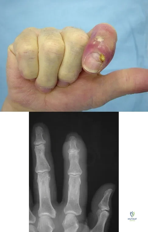
Question 801 High Yield
The condition shown in Figures 9a and 9b is most likely the result of
Detailed Explanation
The clinical photograph and radiograph show gout, which is the result of urate deposition in the joint and soft tissues. Radiographs frequently reveal periarticular erosions. The crystals are intracellular and negatively birefringent under the polarized microscope. Treatment for acute flares include colchicines, indomethacin, and corticosteroids (including injections). Medications such as allopurinol help prevent recurrent flares. Tophi such as that seen in this patient are often confused with and associated with infection. Wortmann RL, Kelley WM: Crystal-induced inflammation: Gout and hyperuricemia, in Harris ED, Budd RC, Firestein GS, et al (eds): Kelley's Textbook of Rheumatology, ed 7. New York, NY, Elsevier Science, 2005, pp 1402-1429. Trumble TE (ed): Hand Surgery Update 3: Hand, Elbow, & Shoulder. Rosemont, IL, American Society for Surgery of the Hand, 2003, pp 433-457.
References:
- Louis DS, Jebson PJ: Mimickers of hand infections. Hand Clin 1998;14:519-529.
<span>Question 802</span> <span>High Yield</span>
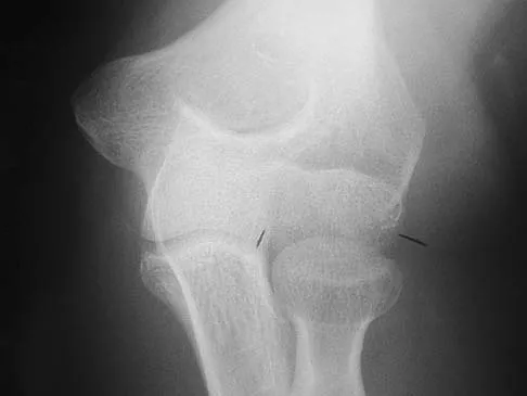
Examination of a 9-year-old girl who injured her left elbow in a fall reveals tenderness and swelling localized to the medial aspect of the elbow. Motor and sensory examinations of the hand are normal, and circulation is intact. A radiograph is seen in Figure 28. Management should consist of
<img alt="General Orthopedics 2026 Practice Questions: Set 9 (Solved) - Figure 3" class="q-img mcq-img" height="393" loading="lazy" onclick="window.open(this.src)" src="/media/mcq-images/25/general-orthopedics-2026-set-9-mcqs-4064-fig-3.webp" title="Click to enlarge" width="280"/>
<button class="opt-btn" data-qid="802" onclick="handleSelect(this, '802', 0)">
<span class="opt-char">A</span>
<span>long arm cast immobilization.</span>
</button>
<button class="opt-btn" data-qid="802" onclick="handleSelect(this, '802', 1)">
<span class="opt-char">B</span>
<span>open reduction and internal fixation, followed by cast immobilization.</span>
</button>
<button class="opt-btn" data-qid="802" onclick="handleSelect(this, '802', 2)">
<span class="opt-char">C</span>
<span>closed reduction and percutaneous pin fixation.</span>
</button>
<button class="opt-btn" data-qid="802" onclick="handleSelect(this, '802', 3)">
<span class="opt-char">D</span>
<span>anterior transposition of the ulnar nerve.</span>
</button>
<button class="opt-btn" data-qid="802" onclick="handleSelect(this, '802', 4)">
<span class="opt-char">E</span>
<span>excision of the loose fragment and repair of the common flexor origin.</span>
</button>
<button onclick="toggleExp('802')" style="background:none; border:none; color:#7f8c8d; text-decoration:underline; cursor:pointer;">Show Explanation</button>
<span class="exp-title">Detailed Explanation</span><div markdown="1">Avulsion fractures of the medial epicondyle are caused by a valgus stress applied to the immature elbow and usually occur in children between the ages of 9 and 14 years. Long-term studies have shown that isolated fractures of the medial epicondyle with between 5 to 15 mm of displacement heal well. Brief immobilization (1 to 2 weeks) in a long arm cast or splint yields results similar to open reduction and internal fixation. Fibrous union of the fragment is not associated with significant symptoms or diminished function. Surgical excision of the fragment yielded the worst results in one study and should be avoided. Open reduction is best reserved for those injuries in which the medial epicondylar fragment becomes entrapped in the elbow joint during reduction and cannot be extracted by closed manipulation. Farsetti P, Potenza V, Caterini R, Ippolito E: Long-term results of treatment of fractures of the medial humeral epicondyle in children. J Bone Joint Surg Am 2001;83:1299-1305.
<strong>References:</strong><ul><li>Josefsson PO, Danielsson LG: Epicondylar elbow fracture in children: 35-year follow-up of 56 unreduced cases. Acta Orthop Scand 1986;57:313-315.</li></ul>
<span>Question 803</span> <span>High Yield</span>
Figure 1 shows the radiograph of an 18-year-old patient who has severe knee pain. Treatment consisting of osteotomy should be perfomed
<img alt="General Orthopedics 2026 Practice Questions: Set 9 (Solved) - Figure 4" class="q-img mcq-img" height="393" loading="lazy" onclick="window.open(this.src)" src="/media/mcq-images/25/general-orthopedics-2026-set-9-mcqs-4064-fig-4.webp" title="Click to enlarge" width="271"/>
<button class="opt-btn" data-qid="803" onclick="handleSelect(this, '803', 0)">
<span class="opt-char">A</span>
<span>above the tibial tubercle.</span>
</button>
<button class="opt-btn" data-qid="803" onclick="handleSelect(this, '803', 1)">
<span class="opt-char">B</span>
<span>at or just below the tibial tubercle.</span>
</button>
<button class="opt-btn" data-qid="803" onclick="handleSelect(this, '803', 2)">
<span class="opt-char">C</span>
<span>in the tibial diaphysis.</span>
</button>
<button class="opt-btn" data-qid="803" onclick="handleSelect(this, '803', 3)">
<span class="opt-char">D</span>
<span>on both the femur and tibia.</span>
</button>
<button class="opt-btn" data-qid="803" onclick="handleSelect(this, '803', 4)">
<span class="opt-char">E</span>
<span>on the femur alone.</span>
</button>
<button onclick="toggleExp('803')" style="background:none; border:none; color:#7f8c8d; text-decoration:underline; cursor:pointer;">Show Explanation</button>
<span class="exp-title">Detailed Explanation</span><div markdown="1">Very large corrections of tibial deformity can be achieved at or just below the tibial tubercle. This level of osteotomy maintains the relationship between the tubercle and the rest of the joint, does not alter patellofemoral mechanics, and avoids complicating possible future conversion to total knee arthroplasty. High tibial osteotomy is contraindicated for large corrections because of excessive elevation of the tibial tubercle and overhang of the lateral plateau. Correction in the tibial diaphysis creates a zig zag pattern in the tibia by correcting below the deformity and risks nonunion in cortical bone. There is no evidence that the femur is deformed; therefore, femoral osteotomy is not indicated.
<strong>References:</strong><ul><li>Murphy SB: Tibial osteotomy for genu varum: Indications, preoperative planning, and technique. Orthop Clin North Am 1994;25:477-482.</li></ul>
