العربية
Part of the Master Guide

Orthopedic Surgery Board Prep: Set 403 (100 MCQs on Fractures & Casting)

Orthopedic Trauma 2026 MCQs: Board Review Questions & Answers (Part 4)

23 Apr 2026 95 min read 81 Views
Figure for Trauma 2006 MCQs - Part 4 - Question 78

Key Takeaway

Here are the crucial details you must know about Orthopedic Trauma 2026 MCQs: Board Review Questions & Answers (Part 4). Top-rated Orthopedic Trauma 2026 MCQs bank. Practice with clinical case questions, orthopedic surgery board review, and evidence-based answers updated for 2026.

Orthopedic Trauma 2026 MCQs: Board Review Questions & Answers (Part 4)

Comprehensive 100-Question Exam


00:00

Start Quiz

Question 1

A 30-year-old woman sustains a transverse amputation of the distal phalanx of the index finger, leaving exposed bone. What is the most appropriate management of the soft-tissue defect?


Explanation

V-Y advancement flaps are ideal for fingertip amputations that are transverse or dorsal oblique in nature. Healing by secondary intention is contraindicated with exposed bone. Shortening of exposed bone to allow primary skin closure is a possible alternative, as long as significant shortening of the index finger is avoided. A Moberg flap is useful only for distal amputations of the thumb. The first dorsal metacarpal artery-island pedicled flap uses tissue from the dorsum of the proximal index finger, and is typically used to resurface defects of the thumb. Fassler PR: Fingertip injuries: Evaluation and treatment. J Am Acad Orthop Surg 1996;4:84-92.

Question 2

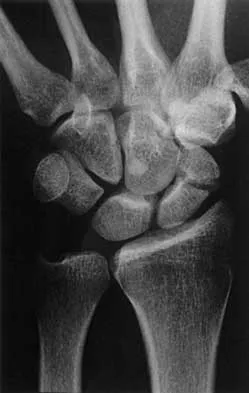
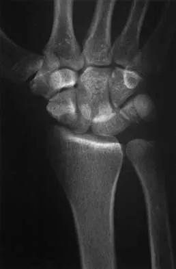
What is the best approach to reduce and stabilize a displaced volar lunate facet fracture of the wrist?


Explanation

A volar lunate fragment of a distal radial fracture is considered a critical component to overall joint stability and function. Obtaining a reduction is difficult through a standard volar approach to the radius between the flexor carpi radialis and radial artery. Visualization and reduction of the ulnar volar facet is not possible from this approach. An extended carpal tunnel incision provides access to the entire articular surface, except for the distal radial styloid component. Hanel DP, Jones MD, Trumble TE: Wrist fractures. Orthop Clin North Am 2002;33:35-57.

Question 3

A 17-year-old man sustained a 5-mm laceration on the lateral aspect of the hindfoot while working on a farm. Examination in the emergency department revealed no fractures. Twenty-four hours later, he returns to the emergency department with increasing foot pain. Thin brown drainage is seen emanating from the wound. He has a temperature of 102.0 degrees F (38.9 degrees C), a pulse rate of 120, and a blood pressure of 80/40 mm Hg. Examination of the foot reveals diffuse swelling, ecchymosis, tenderness, and crepitus with palpation. Current radiographs are shown in Figures 40a and 40b. Management should now consist of


Explanation

40b The mechanism and environment in which the injury occurred, the clinical picture, and the radiographic findings of gas in the tissues suggest an anaerobic Gram-positive bacterial infection. This can be a life- and limb-threatening infection. Treatment should consist of wide debridement of all devitalized tissue, and intravenous antibiotics should be started. Wounds should be left open to allow bacterial effluent and increase oxygen tension in the wound. Hyperbaric oxygen may be used as an adjuvant but is no substitute for debridement. Pellegrini VD, Reid JS, Evarts CM: Complications, in Rockwood CA, Green DP, Bucholz RW, et al (eds): Rockwood and Green's Fractures in Adults, ed 4. Philadelphia, PA, Lippincott-Raven, 1996, vol 1, pp 458-463.

Question 4

A healthy, active, independent 74-year-old woman fell and sustained the elbow injury shown in Figures 41a and 41b. Management should consist of


Explanation

41b Open reduction and internal fixation of distal humeral fractures in elderly patients often fails. These fractures characteristically have a very small distal segment and poor bone quality, resulting in failure of fixation and nonunion. Nonunion is often painful and functionally debilitating. Total elbow arthroplasty provides good results when used for distal humeral fractures in elderly patients with osteopenic bone and fracture patterns thought to be irreconstructable. Long arm casting may result in union, but the resulting stiffness is unacceptable for an active patient. Elbow arthrodesis has few indications. A sling and range-of-motion exercises will often result in a painful and debilitating nonunion at the fracture site. Frankle MA, Herscovici D Jr, DiPasquale TG, et al: A comparison of open reduction and internal fixation and primary total elbow arthroplasty in the treatment of intra-articular distal humerus fractures in women older than 65. J Orthop Trauma 2003;17:473-480. Cobb TK, Morrey BF: Total elbow arthroplasty as primary treatment for distal humerus fractures in elderly patients. J Bone Joint Surg Am 1997;79:826-832.

Question 5

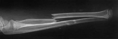
A 35-year-old man sustains a closed Monteggia fracture. Examination reveals that sensation, vascular status, and finger flexion are normal. When he extends his wrist, it deviates radially, and he is unable to extend his fingers or thumb. After reduction of the fracture, what is the next step in treatment for the extensor deficits of the thumb and fingers?


Explanation

The posterior interosseous nerve is located adjacent to the radial neck, placing it at risk for a traction injury with a dislocation of the proximal radius. The typical neurapraxia that results can be expected to resolve with observation within the first 6 to 12 weeks. If recovery is not clinically evident by 3 months, neurophysiologic studies are indicated. Jessing P: Monteggia lesions and their complicating nerve damage. Acta Orthop Scand 1975;46:601-609.

Question 6

A 25-year-old man is brought to the emergency department following a motor vehicle accident. Extrication time was 2 hours, and in the field he had a systolic blood pressure by palpation of 90 mm Hg. Intravenous therapy was started, and on arrival in the emergency department he has a systolic blood pressure of 90 mm Hg with a pulse rate of 130. Examination reveals a flail chest and a femoral diaphyseal fracture. Ultrasound of the abdomen is positive. The trauma surgeons take him to the operating room for an exploratory laparotomy. At the conclusion of the procedure, he has a systolic pressure of 100 mm Hg with a pulse rate of 110. Oxygen saturation is 90% on 100% oxygen, and he has a temperature of 95.0 degrees F (35 degrees C). What is the recommended treatment of the femoral fracture at this time?


Explanation

This is a "borderline trauma" patient where serious consideration for damage control orthopaedic surgery is required. His prolonged hypotension, abdominal injury, and chest injury put him at higher risk for serious postinjury complications. Further surgery, such as definitive fracture fixation, adds metabolic load and injury to his system. It is prudent to consider femoral fracture stabilization with an external fixator until he is physiologically recovered as evidenced by a normal base excess and/or lactate acid levels, as well as all other parameters of resuscitation. A borderline patient has been described as polytrauma with an ISS > 20 and thoracic trauma (AIS > 2); polytrauma and abdominal/pelvic trauma (Moore > 3) and hemodynamic shock (initial BP < 90 mm Hg); ISS > 40; bilateral lung contusions on radiographs; initial mean pulmonary arterial pressure > 24 mm Hg; pulmonary artery pressure increase during intramedullary nailing > 6 mm Hg. Factors that worsen the situation following surgery include multiple long bones and truncal injury (AIS > 2), estimated surgery time of more than 6 hours, arterial injury and hemodynamic instability, and exaggerated inflammatory response (eg, Il-6 > 800 pg/mL). It is incumbent on the orthopaedic surgeon who is a member of the trauma team to make sure that he or she is aware of these factors and guides the team to the best patient care. Pape HC, Hildebrand F, Pertschy S, et al: Changes in the management of femoral shaft fractures in polytrauma patients: From early total care to damage control orthopaedic surgery. J Trauma 2002;53:452-461.

Question 7

A 10-year-old girl has a right elbow deformity that is the result of trauma 5 years ago. She has no pain despite the arm deformity. The radiographs in Figures 42a and 42b show complete healing. This radiographic appearance demonstrates what complication?


Explanation

42b Cubitus varus is a common complication of displaced supracondylar humeral fractures that are treated with closed reduction and cast immobilization. Treatment with closed reduction and percutaneous pinning decreases the incidence of this complication. Cubitus varus also can occur in minimally displaced fractures when unrecognized collapse of the medial column of the distal humerus is not corrected with manipulation. This can be detected on physical examination of the carrying angle or on radiographs measuring Baumann's angle, both in comparison to the opposite side. Cubitus varus may result in unacceptable cosmesis and may predispose the patient to fractures of the lateral condyle. The lateral radiograph demonstrates the crescent sign from overlap of the distal humerus with the olecranon seen in patients with cubitus varus. Patients with growth arrest to the medial trochlear physis would have atrophy of the trochlea on radiographs. Flynn JM, Sarwark JF, Waters PM, et al: The surgical management of pediatric fractures of the upper extremity. Instr Course Lect 2003;52:635-45. Papandrea R, Waters PM: Posttraumatic reconstruction of the elbow in the pediatric patient. Clin Orthop 2000;370:115-126.

Question 8

A 64-year-old woman has left wrist pain and deformity after falling on her hand. Examination shows intact skin and no neurologic or vascular injuries. Radiographs are shown in Figures 43a and 43b. What is the most appropriate management for the injury?


Explanation

43b The patient has a volar displaced two-part intra-articular distal radial fracture-dislocation of the wrist. Although a closed reduction is usually easily obtained, it is very difficult to maintain the reduction without internal fixation. The approach is determined by the direction of the dislocation, in this case volar. Stabilization with a buttress plate neutralizes the axial loading forces on the fractured fragment. A dorsal placed angular stable plate will not provide this buttress effect and will make the reduction difficult.

Question 9

A 26-year-old man was thrown from a car and sustained the injury seen in Figures 44a and 44b. Nonsurgical management of this injury is recommended. Which of the following factors increases the risk of nonunion?


Explanation

44b The patient has a displaced comminuted clavicle middle one third fracture from a high-energy mechanism. Recent literature on high-energy clavicular fractures suggests a higher rate of nonunion than previously reported. A nonunion rate of 30% has been reported by Hill and associates when the fracture fragments are displaced more than 1.5 cm. In addition, several patients had neurologic symptoms related to the injury. Robinson and associates reported an increased risk of nonunion in women, elderly patients, comminuted fractures, and injuries with a lack of cortical contact. Hill JM, McGuire MH, Crosby LA: Closed treatment of displaced middle-third fractures of the clavicle gives poor results. J Bone Joint Surg Br 1997;79:537-539. Wick M, Muller EJ, Kollig E: Midshaft fractures of the clavicle with a shortening of more than 2 cm predispose to nonunion. Arch Orthop Trauma Surg 2001;121:207-211.

Question 10

A 13-year-old girl was riding on an all-terrain vehicle when the driver struck a tree. She sustained the injury shown in Figures 45a through 45d. This injury is best described as what type of acetabular fracture pattern?


Explanation

45b 45c 45d The fracture is a both-column fracture in the Judet/Letournel classification and a C3 in the AO classification. There is extension into the sacroiliac joint along the pelvic brim and comminution along the posterior column above the sciatic notch. Both the anterior and posterior columns are separately broken and displaced. However, the defining feature of a both-column pattern, as seen in this patient, is that all articular fragments are on fracture fragments and no joint surface is left intact to the axial skeleton above. The use of three-dimensional images makes it easier to view the location of the fracture fragments and the amount and direction of displacement. Helfet DL, Beck M, Gautier E, et al: Surgical techniques for acetabular fractures, in Tile M, Helfet DL, Kellam JF (eds): Fractures of the Pelvis and Acetabulum. Philadelphia, PA, Lippincott Williams & Wilkins, 2003, pp 533-603. Tile M: Describing the injury: Classification of acetabular fractures, in Tile M, Helfet DL, Kellam JF (eds): Fractures of the Pelvis and Acetabulum, ed 3. Philadelphia, PA, Lippincott Williams & Wilkins, 2003, pp 427-475.

Question 11

A woman injures the metacarpophalangeal (MCP) joint of her thumb while skiing. Examination reveals tenderness along the ulnar aspect of the MCP joint. Radially directed stress of the joint in full extension produces 5 degrees of angulation. When the MCP joint is flexed 30 degrees, a radially directed stress produces 45 degrees of angulation. Radiographs are otherwise normal. Management should consist of


Explanation

Injuries to the ulnar collateral ligament of the MCP joint of the thumb commonly occur in recreational skiers. Historically, this injury has been referred to as "gamekeeper's thumb." The ligament consists of the proper collateral ligament and the more volar accessory collateral ligament. In extension, the accessory ligament is taut, and in flexion, the proper ligament is taut. For a complete tear of the ligament complex to occur, there must be laxity in full extension. Incomplete tears respond well to thumb spica splinting or casting for 2 to 3 weeks and gradual resumption of range of motion. Prolonged immobilization of incomplete injuries leads to higher rates of MCP joint stiffness. Stener B: Displacement of the ruptured ulnar collateral ligament of the metacarpo-phalangeal joint of the thumb: A clinical and anatomical study. J Bone Joint Surg Br 1971;44:869.

Question 12

A 37-year-old laborer falls 12 feet and sustains a comminuted tibial plafond fracture. Three years after treatment using standard techniques, what will be the most likely outcome?


Explanation

Two recent studies by Pollak and associates and Marsh and associates have focused on function after high-energy tibial plafond fractures. Findings are unfavorable even when anatomic reduction is performed in the best centers and patients are provided excellent rehabilitation. Function improves up to 2 years after injury, but even basic walking skills remain adversely affected. Virtually all patients have long-term adverse general health effects compared to their gender and age-matched peers. Posttraumatic degenerative arthritis is present in most ankles. Patients should be told early about the long-term prognosis, and early vocational/psychological counseling should be given. Despite these adverse outcomes, only a minority of patients require fusion or arthroplasty. Pollak AN, McCarthy ML, Bess RS, et al: Outcomes after treatment of high-energy tibial plafond fractures. J Bone Joint Surg Am 2003;85:1893-1900.

Question 13

A 45-year-old woman sustains an injury to her lower leg. Examination reveals that there is a deformity with no neurologic or vascular problems. The skin is intact. Radiographs are shown in Figures 46a and 46b. Which of the following factors would make closed management the least appropriate choice for this injury?


Explanation

46b All the factors listed, with the exception of an ipsilateral femoral fracture, are representative of a low-energy stable tibial shaft fracture that will do well with closed reduction and immobilization in a long leg cast, followed by weight bearing as tolerated and then a functional brace or patellar tendon bearing cast until union is achieved. Shortening will not increase from that seen on these initial radiographs. The spiral fracture provides a broad surface for healing, and the fibular fracture at another level indicates a stable soft-tissue envelope which, with the immobilization device, will stabilize the fracture reduction. An ipsilateral femoral fracture is a strong indication to surgically stabilize both fractures. Trafton PG: Tibial shaft fractures, in Browner BD (ed): Skeletal Trauma, ed 3. Philadelphia, PA, WB Saunders, 2003, pp 2153-2169.

Question 14

Which of the following medications may have a negative effect on bone healing following fracture?


Explanation

Nonsteroidal anti-inflammatory drugs that are COX-1 primary inhibitors have been shown in animal studies to delay or inhibit fracture healing. COX-2 inhibitors also delay healing but to a lesser extent than COX-1 inhibitors. The other medications listed do not alter fracture callus formation. Gerstenfeld LC, Thiede M, Seibert K, et al: Differential inhibition of fracture healing by non-selective and cyclooxygenase-2 selective non-steroidal anti-inflammatory drugs. J Orthop Res 2003;21:670-675.

Question 15

A 16-year-old boy has abdominal and back pain after being involved in a high-velocity head-on motor vehicle accident. He was restrained in the rear of the automobile by a lap belt only. A radiograph and CT scan are shown in Figure 47. The patient has no other injuries. Optimal management should include


Explanation

Pediatric bony Chance fractures occur following severe flexion injuries as seen after motor vehicle accidents with lap belt restraints. There is a high rate of associated intra-abdominal injuries. In the absence of associated injuries, these fractures are best treated with immobilization. Bed rest is not necessary. Surgical fixation usually is not needed. Surgical stabilization and two-level fusion may be indicated in select individuals with progressive kyphosis of more than 25 degrees or other conditions that preclude cast or brace immobilization. Greenwald TA, Mann DC: Pediatric seatbelt injuries: Diagnosis and treatment of lumbar flexion-distraction injuries. Paraplegia 1994;32:743-751. Glassman SD, Johnson JR, Holt RT: Seatbelt injuries in children. J Trauma 1992;33:882-886.

Question 16

What inflammatory mediator has been most closely associated with the magnitude of the systemic inflammatory response to trauma and with the development of multiple organ dysfunction syndrome (MODS)?


Explanation

Multiple cytokines (inflammatory mediators) are released following trauma, and their levels can be measured in serum. Persistent elevated levels of IL-6 (> 800 pg/mL) indicate an exaggerated systemic inflammatory response to trauma and have been associated with the development of MODS. Recent work has shown that extensive surgical procedures should be avoided when IL-6 levels remain elevated to prevent the precipitation of MODS. In the future, it is likely that this mediator and possibly others will be used to determine timing and techniques of future treatment. Patrick DA, Moore FA, Moore EE, et al: Jack A. Barney Resident Research Award winner: The inflammatory profile of interleukin-6, interleukin-8, and soluble intercellular adhesion molecule-1 in postinjury multiple organ failure. Am J Surg 1996;172:425-429. Pape HC, van Griesven M, Rice J, et al: Major secondary surgery in blunt trauma patients and perioperative cytokine liberation: Determination of the clinical relevance of biochemical markers. J Trauma 2001;50:989-1000.

Question 17

An 8-year-old girl sustained a displaced fracture at the base of the femoral neck in a motor vehicle accident. Management should consist of


Explanation

Pediatric intracapsular hip fractures are challenging because of the high rates of complications, including osteonecrosis and varus malunion. These patients should be treated as emergencies. Principles of treatment include anatomic reduction with internal fixation. Screw fixation short of the physis is preferred and may need to be supplemented with spica cast immobilization. Fixation may be achieved with smooth pins across the physis when little metaphyseal bone is available. Fixation across the physis with threaded screws is acceptable only when the patient is close to skeletal maturity. Pediatric hip screws are appropriate if immediately available. Emergent open reduction, capsulotomy, or joint aspiration may decrease the rate of osteonecrosis. Cheng JC, Tang N: Decompression and stable internal fixation of femoral neck fractures in children can affect the outcome. J Pediatr Orthop 1999;19:338-343. Azouz EM, Karamitsos C, Reed MH, et al: Types and complications of femoral neck fractures in children. Pediatr Radiol 1993;23:415-420. Song KS, Kim YS, Sohn SW, et al: Arthrotomy and open reduction of the displaced fracture of the femoral neck in children. J Pediatr Orthop B 2001;10:205-210.

Question 18

The plate seen in Figure 48a was applied to the fracture seen in Figure 48b, and is functioning in what capacity?


Explanation

48b A Weber type B ankle fracture occurs with a supination external rotation mechanism of injury. The fibula generally fails with a spiral fracture pattern. The lag screws provide compression, and the plate acts to neutralize rotational and angular bending forces. A buttress plate resists vertical shear forces. A tension band is used over areas that may fail in tension, such as an olecranon fracture. Compression is provided by the lag screws, and distraction is again resisted by the lag screws.

Question 19

Which of the following findings is considered the strongest indication for surgical treatment of a mallet fracture of the distal phalanx?


Explanation

The majority of mallet fractures can be treated nonsurgically with a distal interphalangeal joint extension splint. Excellent results can be obtained in most patients with splinting alone. The fragment size, amount of displacement, and degree of articular incongruity usually do not affect final outcome, as long as the joint is reduced. Surgical fixation takes on several forms but is fraught with complications including skin/wound problems, loss of fixation, nonunion, and stiffness of the distal interphalangeal joint. Volar subluxation of the distal phalanx remains the primary indication for surgical treatment. Green DP, Butler TE Jr: Fractures and dislocations in the hand, in Rockwood CA, Green DP, Bucholz RW, Heckman JD (eds): Rockwood and Green's Fractures in Adults, ed 4. Philadelphia, PA, Lippincott-Raven, 1996, pp 621-623.

Question 20

A 20-year-old woman sustained the closed injury shown in Figures 49a and 49b in a motor vehicle accident. Examination reveals that this is an isolated injury; however, she has a complete radial nerve palsy. Management should consist of


Explanation

49b Lacerated radial nerves are associated with open humeral fractures. All open humeral fractures with radial nerve palsy should be managed with radial nerve exploration and skeletal stabilization. Closed humeral fractures with associated radial nerve palsy usually have an intact nerve with neurapraxia. Most of these patients recover without surgical treatment. If the patient has multiple injuries, skeletal stabilization may be indicated to improve mobilization. For an isolated closed humeral fracture with a radial nerve palsy, the treatment of choice is splinting for 1 to 2 weeks, followed by a humeral fracture brace. Ring D, Chin K, Jupiter JB: Radial nerve palsy associated with high-energy humeral shaft fractures. J Hand Surg Am 2004;29:144-147.

Question 21

The fracture shown in Figure 50 is most reliably treated with what form of fixation?


Explanation

The radiograph shows a comminuted proximal ulnar fracture. The most reliable fixation is a posterior plate, acting as a tension band plate. The fracture involves the proximal shaft of the ulna; therefore, a 3.5-mm compression plate or one of similar size should be used to provide adequate stability. Kirschner wires and tension band wires do not provide axial stability of the comminution of the ulna. Compression screws alone will most likely fail and will not provide axial rotational stability to the construct. A medial plate will not resist the distraction forces across this fracture. McKee MD, Seiler JG, Jupiter JB: The application of the limited contact dynamic compression plate in the upper extremity: An analysis of 114 consecutive cases. Injury 1995;26:661-666.

Question 22

A man sustained the injury shown in Figures 51a and 51b. He underwent closed reduction of the radial head dislocation and open reduction and internal fixation of the ulnar fracture. What is the most common cause of persistent radial head subluxation?


Explanation

51b The radiographs reveal a Monteggia injury, with a proximal ulnar shaft fracture and a radial head dislocation. Treatment involves open reduction and internal fixation of the ulnar fracture. With correct reduction of the ulna, the radial head is reducible and remains stable, despite an obvious soft-tissue injury around the elbow. Problems with persistent radial head subluxation are almost always attributed to malreduction of the ulnar fracture. Rare causes of persistent radial head subluxation are interposition of soft tissues in the joint and lateral ligamentous injuries. Jupiter JB, Kellam JF: Diaphyseal fractures of the forearm, in Browner B, Jupiter J, Levine A, Trafton P (eds): Skeletal Trauma, ed 2. Philadelphia, PA, WB Saunders, 1992, pp 1421-1454.

Question 23

Which of the following is an indication for surgical management of a Weber type B distal fibular fracture?


Explanation

A widened medial clear space indicates instability caused by an associated tear of the deltoid ligament; therefore, nonsurgical management is not warranted. Weber type B distal fibular fractures without a deltoid tear have a medial clear space of < 4 mm, even under stress, and may be successfully treated nonsurgically. The presence of medial hindfoot ecchymosis and medial ankle tenderness is not fully indicative of medial soft-tissue instability; however, these findings may indicate a deltoid injury and should raise suspicion of an unstable fracture injury pattern. Stress testing is necessary to demonstrate the presence or absence of instability. A small amount of comminution is also cause for increased suspicion of an unstable pattern; however, it is not a direct contraindication when considering nonsurgical management. Two millimeters of fibular displacement without lateral shift of the talus is an acceptable position when considering nonsurgical management of Weber type B distal fibular fractures. Michelson JD, Magid D, Ney DR, et al, Examination of the pathologic anatomy of ankle fractures. J Trauma 1992;32:65-70.

Question 24

Locked plating techniques have been shown to have biomechanical advantages over standard plating in which of the following scenarios?


Explanation

Locked plating is becoming more common. Some biomechanical data comparing locked plating to standard plating have been reported for osteoporotic distal femoral fractures and humeral shaft fractures. Significant differences were seen mainly for osteoporotic fractures without cortical contact. Not all osteoporotic fractures and all comminuted fractures have been shown to demonstrate significant mechanical improvement with locked plating compared to standard plating. Spiral fractures often can be repaired with a lag screw, obtaining adequate cortical contact. Osteoporotic fractures with a torsion mode of failure failed earlier with locked plating systems than with standard plating systems. Zlowodzki M, Williamson S, Cole PA, et al: Biomechanical evaluation of the less invasive stabilization system, angled blade plate, and retrograde intramedullary nail for the internal fixation of distal femur fractures. J Orthop Trauma 2004;18:494-502.

Question 25

A 25-year-old man sustained a head injury after being ejected from his car. Examination reveals a Glasgow Coma Scale score of 7 and a swollen right knee. Clinical examination shows that the knee is very unstable, suggesting tears of the medial collateral and anterior and posterior cruciate ligaments, as well as the posterior lateral corner. What is the most appropriate first step to rule out a vascular injury?


Explanation

A knee dislocation carries the potential for an arterial injury and has always brought up the question of need for arteriography to rule out this limb-threatening injury. However, arteriography has an inherent complication rate that may compromise the general care of the patient. In over 240 published cases with documented knee dislocations that were evaluated for vascular injury by physical examination (without imaging studies), not a single missed injury was reported, for a 100% negative predictive value (0% false-negative rate). This degree of accuracy at excluding major vascular injury is unsurpassed by the results obtained with arteriography but with no risk involved and a marked savings in time, equipment, and costs. Therefore, the most appropriate first step to rule out vascular injury is examination of the pedal pulses. If there is any doubt about an arterial injury, another option is the ankle-brachial index (ABI). If the ABI is greater than 0.9, the chance of arterial injury is again nonexistent. However, a positive physical examination or an ABI of less than 0.9 is not 100% predictive of an arterial injury; therefore, arteriography is recommended. Miranda FE, Dennis JW, Veldenz HC, et al: Confirmation of the safety and accuracy of physical examination in the evaluation of knee dislocation for injury of the popliteal artery: A prospective study. J Trauma 2002;52:247-252.

Question 26

A 35-year-old male is brought to the trauma bay after a high-speed motorcycle collision. His blood pressure is 80/50 mm Hg and heart rate is 125 beats/min. A FAST exam is negative for intra-abdominal fluid. An AP pelvic radiograph reveals an AP compression type III (APC-III) injury with severe symphyseal diastasis and disruption of both sacroiliac joints. A pelvic binder is appropriately applied. Following the administration of 2 liters of crystalloid and 2 units of uncrossmatched packed red blood cells, his blood pressure remains 85/55 mm Hg. What is the most appropriate next step in management?


Explanation

In a hemodynamically unstable patient with a pelvic ring injury and a negative FAST (no major intra-abdominal bleeding source), the first step in mechanical stabilization is a pelvic binder. If the patient remains hemodynamically unstable despite resuscitation and mechanical stabilization, the shock is presumed to be from venous or arterial pelvic bleeding. The most appropriate next step is preperitoneal pelvic packing and/or pelvic angiography with embolization. External fixation provides mechanical stability similar to a binder but delays the necessary hemorrhage control. Exploratory laparotomy is not indicated with a negative FAST and releases the tamponade effect in the retroperitoneum.

Question 27

A 65-year-old right-hand-dominant woman presents to the clinic 6 months after undergoing volar locking plate fixation for a displaced distal radius fracture. The fracture has healed well radiographically. She now reports a sudden inability to actively flex the interphalangeal (IP) joint of her thumb, though she has no pain. Which of the following technical errors during her index procedure is the most likely cause of this complication?


Explanation

The patient has sustained an iatrogenic rupture of the flexor pollicis longus (FPL) tendon. The FPL tendon is highly vulnerable to attrition over prominent hardware on the volar aspect of the distal radius. Placement of a volar plate distal to the 'watershed line' (the distinct ridge on the volar margin of the distal radius) positions the hardware prominently within the floor of the flexor tendon sheath, leading to FPL tendinopathy and eventual rupture. Screws that are too long dorsally cause extensor tendon irritation or rupture (commonly EPL). Repair of the pronator quadratus has not been consistently shown to prevent FPL rupture if the plate is malpositioned.

Question 28

A 28-year-old male sustains a completely displaced transcervical femoral neck fracture following a fall from height. Radiographs demonstrate a vertical fracture line measuring 65 degrees relative to the horizontal (Pauwels type III). He undergoes closed reduction and internal fixation. Compared to a Pauwels type I fracture, what biomechanical environment predominates at the fracture site in this patient's injury?


Explanation

The Pauwels classification of femoral neck fractures is based on the angle of the fracture line relative to the horizontal plane. Pauwels type III fractures have an angle greater than 50 degrees (highly vertical). This vertical orientation subjects the fracture to immense shear forces and significant bending moments rather than the stable compressive forces seen in horizontal (Pauwels I) fractures. Because of these shear forces, Pauwels III fractures have a notoriously high rate of loss of fixation, varus collapse, and nonunion, especially when treated with parallel cannulated screws alone. Fixed-angle constructs (e.g., sliding hip screws) are frequently favored biomechanically.

Question 29

A 42-year-old skier sustains a high-energy Schatzker VI tibial plateau fracture. On presentation, the leg is tense and markedly swollen. Compartment pressures are measured, yielding a delta P (diastolic blood pressure minus compartment pressure) of 15 mm Hg. A decision is made to perform a two-incision, four-compartment fasciotomy. During the anterolateral incision to release the anterior and lateral compartments, which of the following nerves is at greatest risk of iatrogenic injury?


Explanation

A delta P of less than 30 mm Hg is an absolute indication for emergency fasciotomy. In a standard two-incision technique for the leg, the anterolateral incision accesses the anterior and lateral compartments. The superficial peroneal nerve is at significant risk during this approach, particularly in the middle to distal third of the leg where it pierces the deep fascia to become subcutaneous. The surgeon must carefully identify and protect this nerve when incising the fascia of the lateral compartment. The saphenous nerve is medial, the sural nerve is posterior, and the deep peroneal nerve is protected deep within the anterior compartment along the interosseous membrane.

Question 30

A 68-year-old woman presents with a 4-part proximal humerus fracture. In evaluating the initial plain radiographs and CT scan, the surgeon assesses the risk of subsequent humeral head ischemia. According to the Hertel criteria, which of the following combinations of features is the most reliable predictor of avascular necrosis (AVN) of the humeral head?


Explanation

Hertel et al. identified specific criteria that predict ischemia and subsequent AVN of the humeral head following proximal humerus fractures. The most highly predictive factors include: (1) an anatomic neck fracture (rather than surgical neck), (2) a short calcar segment attached to the articular fragment (metaphyseal extension < 8 mm), and (3) disruption of the medial hinge. When these criteria are present, the risk of AVN is exceedingly high due to the disruption of the anterior circumflex humeral artery and the intraosseous blood supply. Conversely, an intact medial hinge and a long calcar segment (> 8 mm) are protective against ischemia.

Question 31

A 24-year-old farm worker caught his leg in a tractor mechanism, sustaining a severely contaminated open diaphyseal tibia fracture with a 12-cm soft tissue defect and exposed bone (Gustilo-Anderson IIIB). Soil and organic matter are heavily ground into the wound. He is brought to the trauma center within 1 hour. According to evidence-based guidelines for initial antibiotic prophylaxis in this specific scenario, what is the most appropriate empiric intravenous regimen?


Explanation

This is a highly contaminated Gustilo-Anderson IIIB open fracture occurring in an agricultural setting. Standard protocol for severe open fractures (Type III) includes a first-generation cephalosporin (Cefazolin) to cover Gram-positive organisms, plus an aminoglycoside (Gentamicin) or third-generation cephalosporin to cover Gram-negative organisms. Additionally, in the setting of farm/agricultural injuries or profound soil/fecal contamination, there is a distinct risk for Clostridium perfringens (gas gangrene) infection. High-dose Penicillin G is explicitly added to the regimen to provide coverage against these anaerobic, spore-forming organisms.

Question 32

A 32-year-old male is 8 weeks post-operative from an open reduction and internal fixation of a displaced talar neck fracture (Hawkins type II). Routine follow-up radiographs demonstrate a subchondral radiolucent band in the dome of the talus on the AP view. What is the clinical significance of this radiographic finding?


Explanation

The finding described is the 'Hawkins sign'. It is a subchondral radiolucent band in the dome of the talus that typically appears 6 to 8 weeks after a talus fracture. This radiolucency represents subchondral osteopenia (bone resorption) secondary to disuse and active hyperemia. For active hyperemia and bone resorption to occur, the talar body must have an intact blood supply. Therefore, the presence of a Hawkins sign is a highly reliable indicator of intact vascularity and implies that avascular necrosis (AVN) of the talar body will not occur.

Question 33

A 45-year-old male falls from a ladder and sustains an acetabular fracture. The CT scan demonstrates a transverse fracture line across the acetabulum with a large, comminuted posterior wall fragment. The femoral head is subluxated posteriorly. The surgeon plans for open reduction and internal fixation. Which surgical approach provides the most direct access for anatomic reduction and plating of the involved columns in this fracture pattern?


Explanation

The patient has a transverse + posterior wall acetabular fracture. The Kocher-Langenbeck approach is the gold standard for accessing the posterior column and posterior wall of the acetabulum. Because the posterior wall fragment must be directly visualized, reduced anatomically, and buttressed with a plate to ensure hip stability, a posterior approach is mandated. The transverse component can often be reduced indirectly through the posterior exposure using specific clamps (e.g., Jungbluth or Weber clamps) placed into the ischium and intact ilium. The ilioinguinal and modified Stoppa approaches are reserved for anterior column, anterior wall, and associated both-column fractures where anterior access is paramount.

Question 34

A 30-year-old male presents to the emergency department after a direct blow to the leg during a rugby match. Radiographs show a closed, comminuted midshaft tibia fracture. He is complaining of agonizing pain despite receiving intravenous opioids. The nurse notes that his foot is swollen. Which of the following physical examination findings is the most sensitive early clinical indicator of acute compartment syndrome?


Explanation

Pain out of proportion to the injury and pain with passive stretch of the muscles traversing the affected compartment are the earliest, most sensitive clinical signs of acute compartment syndrome. While the '5 Ps' (Pain, Pallor, Pulselessness, Paresthesias, Paralysis) are classically taught, pallor, pulselessness, and paralysis are late findings that indicate severe, often irreversible, tissue ischemia. Waiting for the absence of pulses to diagnose compartment syndrome will result in catastrophic muscle necrosis and nerve damage.

Question 35

A 78-year-old woman with a history of a cemented left total hip arthroplasty performed 15 years ago falls from standing. Radiographs reveal a spiral periprosthetic femur fracture originating at the tip of the femoral stem and extending distally. The cement mantle is cracked, and the stem has subsided 1.5 cm into the canal, demonstrating gross radiographic loosening. However, the proximal femoral bone stock remains robust and circumferentially intact. According to the Vancouver classification, what is the classification of this fracture and the standard accepted treatment?


Explanation

The Vancouver classification for periprosthetic proximal femur fractures is highly tested. Type A is in the trochanteric region. Type B is around or just distal to the stem tip. Type C is well below the stem tip. Type B is subdivided by stem stability and bone stock: B1 (stable stem, good bone) treated with ORIF; B2 (loose stem, good bone) treated with revision arthroplasty bypassing the fracture; B3 (loose stem, poor bone) treated with revision and proximal femoral replacement or allograft. This patient has a fracture at the stem tip (Type B) with a loose stem (subsided 1.5 cm) but good proximal bone stock, classifying it as a Vancouver B2. The standard of care is revision arthroplasty using a long-stem prosthesis that bypasses the fracture by at least two cortical diameters.

Question 36

A 35-year-old male is brought to the emergency department after a high-speed motorcycle collision. His blood pressure is 70/40 mm Hg and heart rate is 135 bpm. A FAST exam is negative. An anteroposterior pelvic radiograph shows a widened pubic symphysis of 4 cm and widened bilateral sacroiliac joints. A pelvic binder is placed, and he receives 2 units of uncrossmatched packed red blood cells. His blood pressure improves transiently to 85/50 mm Hg. What is the next most appropriate step in management?


Explanation

This patient has a hemodynamically unstable pelvic ring injury (APC-III equivalent). The initial step is stabilization with a pelvic binder and resuscitation. With a negative FAST exam, the abdomen is less likely to be the source of major hemorrhage, pointing toward retroperitoneal bleeding from the pelvis. If the patient remains unstable or transiently responds, definitive hemorrhage control via preperitoneal pelvic packing or angiography with embolization is the standard of care. CT scanning is contraindicated in a hemodynamically unstable patient. Exploratory laparotomy is not indicated for isolated retroperitoneal pelvic bleeding and releases the tamponade effect.

Question 37

A 28-year-old man sustains a completely displaced, vertically oriented (Pauwels type III) femoral neck fracture after falling from a roof. He is taken to the operating room for closed reduction and internal fixation. Which of the following biomechanical constructs provides the most stable fixation for this specific fracture pattern?


Explanation

Pauwels type III femoral neck fractures have a highly vertical orientation (angle > 50 degrees), which creates large shear forces across the fracture site. This makes them highly unstable and prone to varus collapse and nonunion. Biomechanical studies have demonstrated that a fixed-angle device, such as a sliding hip screw (often supplemented with a derotational screw to prevent rotation of the femoral head), provides superior biomechanical stability and resistance to vertical shear compared to multiple parallel cannulated screws.

Question 38

A 42-year-old woman sustains a high-energy distal femur fracture. A CT scan of the knee reveals a displaced coronal plane fracture of the lateral femoral condyle. What is the most appropriate fixation strategy for this specific articular fragment?


Explanation

A coronal plane fracture of the femoral condyle is known as a Hoffa fracture, most commonly involving the lateral condyle. Because the fragment is located posteriorly, the standard surgical technique for fixation involves placing lag screws in an anterior-to-posterior direction to compress the fracture fragment against the intact anterior condyle. Screws are often countersunk within the articular cartilage if placed through the weight-bearing zone, or placed outside the articular margin when possible.

Question 39

A 33-year-old man sustains a Gustilo-Anderson Type IIIB open tibia fracture involving the distal third of the tibial diaphysis with a large 10x12 cm anterior soft tissue defect. After aggressive serial debridements and skeletal stabilization, he requires soft tissue coverage. Which of the following options is the most reliable soft tissue flap for this specific defect?


Explanation

The lower extremity is traditionally divided into thirds when planning soft tissue coverage for open tibia fractures. The proximal third is typically covered by a gastrocnemius rotational flap, while the middle third is usually covered by a soleus rotational flap. The distal third of the tibia lacks adequate local muscle bulk for reliable rotational coverage of large defects; therefore, a free tissue transfer (e.g., anterolateral thigh flap, latissimus dorsi, or rectus abdominis) is the gold standard for robust coverage.

Question 40

A 25-year-old snowboarder sustains a hyperdorsiflexion injury to his right ankle. Radiographs reveal a displaced fracture of the talar neck with subluxation of the subtalar joint. The tibiotalar and talonavicular joints remain congruent. According to the Hawkins classification, what is the approximate expected rate of avascular necrosis (AVN) of the talar body?


Explanation

This injury describes a Hawkins Type II talar neck fracture, which is characterized by a talar neck fracture with subluxation or dislocation of the subtalar joint, while the ankle joint remains normally aligned. The historical rate of avascular necrosis (AVN) for a Type II fracture is approximately 20% to 50%. In contrast, Type I (nondisplaced) fractures have an AVN rate of 0-10%, Type III (subtalar and tibiotalar dislocation) have an AVN rate of 70-90%, and Type IV (involving talonavicular dislocation as well) approach a 100% AVN rate.

Question 41

A 45-year-old male undergoes open reduction and internal fixation of a Schatzker VI tibial plateau fracture. Postoperatively, he requires rapidly increasing amounts of intravenous opioids. On examination, his leg is tense, and he experiences excruciating pain with passive stretch of his great toe. His dorsalis pedis pulse is palpable. Intracompartmental pressure testing shows an absolute pressure of 45 mm Hg in the anterior compartment, and his diastolic blood pressure is 65 mm Hg. What is the most appropriate next step?


Explanation

The patient exhibits classic clinical signs of acute compartment syndrome (pain out of proportion, pain with passive stretch, tense compartments) and has an objective Delta P (diastolic blood pressure minus absolute compartment pressure) of 20 mm Hg (65 - 45 = 20 mm Hg). A Delta P of less than 30 mm Hg is an absolute indication for emergency fasciotomy. The presence of a palpable pulse does not rule out compartment syndrome, as arterial pressure remains higher than compartment pressure until late in the disease process. Immediate four-compartment fasciotomy is required.

Question 42

A 62-year-old woman is evaluated 6 months after undergoing volar locked plating of a distal radius fracture. She reports the sudden inability to actively flex the interphalangeal joint of her thumb. She denies any new trauma. Lateral radiographs show that the distal edge of the volar plate is positioned prominent and distal to the watershed line. Which of the following is the most likely cause of her presentation?


Explanation

The patient presents with an inability to actively flex the thumb interphalangeal joint, indicating a failure of the flexor pollicis longus (FPL) tendon. Volar plates placed distal to the watershed line of the distal radius can irritate and eventually cause attritional rupture of the flexor tendons. The FPL tendon is most commonly affected due to its close anatomical proximity to the volar surface of the distal radius. EPL ruptures present as an inability to extend the thumb and are associated with nondisplaced fractures or prominent dorsal screws.

Question 43

A 78-year-old woman with a history of a cemented total hip arthroplasty performed 10 years ago falls and sustains a periprosthetic femur fracture. Radiographs demonstrate a fracture around the tip of the stem. The stem appears to be well-fixed with no evidence of cement mantle fracture or subsidence. According to the Vancouver classification, what is the most appropriate management for this injury?


Explanation

This is a Vancouver B1 periprosthetic fracture, defined as a fracture around or just distal to a well-fixed femoral stem. The standard treatment for a Vancouver B1 fracture is open reduction and internal fixation (ORIF), typically utilizing a locked lateral plate with cables or unicortical screws proximally around the stem and bicortical screws distally. Revision arthroplasty is indicated for Vancouver B2 fractures (loose stem with adequate bone stock) or B3 fractures (loose stem with poor bone stock).

Question 44

A 22-year-old man presents to the emergency department after sustaining a single gunshot wound to the right knee. Radiographs reveal a retained bullet lodged entirely within the intra-articular space of the knee joint. There is no associated fracture. After appropriate initial tetanus prophylaxis and administration of antibiotics, what is the most appropriate definitive management of the retained bullet?


Explanation

Bullets lodged within an intra-articular space (such as the knee joint) must be surgically removed, either arthroscopically or via arthrotomy. The synovial fluid within the joint acts as a solvent for lead, which can lead to systemic lead toxicity (plumbism). Additionally, a retained intra-articular bullet acts as a third body, causing rapid mechanical destruction of the articular cartilage. Extra-articular bullets embedded in muscle or soft tissue without neurovascular compromise can generally be left in place.

Question 45

A 40-year-old construction worker sustains a displaced intra-articular calcaneus fracture (Sanders Type III). He is scheduled for open reduction and internal fixation utilizing a standard extensile lateral approach. Which of the following neurological structures is at greatest risk of iatrogenic injury during the creation and full-thickness elevation of this surgical flap?


Explanation

The extensile lateral approach to the calcaneus involves creating an L-shaped full-thickness fasciocutaneous flap to expose the lateral wall and subtalar joint. The sural nerve courses along the posterolateral aspect of the calf and lateral hindfoot. It is at significant risk of injury—either through direct transection during the incision or via traction neuritis during flap retraction. Retracting the flap using 'no-touch' techniques with K-wires placed into the talus and fibula helps minimize soft tissue and nerve damage.

Question 46

A 45-year-old man is brought to the emergency department after a high-speed motorcycle collision. He is hemodynamically unstable with a blood pressure of 80/40 mm Hg and a heart rate of 125 beats/min. A FAST examination is negative. The anteroposterior pelvic radiograph reveals an Anteroposterior Compression Type III (APC-III) pelvic ring injury. A pelvic binder is ordered to assist with hemodynamic stabilization. What is the most appropriate anatomical landmark for the optimal placement of the pelvic binder to effectively reduce the pelvic volume?


Explanation

Pelvic binders are most effective in reducing pelvic volume and controlling venous hemorrhage when placed centered over the greater trochanters. Placement over the iliac crests is a common error and is less effective; in certain fracture patterns (such as some lateral compression injuries), high placement can paradoxically exacerbate the deformity or internal bleeding.

Question 47

A 38-year-old man sustains a closed, high-energy injury to his right knee. A computed tomography (CT) scan reveals a bicondylar tibial plateau fracture with a large, displaced posteromedial shear fragment. What is the most appropriate surgical approach to achieve anatomical reduction and stable buttress fixation of this specific posteromedial fragment?


Explanation

The posteromedial shear fragment in tibial plateau fractures (often part of Schatzker IV or VI patterns) is difficult or impossible to effectively reduce and buttress from an anterior or anterolateral approach. The optimal approach is a posteromedial approach. This typically utilizes the interval between the medial head of the gastrocnemius (which is retracted laterally) and the pes anserinus tendons (which are retracted medially or anteriorly) to place a posterior buttress plate directly opposing the deforming forces.

Question 48

A 75-year-old right-hand-dominant woman with a history of osteoporosis falls onto her shoulder. Radiographs demonstrate a displaced 4-part proximal humerus fracture with head-splitting components, severe comminution of the tuberosities, and a disrupted medial calcar hinge. Her pre-injury baseline was active and independent. Which of the following surgical options is associated with the most predictable restoration of forward elevation and pain relief in this patient?


Explanation

In elderly patients with complex, displaced 3- or 4-part proximal humerus fractures, particularly those with poor bone quality, head-splitting components, and disrupted medial hinges, reverse total shoulder arthroplasty (RTSA) provides more predictable outcomes regarding pain relief and functional restoration (especially forward elevation) compared to ORIF or hemiarthroplasty. Hemiarthroplasty outcomes are heavily dependent on anatomical tuberosity healing, which is highly unpredictable in the osteoporotic elderly population.

Question 49

A 25-year-old man sustains a vertical, displaced femoral neck fracture (Pauwels type III) after a fall from a height. He is taken to the operating room for closed reduction and internal fixation. Which of the following fixation constructs provides the greatest biomechanical stability against the predominant deforming forces for this specific fracture pattern?


Explanation

Pauwels type III femoral neck fractures are vertically oriented (>50 degrees) and are subject to high shear forces. Biomechanical studies have consistently demonstrated that a fixed-angle construct, such as a sliding hip screw (dynamic hip screw), provides superior stability against vertical shear stress compared to multiple cancellous lag screws. A supplemental derotation screw is often added to control rotational forces.

Question 50

A 32-year-old construction worker sustains a Gustilo-Anderson Type IIIB open tibia fracture. He undergoes immediate irrigation and debridement, and skeletal stabilization with an external fixator. Following sequential debridements, the wound bed is deemed clean, but there is exposed diaphyseal bone devoid of periosteum requiring a free tissue transfer. To minimize the risk of deep infection and flap failure, soft-tissue coverage should ideally be performed within what timeframe from the initial injury?


Explanation

Based on classic principles established by Godina and supported by subsequent orthoplastic literature, early soft-tissue coverage of open tibia fractures requiring flaps should ideally be performed within 72 hours, and generally no later than 7 days from the injury. Delayed coverage beyond 7 days significantly increases the rates of deep infection, flap failure, and eventual nonunion.

Question 51

A 28-year-old man sustains a closed diaphyseal fracture of the tibia. Eight hours post-admission, he complains of worsening leg pain that is out of proportion to the injury and not relieved by intravenous opioids. Examination reveals tense calf compartments and excruciating pain with passive dorsiflexion of the hallux. His blood pressure is 110/70 mm Hg. Intracompartmental pressure monitoring is obtained. Which of the following pressure readings provides the strongest absolute indication for emergent fasciotomy?


Explanation

The most reliable and universally accepted threshold for diagnosing acute compartment syndrome in a borderline or uncooperative patient is the delta pressure (Delta P), calculated as the diastolic blood pressure minus the measured intracompartmental pressure. A Delta P of less than 30 mm Hg indicates critically impaired tissue perfusion and is an absolute indication for emergent fasciotomy. In this scenario, a Delta P of 20 mm Hg (indicating a compartment pressure of 50 mm Hg) clearly mandates surgical release.

Question 52

A 40-year-old woman undergoes open reduction and internal fixation for an unstable pronation-external rotation ankle fracture. Intraoperatively, the external rotation stress test demonstrates widening of the medial clear space and the distal tibiofibular articulation. Syndesmotic fixation is planned. Which of the following statements is true regarding syndesmotic reduction and fixation?


Explanation

Malreduction of the syndesmosis is a frequent and detrimental complication, occurring in up to 50% of cases when relying on 2D fluoroscopy alone. Direct open visualization of the syndesmosis (specifically the anterior tibiofibular articulation) or the use of intraoperative 3D imaging significantly improves the accuracy of reduction. The classic teaching of maximal dorsiflexion has been largely debunked. Suture-buttons have similar or lower malreduction rates compared to screws, and routine screw removal is no longer considered universally mandatory.

Question 53

An 82-year-old woman with severe rheumatoid arthritis and baseline limited household mobility sustains a highly comminuted, intra-articular distal femur fracture (OTA/AO 33-C3) after a fall. Her bone stock is extremely osteopenic. What is the primary advantage of choosing a distal femoral replacement (DFR) over open reduction and internal fixation (ORIF) with a lateral locking plate in this specific patient?


Explanation

In elderly, osteoporotic patients with complex, comminuted intra-articular distal femur fractures, achieving stable fixation for early mobilization via ORIF is challenging and often requires a period of restricted weight-bearing. Distal femoral replacement (DFR) bypasses the fracture and allows for immediate full weight-bearing. This faster mobilization is critical in the frail elderly population to prevent the severe medical complications of prolonged immobility. DFR generally carries a higher risk of blood loss and infection compared to ORIF.

Question 54

A 19-year-old competitive cyclist falls onto his left shoulder. Radiographs reveal a completely displaced midshaft clavicle fracture. Which of the following radiographic findings is the strongest relative indication for operative intervention (ORIF) over nonoperative management to optimize functional recovery and minimize the risk of symptomatic nonunion?


Explanation

Complete displacement, particularly with shortening greater than 20 mm (or 2 cm), is a strong relative indication for operative fixation of midshaft clavicle fractures. Multiple prospective studies have shown that shortening >2 cm treated nonoperatively is associated with significantly higher rates of nonunion, symptomatic malunion, altered shoulder kinematics, and residual weakness, particularly in highly active patients or laborers.

Question 55

A 22-year-old man sustains a low-velocity handgun wound to the right thigh. His hemodynamics are stable, and neurovascular examination is entirely intact. Radiographs show a midshaft femur fracture with a retained bullet fragment situated within the vastus intermedius muscle belly. There is no evidence of intra-articular extension or compartment syndrome. What is the most appropriate initial management?


Explanation

For low-velocity gunshot wounds resulting in a fracture without vascular compromise, compartment syndrome, or intra-articular extension, standard management includes local wound care (superficial debridement of the entry and exit sites), a short course of systemic antibiotics (often a first-generation cephalosporin), and appropriate fracture stabilization (which can be delayed or immediate depending on the fracture pattern and hospital protocol). The bullet fragment itself does not routinely need to be extracted unless it is intra-articular (risk of lead arthropathy), compressing a neurovascular structure, or superficial enough to cause skin irritation.

Question 56

A 45-year-old man is brought to the trauma bay after a high-speed motorcycle collision. His blood pressure is 80/40 mm Hg and his heart rate is 135 bpm. Despite receiving 2 units of uncrossmatched packed red blood cells, he remains hemodynamically unstable. A pelvic binder has been applied. The FAST exam is negative. An AP pelvis radiograph reveals an APC-III (open-book) pelvic ring injury with wide disruption of the pubic symphysis and bilateral sacroiliac joints. What is the most appropriate next step in management?


Explanation

In a hemodynamically unstable patient with an open-book pelvic ring injury and a negative FAST exam, the primary source of bleeding is presumed to be the retroperitoneal venous plexus or cancellous bone from the pelvic fracture. Current advanced trauma guidelines recommend mechanical stabilization (e.g., pelvic binder or external fixator) followed by immediate pre-peritoneal pelvic packing and/or pelvic angioembolization. A CT scan is contraindicated in an unstable patient, and laparotomy is generally not indicated for retroperitoneal pelvic bleeding unless there is concurrent intra-abdominal injury.

Question 57

A 28-year-old male sustains a vertically oriented, displaced femoral neck fracture (Pauwels type III) in a motor vehicle accident. He is scheduled for surgical fixation. According to biomechanical studies, which of the following constructs provides the greatest stability for this specific fracture pattern?


Explanation

Pauwels type III femoral neck fractures are characterized by a vertically oriented fracture line, which exposes the fracture to high shear forces and a greater risk of varus collapse and nonunion. Biomechanical studies have consistently shown that a fixed-angle construct, such as a sliding hip screw (SHS), provides superior biomechanical stability and resistance to shear forces compared to multiple cancellous screws. Adding a superior derotational screw helps control rotation of the femoral head during insertion and weight-bearing.

Question 58

A 35-year-old construction worker sustains a highly comminuted midshaft tibia fracture with a 12 cm anterior soft-tissue defect exposing bone (Gustilo-Anderson type IIIB) after a crush injury. He undergoes emergent irrigation, debridement, and placement of an external fixator. Serial debridements render the wound bed clean. To minimize the risk of deep infection and flap failure, what is the optimal timing for definitive soft-tissue coverage?


Explanation

The timing of soft-tissue coverage for Gustilo type IIIB open tibia fractures is critical. The LEAP (Lower Extremity Assessment Project) study and subsequent meta-analyses have demonstrated that definitive soft-tissue coverage performed within 7 days (and ideally within 72 hours) of the injury is associated with significantly lower rates of deep infection, flap failure, and nonunion compared to coverage performed after 7 days, provided the wound has been adequately debrided.

Question 59

A 65-year-old woman undergoes open reduction and internal fixation of a 3-part proximal humerus fracture using a lateral deltoid-splitting approach and a locking plate. Postoperatively, she demonstrates a positive Hornblower's sign (inability to actively maintain external rotation of the arm in 90 degrees of abduction). Which nerve is most likely to have been injured?


Explanation

Hornblower's sign is highly specific for weakness or absence of the teres minor muscle. The teres minor, along with the deltoid, is innervated by the axillary nerve. The axillary nerve is particularly vulnerable to injury during a lateral deltoid-splitting approach to the proximal humerus if the dissection extends more than 5 cm distal to the acromion, or it can be injured at the time of the initial fracture.

Question 60

A 32-year-old male is admitted with a closed diaphyseal tibia fracture. Twelve hours later, he complains of severe, unrelenting leg pain that is exacerbated by passive stretch of his toes. The clinical suspicion for acute compartment syndrome is high, and continuous compartment pressure monitoring is initiated. Which of the following pressure measurements is the most widely accepted threshold indicating the need for an emergent fasciotomy?


Explanation

McQueen et al. established that utilizing a delta pressure—calculated as the diastolic blood pressure minus the absolute compartment pressure—is the most reliable threshold for diagnosing acute compartment syndrome. A delta pressure of less than 30 mm Hg indicates critically impaired tissue perfusion and is a universally accepted threshold for proceeding with an emergent fasciotomy. Absolute pressure alone can lead to unnecessary fasciotomies or missed diagnoses depending on the patient's systemic blood pressure.

Question 61

An 80-year-old woman with severe osteoporosis presents to the emergency department after a fall. Radiographs reveal a highly comminuted, intra-articular distal femur fracture (AO/OTA 33-C3) with profoundly poor bone stock. Prior to the injury, she used a walker for short-distance ambulation. Which of the following surgical interventions will best allow for immediate, full weight-bearing in this patient?


Explanation

In elderly, osteoporotic patients presenting with severely comminuted intra-articular distal femur fractures, achieving stable internal fixation is challenging, and postoperative restricted weight-bearing is usually necessary with ORIF. Distal femoral replacement (megaprosthesis) bypasses the compromised bone, providing immediate stability and allowing the patient to weight-bear fully right after surgery. This significantly reduces the risks associated with prolonged immobility in the geriatric population.

Question 62

A 34-year-old male who sustained a Hawkins type II talar neck fracture 8 weeks ago returns for a follow-up clinic visit. His AP ankle radiograph reveals a distinct subchondral radiolucent band in the dome of the talus. What does this radiographic finding indicate?


Explanation

The presence of a subchondral radiolucent band in the talar dome, visible on an AP or mortise radiograph 6 to 8 weeks after a talar neck fracture, is known as the Hawkins sign. This radiolucency represents subchondral bone resorption (osteopenia) secondary to disuse. Because bone resorption requires an active blood supply, a positive Hawkins sign is a highly reliable prognostic indicator that the vascularity to the talar body is intact, making the development of avascular necrosis highly unlikely.

Question 63

A 22-year-old collegiate football player sustains a high-energy foot injury. Advanced imaging reveals a purely ligamentous Lisfranc injury with complete disruption of the Lisfranc ligament complex and dorsal subluxation of the 1st, 2nd, and 3rd tarsometatarsal (TMT) joints. Based on recent prospective evidence, which of the following is the most appropriate primary surgical management?


Explanation

For purely ligamentous Lisfranc injuries, multiple prospective randomized studies (e.g., Ly and Coetzee) have demonstrated that primary arthrodesis of the involved medial column TMT joints yields significantly better functional outcomes, decreased pain scores, and a much lower rate of revision surgeries compared to open reduction and internal fixation (ORIF). ORIF remains a standard option for bony Lisfranc fracture-dislocations.

Question 64

A 28-year-old farmer sustains a Gustilo-Anderson Type IIIA open tibia fracture after his leg is caught in a tractor implement. The wound is heavily contaminated with soil and manure. He is brought to the emergency department for initial resuscitation. In addition to prompt surgical debridement, which of the following prophylactic antibiotic regimens is classically recommended for this specific injury pattern?


Explanation

Open fractures in agricultural environments or those with heavy soil contamination are at high risk for infection with anaerobic organisms, most notably Clostridium perfringens, which can cause gas gangrene. The classic board-tested prophylactic antibiotic regimen for such injuries involves a first-generation cephalosporin (Cefazolin) to cover Gram-positives, an aminoglycoside (Gentamicin) for Gram-negatives, and the addition of high-dose Penicillin G to specifically target anaerobic clostridial species.

Question 65

A 35-year-old man falls from a roof and sustains a severely comminuted, joint-depressed intra-articular calcaneus fracture (Sanders Type IV). Due to his heavy smoking history and poorly controlled diabetes, a shared decision is made to proceed with nonoperative management. What is the most common long-term clinical consequence of managing this displaced intra-articular fracture nonoperatively?


Explanation

The most common and significant long-term complication of a displaced intra-articular calcaneus fracture, particularly when managed nonoperatively, is the development of post-traumatic subtalar arthritis. This results from the residual incongruity of the posterior facet of the subtalar joint and altered hindfoot biomechanics (loss of calcaneal height, increased width, and varus malalignment). Patients frequently present with chronic lateral hindfoot pain and stiffness, eventually requiring a subtalar arthrodesis.

Question 66

A 45-year-old man is brought to the trauma bay after a high-speed motorcycle collision. He has a heart rate of 130 bpm and a blood pressure of 80/50 mm Hg. A pelvic radiograph demonstrates an anteroposterior compression type III (APC-III) pelvic ring injury. A pelvic binder is applied, but his blood pressure remains 85/50 mm Hg after initial fluid resuscitation. A Focused Assessment with Sonography for Trauma (FAST) examination is negative. What is the next best step in management?


Explanation

In a hemodynamically unstable patient with a pelvic ring injury and a negative FAST exam (which rules out massive intra-abdominal hemorrhage), the source of bleeding is presumed to be the pelvis (venous plexus or arterial injury). Management dictates immediate mechanical stabilization (e.g., pelvic binder), followed by interventions to control the hemorrhage directly, such as preperitoneal pelvic packing or angioembolization. A CT scan is contraindicated in a hemodynamically unstable patient.

Question 67

A 35-year-old woman sustains a high-energy Schatzker VI tibial plateau fracture. Upon admission, her leg is severely swollen and tense. Her current diastolic blood pressure is 60 mm Hg. Intracompartmental pressure measurement of the anterior compartment of the lower leg is 35 mm Hg. What is the most appropriate management of her lower extremity?


Explanation

The diagnosis of acute compartment syndrome is primarily clinical, but absolute pressure or delta pressure measurements are valuable, especially in polytraumatized or obtunded patients. A delta pressure (Diastolic BP - Compartment Pressure) of less than 30 mm Hg is a well-established threshold that is highly suggestive of acute compartment syndrome. In this patient, the delta pressure is 25 mm Hg (60 - 35). Immediate four-compartment fasciotomy is indicated to prevent irreversible muscle necrosis and nerve ischemia.

Question 68

A 28-year-old man sustains a displaced, vertically oriented (Pauwels type III) femoral neck fracture after a fall from a height. He is an otherwise healthy construction worker. To minimize the risk of shear-related fixation failure and subsequent nonunion, which of the following is the most biomechanically appropriate surgical fixation construct?


Explanation

Pauwels type III femoral neck fractures in young adults are highly unstable due to the vertical orientation of the fracture line, which subjects the fracture site to significant shear forces during weight-bearing. Biomechanical studies have shown that a fixed-angle construct, such as a sliding hip screw, provides superior stability against vertical shear forces compared to multiple cancellous screws. In young adults, head-preserving surgery is prioritized over arthroplasty.

Question 69

A 42-year-old agricultural worker sustains a severe open tibia fracture (Gustilo-Anderson IIIB) that is heavily contaminated with soil and manure. He arrives at the emergency department within 1 hour of the injury. According to current trauma guidelines, what is the most appropriate initial intravenous antibiotic regimen?


Explanation

Standard antibiotic prophylaxis for severe open fractures (Gustilo Type III) includes a first-generation cephalosporin (for Gram-positive coverage) and an aminoglycoside or third-generation cephalosporin (for Gram-negative coverage). In the setting of gross agricultural or soil contamination, there is a high risk of anaerobic infection, particularly Clostridium species, which can cause gas gangrene. Therefore, high-dose penicillin (or metronidazole/ampicillin) must be added to the regimen to provide adequate anaerobic coverage.

Question 70

A 22-year-old collegiate football player sustains a hyperplantarflexion injury to his midfoot. Radiographs demonstrate widening of the interval between the bases of the first and second metatarsals. Weight-bearing views confirm a 4-mm diastasis. MRI reveals complete disruption of the Lisfranc ligament complex without associated fractures. What is the most appropriate definitive surgical management?


Explanation

For purely ligamentous Lisfranc injuries in young, active patients, strong clinical evidence (including prospective randomized trials) demonstrates that primary arthrodesis of the medial column (1st, 2nd, and 3rd TMT joints) results in superior functional outcomes, a lower rate of hardware-related complications, and a significantly lower need for revision surgery compared to open reduction and internal fixation (ORIF). ORIF is generally preferred for cases with significant bony involvement.

Question 71

A 60-year-old woman presents with persistent lower thigh pain 9 months after undergoing retrograde intramedullary nailing for a supracondylar distal femur fracture. Radiographs reveal a hypertrophic nonunion at the fracture site with abundant callus formation but a persistent fracture line. Laboratory markers for infection (CRP, ESR) are strictly normal. What is the most appropriate management?


Explanation

A hypertrophic nonunion indicates that the fracture site has adequate biological healing potential (excellent blood supply leading to callus formation) but lacks sufficient mechanical stability to bridge the gap. The treatment of choice for a hypertrophic nonunion is to improve the mechanical stability of the construct. Revision to a stiffer construct, such as exchanging to a larger nail or applying a fixed-angle locking plate, limits the excessive micro-motion and allows the fracture to heal. Bone grafting is unnecessary as the biology is already active.

Question 72

A 34-year-old man sustains a Hawkins Type III fracture of the talar neck following a severe motor vehicle collision. In this specific injury pattern, which of the following blood vessels typically represents the ONLY remaining source of perfusion to the talar body?


Explanation

A Hawkins Type III fracture involves a fracture of the talar neck with dislocation of both the subtalar and tibiotalar joints. The blood supply to the talus is extremely precarious. The artery of the tarsal canal and the artery of the tarsal sinus are disrupted by the subtalar dislocation. The dorsalis pedis branches supplying the dorsal neck are disrupted by the fracture itself. The deltoid branch of the posterior tibial artery, which enters the medial aspect of the talar body, is often the only remaining intact blood supply. Preservation of the deltoid ligament during surgical approaches is therefore critical.

Question 73

A 45-year-old man falls onto an outstretched hand and sustains a distal radius fracture. CT imaging demonstrates a displaced volar marginal articular shear fracture (volar Barton's fracture) with associated volar subluxation of the carpus. What is the most appropriate surgical approach and fixation strategy?


Explanation

Volar shear fractures (volar Barton's fractures) are inherently unstable because the carpus follows the displaced volar articular fragment. To mechanically neutralize the volar shear forces, the gold standard treatment is a volar surgical approach and the application of a volar plate acting in a buttress mode. Dorsal plating, external fixation, or percutaneous pinning are biomechanically inadequate to resist the continuous volar translation forces and frequently lead to secondary displacement.

Question 74

A 72-year-old woman, who has been on alendronate therapy for 8 years, presents with atraumatic, persistent lateral thigh pain for the past 3 weeks. Radiographs reveal generalized cortical thickening and a transverse radiolucent line on the lateral cortex of the subtrochanteric femur. What is the most appropriate management to prevent a complete fracture?


Explanation

This patient presents with a symptomatic impending atypical femur fracture (AFF) associated with long-term bisphosphonate use. The classic radiographic signs include lateral cortical thickening and the 'dreaded black line' (a transverse radiolucent stress fracture). Because symptomatic impending AFFs have a very high rate of progressing to complete fractures with notoriously poor healing rates, prophylactic intramedullary nailing is the gold standard of care. Medical management (stopping bisphosphonates) must also occur, but mechanical stabilization is urgently required.

Question 75

A 50-year-old construction worker falls from a ladder, sustaining a severely comminuted, displaced intra-articular calcaneus fracture with a 3-cm laceration on the medial hindfoot exposing the fracture site. After immediate irrigation and debridement in the operating room, what is the most appropriate strategy for definitive fracture stabilization once the soft tissues permit?


Explanation

Open calcaneus fractures, particularly those with medial wounds resulting from high-energy shear or inside-out forces, are associated with an exceedingly high rate of deep infection, osteomyelitis, and amputation. An extensile lateral approach in this setting further compromises the delicate soft tissue envelope and dramatically increases the risk of catastrophic wound complications. Therefore, definitive management relies on minimizing further soft tissue injury via minimally invasive percutaneous fixation, limited open reduction, or external fixation.

Question 76

A 28-year-old male presents with a hemodynamically unstable APC III pelvic ring injury following a high-speed motorcycle collision. Despite application of a pelvic binder and initiation of a massive transfusion protocol, he remains hypotensive. A Focused Assessment with Sonography for Trauma (FAST) exam is negative. He is taken emergently to the operating room for preperitoneal pelvic packing. Which of the following is the most likely anatomic source of his hemorrhage?


Explanation

Up to 80-85% of massive bleeding in severe pelvic ring injuries is venous in origin, most commonly arising from the presacral venous plexus and prevesical veins. Arterial bleeding (such as from the superior gluteal or internal pudendal arteries) accounts for only 10-15% of cases. Preperitoneal pelvic packing is specifically highly effective in tamponading this diffuse venous bleeding.

Question 77

A 32-year-old female undergoes open reduction and internal fixation of a displaced talar neck fracture (Hawkins Type II). At her 8-week postoperative follow-up, an anteroposterior radiograph of the ankle is obtained. Which of the following radiographic findings serves as a reliable prognostic indicator that she will not develop clinically significant avascular necrosis (AVN) of the talar body?


Explanation

A subchondral radiolucency of the talar dome, known as the Hawkins sign, indicates active resorption of subchondral bone secondary to disuse osteopenia. The presence of this sign at 6 to 8 weeks post-injury confirms that the vascular supply to the talar body is intact, effectively ruling out the development of widespread avascular necrosis.

Question 78

A 42-year-old man sustains a complex acetabular fracture in a motor vehicle collision. Radiographs include an anteroposterior pelvis and Judet views. On the obturator oblique radiograph, a prominent "spur sign" is visualized. This pathognomonic finding is diagnostic of which fracture pattern, and what anatomical structure does the spur represent?


Explanation

The "spur sign" on an obturator oblique radiograph is a classic finding of a both-column acetabular fracture. It represents the lowest extent of the intact posterior ilium that remains attached to the axial skeleton, while the articular segments of the acetabulum are displaced medially and anteriorly.

Question 79

A 50-year-old woman is scheduled for open reduction and internal fixation of a medial tibial plateau fracture with posterior extension (Moore type I). The surgeon elects to use a posteromedial approach to the knee. The standard surgical interval for this approach is developed between which of the following two structures?


Explanation

The posteromedial approach to the tibial plateau classically utilizes the internervous/intermuscular interval between the pes anserinus tendons (sartorius, gracilis, semitendinosus) anteriorly and the medial head of the gastrocnemius posteriorly. Retracting the pes anteriorly and the gastrocnemius posteriorly protects the neurovascular bundle and provides direct access to the posteromedial tibia.

Question 80

A 25-year-old male is admitted with a severe closed midshaft tibia fracture. Twelve hours later, he reports severe, escalating leg pain that is unresponsive to intravenous opioids and is exacerbated by passive stretch of his great toe. His compartment pressures are measured. Which of the following parameters represents an absolute indication for emergency four-compartment fasciotomy?


Explanation

The differential pressure (Delta P), calculated as the diastolic blood pressure minus the absolute compartment pressure, is the most reliable diagnostic metric for acute compartment syndrome. A Delta P of less than 30 mmHg strongly indicates impaired capillary perfusion and represents an absolute indication for emergent fasciotomy.

Question 81

A 25-year-old healthy male sustains a vertically oriented basicervical femoral neck fracture (Pauwels type III) after a fall from a roof. The fracture is successfully closed reduced. Which of the following internal fixation constructs offers the highest biomechanical stability against vertical shear forces for this specific fracture pattern?


Explanation

Pauwels type III (vertically oriented) femoral neck fractures in young adults are subjected to massive vertical shear forces, leading to a high rate of nonunion and varus collapse. Biomechanical studies demonstrate that fixed-angle devices, such as a sliding hip screw (DHS), offer superior resistance to these shear forces compared to multiple cancellous screws. An anti-rotation screw is often added to control rotational forces.

Question 82

A 22-year-old collegiate football player sustains a hyperplantarflexion injury to his midfoot. Weight-bearing radiographs demonstrate a 3 mm diastasis between the base of the first and second metatarsals. Operative stabilization is planned. The primary stabilizing ligament of this articulation (the Lisfranc ligament) is correctly described by which of the following anatomic paths?


Explanation

The Lisfranc ligament is a robust interosseous ligament that provides vital stability to the midfoot. It runs obliquely from the lateral aspect of the medial cuneiform to the medial aspect of the base of the second metatarsal. There is no transverse ligament directly connecting the bases of the first and second metatarsals, making this ligament critical.

Question 83

A 45-year-old male with poorly controlled type 2 diabetes presents with a highly comminuted, displaced tibial pilon fracture (OTA/AO 43-C3). On presentation, he has severe soft tissue swelling, skin tension, and fracture blisters. He undergoes initial spanning external fixation with fibular stabilization. What is the optimal clinical indicator that dictates the appropriate timing for definitive open reduction and internal fixation (ORIF) of the pilon?


Explanation

High-energy pilon fractures are fraught with severe soft-tissue complications, including catastrophic wound dehiscence and deep infection. The standard of care is staged management. Definitive ORIF should be delayed until the soft-tissue envelope has sufficiently recovered, which is clinically indicated by the resolution of edema and the appearance of the "wrinkle sign" (return of normal skin wrinkles), typically taking 10 to 21 days.

Question 84

A 28-year-old male presents with chronic radial-sided wrist pain 18 months after an untreated fall onto his outstretched hand. Imaging reveals a scaphoid waist nonunion with avascular necrosis of the proximal pole. He has an associated humpback deformity with a dorsal intercalated segment instability (DISI) pattern, but no radiocarpal arthritis. Which of the following surgical management strategies is most appropriate to restore carpal kinematics and achieve union?


Explanation

In the setting of a scaphoid nonunion with avascular necrosis of the proximal pole AND a humpback deformity (structural collapse), the 1,2-ICSRA pedicled graft is insufficient because it lacks structural integrity. A free vascularized bone graft from the medial femoral condyle provides both robust vascularity for the AVN and structural corticocancellous support to correct the DISI/humpback deformity.

Question 85

During a standard volar (Henry) approach for open reduction and internal fixation of a distal radius fracture, the surgeon must carefully position the volar locking plate to minimize postoperative complications. Which of the following technical errors in plate placement is most strongly associated with late iatrogenic rupture of the flexor pollicis longus (FPL) tendon?


Explanation

The watershed line is the distal margin of the pronator fossa on the volar radius. Placing a volar plate distal to the watershed line causes the implant to protrude anteriorly, placing it in direct mechanical contact with the overlying flexor tendons. This prominence causes mechanical attrition, most commonly leading to late rupture of the flexor pollicis longus (FPL) tendon.

Question 86

A 45-year-old male is brought to the trauma bay following a high-speed motorcycle collision. He is diagnosed with an APC III (Anteroposterior Compression type III) pelvic ring injury. Despite the application of a pelvic binder, 2 liters of warmed crystalloid, and 2 units of uncrossmatched packed red blood cells, his blood pressure remains 80/50 mm Hg with a heart rate of 130 bpm. A FAST (Focused Assessment with Sonography for Trauma) exam is negative. What is the most appropriate next step in the management of this patient?


Explanation

This patient has a hemodynamically unstable pelvic ring injury with a negative FAST exam, ruling out major intra-abdominal hemorrhage as the primary cause of shock. According to advanced trauma life support (ATLS) and orthopedic trauma protocols, after mechanical stabilization with a binder (or sheet), patients who remain hemodynamically unstable require immediate hemorrhage control. This is best achieved through preperitoneal pelvic packing (PPP) and/or pelvic angiography with embolization. CT scanning is contraindicated in a hemodynamically unstable patient. Exploratory laparotomy is not indicated with a negative FAST, as entering the peritoneum can release the tamponade effect on a retroperitoneal pelvic hematoma.

Question 87

A 28-year-old female sustains a vertical, displaced femoral neck fracture (Pauwels type III) during a fall from a height. She is taken to the operating room for definitive fixation. Which of the following fixation constructs provides the greatest biomechanical stability and highest load-to-failure for this specific fracture pattern?


Explanation

Pauwels type III femoral neck fractures are characterized by a highly vertical fracture line (angle > 50 degrees), which subjects the fracture site to tremendous shear forces during weight-bearing. Biomechanical studies have consistently demonstrated that a fixed-angle construct, such as a sliding hip screw (SHS), provides superior biomechanical stability against vertical shear compared to multiple cancellous screws. To prevent rotation of the femoral head during insertion and load-bearing, a supplemental derotational screw is typically placed proximal to the SHS. Cancellous screws alone have a high rate of failure in vertical fracture patterns.

Question 88

A 78-year-old independent female with a 15-year history of severe rheumatoid arthritis falls onto her flexed elbow. Radiographs reveal a highly comminuted, intra-articular distal humerus fracture (OTA/AO type 13C3) with severe osteopenia. What is the most appropriate definitive management to maximize early function and minimize the need for revision surgery?


Explanation

In elderly patients with poor bone stock (osteopenia/osteoporosis) and pre-existing inflammatory arthritis (such as rheumatoid arthritis), highly comminuted distal humerus fractures (OTA/AO 13C3) are notoriously difficult to reconstruct securely with Open Reduction and Internal Fixation (ORIF). Total Elbow Arthroplasty (TEA) is the treatment of choice in this demographic. Evidence shows that TEA in this specific patient population provides more predictable clinical outcomes, allows for immediate postoperative mobilization, and has a lower rate of reoperation and failure compared to ORIF.

Question 89

A 32-year-old man presents to the emergency department with severe ankle pain after an axial load injury. Radiographs show a displaced talar neck fracture with associated dislocation of both the subtalar and tibiotalar joints (Hawkins type III). What is the approximate reported rate of avascular necrosis (AVN) of the talar body associated with this specific injury pattern?


Explanation

Talar neck fractures are classified by the Hawkins classification. A Hawkins type I is nondisplaced (AVN risk 0-10%). A type II involves displacement of the talar neck with subtalar subluxation/dislocation (AVN risk 20-50%). A type III involves displacement of the talar neck with dislocation of both the subtalar and tibiotalar joints, completely disrupting all three major sources of blood supply to the talus (the artery of the tarsal canal, deltoid branches, and dorsalis pedis branches). The rate of AVN for a Hawkins type III fracture is exceptionally high, reported between 70% and 100%.

Question 90

A 65-year-old woman with a 10-year history of alendronate use presents with a 3-month history of dull, aching right thigh pain. Plain radiographs reveal localized periosteal thickening of the lateral cortex of the subtrochanteric femur with a subtle transverse radiolucent line, but no complete fracture. What is the most appropriate prophylactic management?


Explanation

This patient presents with signs of an impending atypical femur fracture (AFF), which is strongly associated with long-term bisphosphonate use. Radiographic criteria include lateral cortical thickening (the 'beak') and a transverse radiolucent line. Because she has prodromal thigh pain and radiographic evidence of an incomplete fracture (radiolucent line), the risk of completion is extremely high. The standard of care is prophylactic intramedullary nailing to prevent a complete fracture. Medical management includes discontinuing the bisphosphonate; initiating anabolic agents like teriparatide may help healing, but mechanical stabilization with a cephalomedullary nail is the primary required intervention for symptomatic incomplete AFFs.

Question 91

A 40-year-old pedestrian is struck by a vehicle and sustains a severe, high-energy bicondylar tibial plateau fracture with metaphyseal-diaphyseal dissociation (Schatzker VI). Clinical examination reveals tense, severe swelling and extensive fracture blisters over the proximal leg. What is the most appropriate initial management?


Explanation

High-energy tibial plateau fractures (Schatzker VI) are associated with massive soft-tissue trauma. Early definitive open reduction and internal fixation (ORIF) through compromised soft tissues (swelling, fracture blisters) is associated with an unacceptably high rate of catastrophic complications, including deep infection and wound dehiscence. The standard 'damage control orthopedics' approach is the application of a knee-spanning external fixator to restore length, alignment, and joint distraction. Definitive ORIF is delayed until the soft tissue envelope recovers, which is typically indicated by the resolution of swelling and the appearance of skin wrinkling (often 10 to 21 days later).

Question 92

A 25-year-old farmer sustains a severe open tibia fracture after a tractor rollover. The wound is 12 cm long with extensive muscle damage, heavy soil contamination, and exposed bone stripped of periosteum requiring a rotational flap for coverage (Gustilo-Anderson IIIB). Upon arrival in the emergency department, which of the following prophylactic antibiotic regimens is most traditionally appropriate?


Explanation

For severe open fractures (Gustilo-Anderson type III), coverage for both Gram-positive and Gram-negative organisms is required, typically achieved with a first-generation cephalosporin (e.g., cefazolin) and an aminoglycoside (e.g., gentamicin). Because this injury occurred in a farm environment and involves heavy soil contamination and devitalized muscle, there is a high risk of anaerobic infection, specifically Clostridium perfringens. Therefore, the addition of high-dose penicillin is required for anaerobic coverage. While contemporary guidelines sometimes utilize ceftriaxone in place of the aminoglycoside, the classic triad of a cephalosporin, aminoglycoside, and penicillin remains the gold standard for farm-related/highly contaminated type III open fractures on board examinations.

Question 93

A 35-year-old construction worker falls from a height of 15 feet, sustaining a closed, displaced, intra-articular calcaneus fracture (Sanders Type II). He is scheduled for ORIF via an extensile lateral approach. Which of the following strategies is most critical for decreasing the risk of postoperative wound complications?


Explanation

Wound complications (dehiscence, infection) are the most frequent major complications associated with the extensile lateral approach for calcaneus fractures, occurring in up to 10-25% of cases. The most critical factor in minimizing this risk is appropriate surgical timing. Surgery must be delayed until the severe soft tissue swelling has subsided, which is clinically indicated by the return of normal skin creases or the 'wrinkle sign' (often 1 to 3 weeks post-injury). The flap created should be a full-thickness, 'no-touch' subperiosteal flap to preserve the precarious vascular supply (calcaneal branches of the peroneal and posterior tibial arteries) to the L-shaped corner.

Question 94

A 55-year-old male presents with a pelvic injury following a motor vehicle collision. Standard AP and Judet views of the pelvis are obtained. On the obturator oblique radiograph, an intact segment of the ilium attached to the axial skeleton is visible, while the articular surface of the acetabulum is completely separated from it, creating a classic 'spur sign'. This radiographic finding is pathognomonic for which type of acetabular fracture?


Explanation

The 'spur sign' is a pathognomonic radiographic sign seen on the obturator oblique view of the pelvis in 'Both Column' acetabular fractures (a type of associated fracture in the Judet-Letournel classification). It represents the inferior-most aspect of the intact ilium that remains attached to the axial skeleton (the 'spur'), while the entire articular surface of the acetabulum (both anterior and posterior columns) is fractured and medially displaced. In a both column fracture, no part of the articular surface remains attached to the intact axial skeleton.

Question 95

A 24-year-old male undergoes intramedullary nailing of a closed diaphyseal tibia fracture. Six hours postoperatively, he complains of agonizing leg pain requiring rapidly escalating doses of IV narcotics. The pain is severely exacerbated by passive extension of the hallux. His blood pressure is 110/75 mm Hg. Intracompartmental pressure monitoring of the anterior compartment reveals a pressure of 55 mm Hg. What is the most appropriate definitive management?


Explanation

This patient exhibits classic signs of acute compartment syndrome: pain out of proportion to the injury, increasing narcotic requirement, pain with passive stretch (hallux extension stretches the extensor hallucis longus in the anterior compartment), and a dangerous Delta pressure. The Delta pressure (Diastolic BP minus compartment pressure) is 75 - 55 = 20 mm Hg. A Delta pressure of less than 30 mm Hg is an absolute indication for surgical decompression. The definitive treatment is immediate four-compartment fasciotomy. Leg elevation is contraindicated as it further decreases arterial perfusion pressure to the ischemic compartment.

Question 96

A 32-year-old male sustains a closed, isolated Pauwels type III (vertical shear) femoral neck fracture following a high-energy fall. You are planning definitive surgical intervention. Which of the following internal fixation constructs provides the greatest biomechanical stability for this specific fracture pattern?


Explanation

Pauwels type III femoral neck fractures are characterized by a vertically oriented fracture line (typically >50 degrees from the horizontal). This vertical orientation subjects the fracture to immense shear forces and a high risk of varus collapse, nonunion, and fixation failure. Biomechanical studies have consistently shown that fixed-angle constructs, such as a sliding hip screw (SHS) combined with a derotation screw to control rotational forces, provide superior biomechanical stability and a higher load-to-failure rate compared to multiple parallel cancellous screws for vertically oriented femoral neck fractures.

Question 97

A 27-year-old male is involved in a high-speed motor vehicle collision and sustains a displaced talar neck fracture with subluxation of the subtalar joint. The tibiotalar and talonavicular joints remain congruent.

He undergoes urgent open reduction and internal fixation. At 8 weeks postoperatively, plain radiographs demonstrate a subchondral radiolucent band in the dome of the talus. What is the clinical significance of this radiographic finding?


Explanation

The radiographic finding described is Hawkins' sign, which presents as a subchondral radiolucent band in the talar dome typically visible 6 to 8 weeks post-injury. This radiolucency is the result of subchondral osteopenia secondary to disuse and active bone resorption. Because osteoclastic bone resorption requires a viable blood supply, the presence of Hawkins' sign is a highly reliable indicator that vascularity to the talar body is intact, thereby predicting a very low risk of developing avascular necrosis (AVN). The fracture described in the vignette is a Hawkins type II talar neck fracture, which generally carries a 20-50% baseline risk of AVN.

Question 98

A 42-year-old construction worker sustains an open middle-third tibia fracture (Gustilo-Anderson IIIB) after being struck by heavy machinery. Following serial thorough surgical debridements, the wound is clean and free of necrotic tissue, but there remains a 7 cm by 5 cm anterior soft tissue defect with exposed tibial bone devoid of periosteum.

What is the most appropriate method for providing soft-tissue coverage of this specific defect?


Explanation

Soft-tissue coverage for exposed tibial bone (Gustilo-Anderson type IIIB injuries) is dictated by the anatomic zone of the defect. The lower extremity is traditionally divided into thirds for flap selection. Proximal-third tibial defects are classically covered using a medial (or lateral) gastrocnemius rotational flap. Middle-third defects are optimally managed with a soleus muscle rotational flap. Distal-third defects generally lack adequate local muscle bulk and therefore typically require a free tissue transfer (e.g., free latissimus dorsi or anterolateral thigh flap). Split-thickness skin grafts require a vascularized bed and cannot survive on bare bone without periosteum.

Question 99

A 38-year-old male falls from a height of 20 feet and sustains a closed, highly comminuted intra-articular calcaneus fracture.

Surgical management with open reduction and internal fixation is planned utilizing a standard extensile lateral approach. During the surgical approach, creation of a full-thickness subperiosteal flap is critical to minimize wound healing complications. Which of the following structures is most at risk of injury and must be carefully identified and protected near the proximal aspect of the vertical limb of this incision?


Explanation

The extensile lateral approach to the calcaneus involves an L-shaped incision, with the vertical limb placed midway between the fibula and the Achilles tendon, and the horizontal limb in line with the base of the fifth metatarsal. The sural nerve and the lesser saphenous vein run posteriorly and laterally in the ankle and are at significant risk of injury, particularly near the proximal aspect of the vertical limb and the distal extent of the horizontal limb. Creating a full-thickness subperiosteal 'no-touch' flap is essential to protect the vascular supply to the lateral skin flap (primarily from the lateral calcaneal artery) and to retract the sural nerve and peroneal tendons safely out of the surgical field.

Question 100

A 72-year-old female with a 20-year history of severe rheumatoid arthritis presents to the emergency department after a mechanical fall onto her left arm. Radiographs demonstrate a closed, severely comminuted intra-articular fracture of the distal humerus (OTA/AO type 13C3) with profound osteopenia.

Given the patient's age, bone quality, and medical comorbidities, what is the most appropriate definitive surgical intervention?


Explanation

While open reduction and internal fixation (ORIF) with dual plating (parallel or orthogonal) remains the standard of care for most displaced distal humerus fractures in younger patients, total elbow arthroplasty (TEA) is the treatment of choice for elderly patients with severe intra-articular comminution (such as OTA 13C3), severely osteoporotic bone, or pre-existing inflammatory arthritis (e.g., rheumatoid arthritis). In this specific demographic, TEA provides a more predictable recovery, allows for immediate postoperative weight-bearing and range of motion, and demonstrates a lower rate of reoperation for fixation failure compared to ORIF, which struggles to obtain adequate purchase in osteoporotic, rheumatoid bone.

None

Clinic OS
Medically Verified Content by
Prof. Clinic OS
Consultant Orthopedic & Spine Surgeon
Chapter Index