العربية
Part of the Master Guide

Orthopedic Surgery Board Prep: Set 403 (100 MCQs on Fractures & Casting)

AAOS Orthopedic MCQs (Set 1): Upper Extremity Trauma & Sports Injuries | Board Review

23 Apr 2026 60 min read 100 Views
Upper Extremity 2005 MCQs - Part 1

Key Takeaway

This high-yield question set, tailored for AAOS and ABOS exams, delves into critical upper extremity topics. It covers the diagnosis, classification, and management of shoulder, elbow, wrist, and hand trauma, including fracture care and common soft tissue injuries relevant to board certification.

AAOS Orthopedic MCQs (Set 1): Upper Extremity Trauma & Sports Injuries | Board Review

Comprehensive 100-Question Exam


00:00

Start Quiz

Question 1

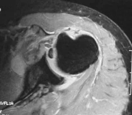
A 36-year-old woman has pain and swelling of the anterior arm after undergoing arthroscopic shoulder surgery 8 months ago. At the time of the procedure, extensive debridement and synovectomy of the anterior aspect of the joint was performed to remove scar tissue that had formed after an open rotator cuff repair. Examination reveals a golf ball-sized swelling just lateral to the coracoid. The area is not warm and shows no other signs of infection. An MRI scan is shown in Figure 1. Management should now consist of


Explanation

Deficiency of the rotator cuff interval may be acquired or congenital. In this patient, extensive debridement of the rotator cuff interval capsule at the time of arthroscopy most likely is the cause of the defect seen on the MRI scan. Surgical closure of the defect is the treatment of choice. During the repair, the shoulder should be placed in 30 degrees of external rotation to avoid overtightening. Care should be taken to include the leading edge of both the supraspinatus and subscapularis tendons in the repair because the rotator cuff interval capsular tissue is likely to be of poor quality. Cole BJ, Rodeo SA, O'Brien SJ, et al: The anatomy and histology of the rotator interval capsule of the shoulder. Clin Orthop 2001;390:129-137.

Question 2

A 45-year-old recreational tennis player underwent arthroscopic decompression and mini-open repair of a small supraspinatus tendon tear 3 weeks ago after nonsurgical management failed to provide relief. He now has pain, swelling about the wound, erythema, and purulent drainage. The patient is returned to the operating room for irrigation, debridement, and cultures. What is the most common organism causing this infection?


Explanation

In a large series of mini-open rotator cuff repairs, an infection rate of at least 2% was found, with the majority of the infections caused by Propionibacterium acnes. To prevent this complication, the shoulder should be re-prepped before the mini-open incision is made to prevent bacterial contamination from the arthroscopic procedure. Herrera MF, Bauer G, Reynolds F, et al: Infection after mini-open rotator cuff repair. J Shoulder Elbow Surg 2002;11:605-608.


Question 3

A paraplegic 32-year-old man was pulling himself up in bed by grasping the headboard rails when he felt a pop and immediate pain. A radiograph and CT scan are shown in Figures 2a and 2b. Based on these findings, management should consist of


Explanation

The coracoid process is an essential component of the superior shoulder suspensory complex and must be maintained. Open reduction and internal fixation is recommended if the fragment is large and displaced more than 1 cm. Froimson AI: Fracture of the coracoid process of the scapula. J Bone Joint Surg Am 1978;60:710-711.


Question 4

A 23-year-old baseball pitcher reports pain in the posterior aspect of his dominant shoulder during the late cocking phase of throwing. With the dominant shoulder positioned in 90 degrees of abduction from the body and with the scapula stabilized, examination reveals 135 degrees of external rotation and 20 degrees of internal rotation. Examination of the opposite shoulder reveals 100 degrees of external rotation and 75 degrees of internal rotation. Both shoulders are stable on examination. Radiographs and MRI scans are unremarkable. What is the primary cause of his pain?


Explanation

Internal impingement of the shoulder is a leading cause of shoulder pain in the throwing athlete. The primary lesion in pathologic internal impingement is excessive tightening of the posterior band of the inferior glenohumeral ligament complex. To obtain an accurate assessment of true glenohumeral rotation, the scapula is stabilized during examination. A loss of 20 degrees or more of internal rotation, as measured with the shoulder positioned in 90 degrees of abduction, indicates excessive tightness of the posterior band of the inferior glenohumeral ligament complex. Burkhart SS, Morgan CD, Kibler WB: The disabled throwing shoulder: Spectrum of pathology. Part I: Pathoanatomy and biomechanics. Arthroscopy 2003;19:404-420.


Question 5

What is the most common indication for revision following unconstrained elbow arthroplasty?


Explanation

Instability following unconstrained elbow arthroplasty occurs in 10% of patients. Subluxation is twice as common as frank dislocation; however, only 20% of these patients undergo revision. Instability following unconstrained elbow arthroplasty can be caused by component malposition or ligament insufficiency. King GJ, Itoi E, Niebur GL, et al: Motion and laxity of the capitellocondylar total elbow prosthesis. J Bone Joint Surg Am 1994;76:1000-1008.


Question 6

What part of the glenoid labrum has the least vascularity?


Explanation

The glenoid labrum receives its blood supply from the suprascapular, posterior humeral circumflex, and circumflex scapular arteries. The labral vessels arise from the capsular and periosteal vessels that penetrate the periphery of the labrum. The bone does not appear to be a source of vascularity. The posterior/superior and inferior labrum have a fairly robust vascular supply, whereas the anterior/superior labrum has relatively poor vascularity, which may influence the success of superior lateral repairs.


Question 7

One of the serious potential complications of repair of distal biceps tendon ruptures is limited pronation and supination as a result of synostosis. What surgical approach and technique presents the highest risk for development of this complication?


Explanation

The risk of synostosis is imminent with any technique for repairing a distal biceps tendon rupture. However, the risk is quite low for all approaches that avoid exposure of the ulna, including the muscle-splitting two-incision technique.


Question 8

A 25-year-old carpenter falls on his outstretched arm. What physical finding best correlates with the lesion seen on the MRI scan shown in Figure 3?


Explanation

The MRI scan shows disruption of the subscapularis muscle. Subscapularis rupture is associated with weakness in internal rotation as shown with a positive lift-off test as described by Gerber and Krushell. The belly press test also has been shown to be a useful clinical test for this problem. Weakness in external rotation and abduction is more consistent with supraspinatus and infraspinatus tears. Deltoid atrophy is associated with an axillary nerve injury. Loss of biceps contour is associated with rupture of the long head of the biceps. Gerber C, Krushell RJ: Isolated rupture of the tendon of the subscapularis muscle: Clinical features in 16 cases. J Bone Joint Surg Br 1991;73:389-394.


Question 9

A 72-year-old woman who sustained a cerebrovascular accident 9 months ago now has a fixed elbow flexion contracture of 80 degrees. Management should consist of


Explanation

A flexion contracture of the elbow is commonly seen in hemiplegic patients following cerebrovascular accidents. Spasticity and myostatic contracture of the joint are both causative factors. In patients with a flexion deformity of less than 90 degrees, musculocutaneous neurectomy is recommended, followed by serial casting to treat any residual deformity. At 9 months after injury, physical therapy will not significantly improve motion. Nerve blocks may be used in the early stages of recovery to facilitate therapy and serial casting.


Question 10

A 44-year-old recreational weight lifter reports chronic deep pain in his left shoulder that is aggravated by any pressing exercises. He also notes a painful catch in the shoulder occurring with rotational movements. Physical therapy and nonsteroidal anti-inflammatory drugs for 3 months have failed to provide relief. Examination reveals pain with O'Brien's test but no signs of instability. MRI scans are shown in Figures 4a and 4b. Treatment should now consist of


Explanation

The MRI scans show a large paralabral ganglion cyst in the spinoglenoid notch that communicates with an extensive tear of the glenoid labrum. Snyder and associates have classified superior labral tears into several subtypes that reflect the location and extent of the injury. Arthroscopic repair of the labral tear and aspiration of the ganglion cyst is the treatment of choice. Open excision of the cyst does not address the underlying problem of the labral tear. Snyder SJ, Karzel RP, Delpizzo W: SLAP lesions of the shoulder. Arthroscopy 1990;6:274-279. Piatt BE, Hawkins RJ, Fritz RC, et al: Clinical evaluation and treatment of spinoglenoid notch cysts. J Shoulder Elbow Surg 2002;11:600-604. McFarland EG, Kim TK, Savino RM: Clinical assessment of three common tests for superior labral anterior-posterior lesions. Am J Sports Med 2002;30:810-815.


Question 11

A 35-year-old carpenter sustained an injury to his dominant shoulder in a fall. He reports that he felt a sharp tearing sensation as he held on to a scaffold to keep from falling. Examination reveals swelling and ecchymosis down the upper arm, weakness to internal rotation, and deformity of the anterior axilla. He has good strength in external rotation and no apprehension with instability testing. Radiographs are normal. Management should consist of


Explanation

The findings are classic for a pectoralis major tendon avulsion. Deformity of the anterior axillary fold is a classic finding, and ecchymosis down the arm suggests that the injury is at the humeral attachment rather than at the musculotendinous junction. Good external rotation strength indicates that function in the supraspinatus and infraspinatus has been preserved. The treatment of choice for a tendon avulsion in a young individual is early surgical repair. Conversely, if the injury is within the muscle or at the musculotendinous junction, initial nonsurgical management is recommended. If the location of the injury cannot be determined by physical examination, then MRI of the pectoralis major can be helpful. Hanna CM, Glenny AB, Stanley SN, et al: Pectoralis major tears: Comparison of surgical and conservative treatment. Br J Sports Med 2001;35:202-206.


Question 12

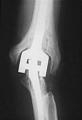
A 48-year-old woman with rheumatoid arthritis reports increasing elbow pain for the past 6 months. History reveals that she underwent total elbow arthroplasty 7 years ago. A peripheral WBC count, erythrocyte sedimentation rate, and C-reactive protein studies are normal. An AP radiograph is shown in Figure 5. What is the next most appropriate step in management?


Explanation

Pain relief is excellent after total elbow arthroplasty and is comparable to the results found with hip and knee arthroplasty. The failure of total elbow arthroplasty in the treatment of rheumatoid arthritis can be the result of infection, aseptic loosening, instability, and bearing surface wear. The radiographic findings shown here are consistent with bushing wear in a linked device. The bushings can be changed before continued wear results in osteolysis and implant loosening. If the implants become loose, then reimplantation is necessary. Resection arthroplasty is not indicated if the components are well fixed. Elbow arthrodesis is not indicated in patients with rheumatoid arthritis. Gill DR, Morrey BF: The Coonrad-Morrey total elbow arthroplasty in patients who have rheumatoid arthritis: A ten to fifteen-year follow-up study. J Bone Joint Surg Am 1998;80:1327-1335.


Question 13

Figure 6a shows the radiograph of a 50-year-old man who sustained an anterior dislocation of the shoulder. He undergoes closed reduction, and the postreduction radiograph is shown in Figure 6b. Management should now consist of


Explanation

Displaced greater tuberosity fractures often will block abduction and/or external rotation by impinging on the underside of the acromion or posterior glenoid. The indications for open reduction and internal fixation are 1 cm of displacement or 45 degrees of rotation of the tuberosity fracture. Surgical treatment has recently been recommended for 0.5 cm of tuberosity displacement. Neer CS II: Displaced proximal humeral fractures: II. Treatment of three-part and four-part displacement. J Bone Joint Surg Am 1970;52:1090-1103.


Question 14

A 42-year-old man sustained a fracture of the distal radius with subsequent stiffness in the ipsilateral shoulder. Despite a 6-month program of range-of-motion exercises, external rotation at the side is limited to 10 degrees. Attempts at closed manipulation are unsuccessful. Treatment should now consist of


Explanation

When external rotation at the side is limited, the most likely diagnosis is contracture of the rotator cuff interval, including the superior glenohumeral and coracohumeral ligaments. Therefore, the treatment of choice is arthroscopic release of the rotator cuff interval.


Question 15

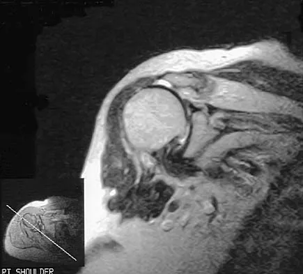
A 72-year-old woman who fell on her right shoulder while using a treadmill is now unable to elevate her right arm. An MRI scan is shown in Figure 7. What is the most likely diagnosis?


Explanation

The MRI scan reveals a large chronic rotator cuff tear with retraction and fatty infiltration atrophy of the supraspinatus and infraspinatus tendons. This tear is responsible for the patient's severe weakness and inability to elevate the arm.


Question 16

A 45-year-old man who underwent an open capsulolabral stabilization procedure 15 years ago now reports pain and has no external rotation on the affected side. Nonsurgical management has failed to provide relief. Examination reveals external rotation to -5 degrees compared with 50 degrees of external rotation on the contralateral side. Radiographs show a small inferior osteophyte and minimal posterior glenoid wear. Which of the following procedures will offer the best chance of restoring motion, decreasing pain, and preserving the native joint?


Explanation

Loss of external rotation following stabilization procedures can result in progressive degenerative joint disease. A tight anterior capsule results in posterior humeral translation and progressive posterior glenoid wear. Patients with early degenerative joint disease and pain can be treated with anterior release to restore more normal glenohumeral biomechanics. This procedure not only improves function but also decreases pain in most patients. Closed manipulation at 15 years after surgery is unlikely to be successful and carries the risk of complications. Acromioplasty, posterior release, and removal of osteophytes do not address the pathology. Arthroscopic releases are favored for intra-articular procedures that have addressed the pathology of instability. Open releases are recommended for nonanatomic extra-articular repairs that include subscapularis tightening procedures. MacDonald PB, Hawkins RJ, Fowler PJ, et al: Release of the subscapularis for internal rotation contracture and pain after anterior repair for recurrent anterior dislocation of the shoulder. J Bone Joint Surg Am 1992;74:734-737.


Question 17

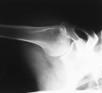
A right-handed 44-year-old construction worker reports pain and limited range of motion in his right elbow that has limited his ability to work for the past year. Examination reveals range of motion from 60 to 90 degrees, and he has pain at the extremes of flexion and extension. Pronation and supination are minimally restricted. Anti-inflammatory drugs have failed to provide relief. A radiograph is shown in Figure 8. Management should now consist of


Explanation

The radiograph reveals primary osteoarthritis of the elbow; therefore, ulnohumeral arthroplasty is the preferred procedure. Patients with severely limited preoperative elbow extension of more than 60 degrees and flexion of less than 100 degrees are at risk for ulnar nerve dysfunction postoperatively and should undergo a concomitant ulnar nerve decompression. Nonsurgical methods are unlikely to improve his chronic condition. Elbow arthroplasty is contraindicated for patients in this age group and with this diagnosis. Antuna SA, Morrey BF, Adams RA, et al: Ulnohumeral arthroplasty for primary degenerative arthritis of the elbow: Long-term outcome and complications. J Bone Joint Surg Am 2002;84:2168-2173.


Question 18

Spontaneous recovery of upper extremtiy motor function after a cerebrovascular accident occurs in which of the following predictable patterns?


Explanation

Recovery of upper extremity motor function after a cerebrovascular accident follows a predictable pattern. The greatest amount of recovery is seen within the first 6 weeks. Return of function proceeds from proximal to distal. Shoulder flexion occurs first, followed by return of flexion to the elbow, wrist, and fingers. Return of forearm supination follows the return of finger flexion.


Question 19

A 65-year-old woman sustained an axial load on the arm followed by an abduction injury after falling on ice. Treatment in the emergency department consisted of reduction of an anterior dislocation. She now has a positive drop arm sign and a positive lift-off test. An MRI scan is shown in Figure 9. Based on these findings, management should consist of


Explanation

Dislocation of the long head of the biceps tendon is the result of a defect in the region of the rotator cuff interval, coracohumeral ligament-superior glenohumeral ligament pulley, or an associated tear of the medial insertion of the subscapularis tendon. In the case of an intra-articular dislocation of the long head of the biceps tendon associated with a tear of the subscapularis tendon, stabilization of the biceps tendon is difficult in this situation; therefore, biceps release or tenodesis and repair of the subscapularis tendon is the treatment of choice. Eakin CL, Faber KJ, Hawkins RJ, et al: Biceps tendon disorders in athletes. J Am Acad Orthop Surg 1999;7:300-310. Sethi N, Wright R, Yamaguchi K: Disorders of the long head of the biceps tendon. J Shoulder Elbow Surg 1999;8:644-654.


Question 20

What type of nerve palsy is most common following elbow arthroscopy?


Explanation

Transient ulnar nerve palsy is the most common palsy following elbow arthroscopy. The ulnar nerve is most frequently affected, followed by the radial nerve. Injury to the other nerves has been reported but less frequently. Kelly EW, Morrey BF, O'Driscoll SW: Complications of elbow arthroscopy. J Bone Joint Surg Am 2001;83:25-34.


Question 21

A 72-year-old man injured his right shoulder after tripping over a chair leg. Radiographs obtained in the emergency department reveal an isolated anterior dislocation. After successful closed reduction, the patient has recurrent anterior instability and is unable to elevate the arm. What is the most likely cause of the recurrent instability?


Explanation

A rotator cuff tear is the most common cause of recurrent instability following a first-time dislocation in patients older than age 40 years. Dislocations occur through a posterior mechanism rather than by an isolated labral avulsion or a Bankart lesion as seen in younger patients. Nevaiser RJ, Nevaiser TJ: Recurrent instability of the shoulder after age 40. J Shoulder Elbow Surg 1995;4:416-418.


Question 22

A 54-year-old woman sustained an elbow injury 3 months ago that was treated with open reduction and internal fixation. She now reports pain and limited elbow motion. Radiographs are shown in Figures 10a and 10b. Treatment should now consist of


Explanation

Radiographs reveal malunion of a Monteggia fracture-dislocation. Dislocation of the posterior radial head is caused by the malunited ulnar fracture. The deformity includes shortening with an apex posterior angulation. In the acute setting, open reduction of the radial head rarely is necessary; however, in chronic dislocations, open reduction is required. Without ulnar osteotomy, recurrent radial head dislocation is likely.


Question 23

Ulnohumeral distraction interposition arthroplasty is considered the most appropriate treatment for which of the following patients?


Explanation

Distraction interposition arthroplasty is indicated for the treatment of both rheumatoid and posttraumatic arthritis and is reserved for younger patients who are not suitable candidates for total elbow arthroplasty. Although less reliable than prosthetic replacement, distraction interposition arthroplasty is a useful option in the treatment of young, high-demand patients with elbow arthritis. It is rarely indicated in the presence of polyarticular inflammatory arthritis but may be of value in those patients in whom the disease is limited primarily to the elbow. Isolated radiocapitellar arthritis can be successfully treated with radial head resection, although caution should be exercised if there is evidence of instability. Osteoarthritis is best treated with ulnohumeral arthroplasty. Cheng SL, Morrey BF: Treatment of the mobile, painful arthritic elbow by distraction interposition arthroplasty. J Bone Joint Surg Br 2000;82:233-238.


Question 24

A 57-year-old man with type I diabetes mellitus has had a tender, erythematous right sternoclavicular joint for the past 2 weeks. Radiographs reveal mild osteolysis without arthritic changes, within normal limits. Management should consist of


Explanation

Sternoclavicular joint sepsis is a rare condition that is most often restricted to patients who are immunocompromised, diabetic, or IV drug abusers. Examination commonly reveals a tender, painful, and possibly swollen sternoclavicular joint. If suspicion remains high following a thorough history, physical examination, radiographs, and routine blood tests, joint aspiration should be performed prior to incision and drainage or administration of antibiotics. Bremner RA: Monarticular noninfected subacute arthritis of the sternoclavicular joint. J Bone Joint Surg Br 1959;41:749-753.


Question 25

A 58-year-old man has persistent pain and weakness of his right shoulder after undergoing primary rotator cuff repair 1 year ago. A clinical photograph is shown in Figure 11. Which of the following factors might make functional improvement problematic with revision rotator cuff surgery?


Explanation

Functional improvement after revision rotator cuff surgery is most likely to occur in patients with an intact deltoid, good-quality rotator cuff tissue, preoperative active elevation alone to 90 degrees, and only one prior rotator cuff repair. In this patient, the compromised deltoid origin might make functional improvement less likely. Djurasovic M, Marra G, Arroyo JS, et al: Revision rotator cuff repair: Factors influencing results. J Bone Joint Surg Am 2001;83:1849-1855. Bigliani LU, Cordasco FA, McIlveen SJ, et al: Operative treatment of failed repairs of the rotator cuff. J Bone Joint Surg Am 1992;74:1505-1515.


Question 26

A 42-year-old man falls from a height and sustains a complex elbow dislocation.

Imaging reveals a posterolateral elbow dislocation, a comminuted radial head fracture, and a type II coronoid fracture. During operative management, what is the most appropriate sequence of reconstruction?


Explanation

The standard surgical sequence for a terrible triad injury is from deep to superficial: coronoid fixation, followed by radial head replacement or fixation, and finally lateral collateral ligament (LCL) repair. This restores the anterior and lateral bony and ligamentous buttresses to prevent recurrent posterior subluxation.

Question 27

A 22-year-old collegiate rugby player presents with recurrent anterior shoulder instability. A 3D CT scan demonstrates 25% anterior glenoid bone loss.

Which of the following is the most appropriate definitive management?


Explanation

Critical glenoid bone loss (typically >20-25%) in a contact athlete is a primary indication for a bony augmentation procedure like the Latarjet. Arthroscopic soft-tissue repairs alone in this setting have an unacceptably high failure rate.

Question 28

A 34-year-old cyclist falls directly onto his shoulder. Radiographs show a displaced distal third clavicle fracture with the fracture line located medial to the intact coracoclavicular ligaments. The proximal fragment is displaced superiorly. This injury is best classified as:


Explanation

Neer Type II distal clavicle fractures occur when the fracture is medial to the coracoclavicular ligaments, and the proximal fragment is displaced superiorly by the pull of the trapezius. They have a high rate of nonunion with conservative management and typically require surgical fixation.

Question 29

A 45-year-old man undergoes a single-incision anterior approach for repair of an acute distal biceps tendon rupture. Postoperatively, he notes numbness along the lateral aspect of his forearm. Which nerve was most likely injured during the exposure?


Explanation

The lateral antebrachial cutaneous nerve (LABC) is the most commonly injured structure during a single-incision anterior approach for distal biceps repair. The posterior interosseous nerve (PIN) is more at risk during a two-incision approach or when deep retractors are placed laterally.

Question 30

A 78-year-old woman with severe osteoporosis sustains a comminuted 4-part proximal humerus fracture. The humeral head is split, and the tuberosities are widely displaced. She is treated with a reverse total shoulder arthroplasty (RTSA). Healing of which structure is most critical to restore active external rotation?


Explanation

In RTSA for proximal humerus fractures, anatomic healing of the greater tuberosity is crucial for restoring active external rotation, as it serves as the attachment point for the infraspinatus and teres minor. Failure of greater tuberosity healing often results in a significant external rotation lag.

Question 31

A 45-year-old man presents with chronic wrist pain. Radiographs demonstrate advanced osteoarthritis of the radioscaphoid and capitolunate joints, while the radiolunate joint is spared. A scaphoid nonunion is also noted. Which of the following surgical interventions is most appropriate?


Explanation

The patient has Stage III Scaphoid Nonunion Advanced Collapse (SNAC). Because the capitate is arthritic, a proximal row carpectomy is contraindicated. Scaphoid excision with four-corner fusion is the most appropriate motion-preserving procedure.

Question 32

A 22-year-old collegiate football player presents with his third anterior shoulder dislocation. Imaging reveals an engaged Hill-Sachs lesion and 25% anterior glenoid bone loss. What is the most appropriate surgical management?


Explanation

In the setting of recurrent anterior instability with critical glenoid bone loss (greater than 20-25%), a soft tissue repair alone has an unacceptably high failure rate. The Latarjet procedure (coracoid transfer) addresses the bony defect and provides a sling effect via the conjoint tendon.

Question 33

A 40-year-old weightlifter felt a sudden pop in his antecubital fossa and presents with weakness in supination. The Hook test is positive. If he undergoes a single-incision anterior approach for distal biceps tendon repair, which nerve is most at risk of injury?


Explanation

The lateral antebrachial cutaneous nerve is the most frequently injured nerve during a single-incision anterior approach for distal biceps repair. The posterior interosseous nerve is more commonly at risk during a two-incision approach.

Question 34

A 25-year-old cyclist falls and sustains a midshaft clavicle fracture. Which of the following is considered an absolute indication for open reduction and internal fixation?


Explanation

Absolute indications for operative fixation of a clavicle fracture include open fractures, neurovascular compromise, and severe skin tenting predicting imminent skin breakdown. Shortening and severe displacement are relative indications depending on patient activity level.

Question 35

A 28-year-old recreational hockey player falls onto the point of his shoulder. Radiographs show a 100% superior displacement of the distal clavicle relative to the acromion. The coracoclavicular distance is increased by 50% compared to the contralateral side. What is the most appropriate initial management?


Explanation

This is a Type III acromioclavicular (AC) joint separation. Initial management for acute Type III AC separations is generally nonoperative with a short period of sling immobilization, followed by early range of motion.

Question 36

A 45-year-old laborer with chronic shoulder pain has failed 6 months of physical therapy. MRI demonstrates a Type II SLAP tear without rotator cuff pathology. Based on recent literature, which surgical intervention provides the most reliable functional outcome in this demographic?


Explanation

In patients older than 40 years with symptomatic Type II SLAP tears, biceps tenodesis provides superior clinical outcomes and a lower revision rate compared to arthroscopic SLAP repair. SLAP repairs in older patients often result in postoperative stiffness and persistent pain.

Question 37

A 35-year-old woman falls on an outstretched hand, sustaining an elbow dislocation with associated fractures of the radial head and the coronoid process. During surgical reconstruction, what is the most widely accepted sequence of repair?


Explanation

The standard surgical sequence for treating a terrible triad injury of the elbow is fixation of the coronoid first (to restore the anterior buttress), followed by radial head replacement or fixation, and finally repair of the lateral ulnar collateral ligament (LUCL).

Question 38

A 65-year-old woman undergoes volar locking plate fixation for a displaced distal radius fracture. Postoperative radiographs show the plate is placed distal to the watershed line. Which structure is at greatest risk of rupture?


Explanation

Placing a volar plate distal to the watershed line of the distal radius places the flexor tendons, most notably the flexor pollicis longus (FPL), at high risk of irritation and subsequent spontaneous rupture. Extensor pollicis longus rupture is more classically associated with nonoperative treatment or dorsal screw prominence.

Question 39

A 25-year-old man sustains a closed, transverse midshaft humerus fracture. Upon examination in the emergency department, he is unable to extend his wrist or fingers, though he had normal function immediately after the injury prior to closed reduction. What is the most appropriate management of this neurologic deficit?


Explanation

A secondary radial nerve palsy (occurring after manipulation or closed reduction) is a classic indication for immediate surgical exploration and fracture fixation, as the nerve may be entrapped in the fracture site. Primary radial nerve palsies are usually observed.

Question 40

A 75-year-old osteoporotic woman sustains a displaced 4-part proximal humerus fracture. The articular surface is subluxated, and the tuberosities are widely displaced. Which treatment modality is associated with the most reliable return of forward elevation?


Explanation

In elderly patients with severe osteoporosis and displaced 4-part proximal humerus fractures, reverse total shoulder arthroplasty (RTSA) provides more reliable pain relief and functional restoration than hemiarthroplasty or ORIF. It relies on deltoid function rather than predictable tuberosity healing.

Question 41

A scaphoid waist fracture is at high risk of nonunion and avascular necrosis due to its tenuous retrograde blood supply. Which vessel provides the dominant blood supply to the proximal pole of the scaphoid?


Explanation

The dorsal carpal branch of the radial artery provides approximately 80% of the blood supply to the scaphoid. It enters the bone distally and provides retrograde flow to the proximal pole, explaining the high risk of avascular necrosis in proximal pole fractures.

Question 42

A 30-year-old man requires tension band wiring for a displaced, non-comminuted transverse olecranon fracture. What is the primary biomechanical principle underlying tension band wiring in this setting?


Explanation

Tension band wiring operates on the principle of converting tensile forces (from the pull of the triceps on the dorsal cortex) into dynamic compressive forces at the articular surface during active elbow flexion.

Question 43

A 72-year-old man presents with chronic shoulder pain and an inability to actively raise his arm above 45 degrees. He has a positive drop arm sign. MRI reveals a massive, retracted, and irreparable posterosuperior rotator cuff tear with fatty infiltration of the infraspinatus and teres minor. The subscapularis is intact. What is the most appropriate definitive management?


Explanation

The patient exhibits pseudoparalysis (inability to actively elevate the arm above 90 degrees) in the setting of an irreparable massive rotator cuff tear. Reverse total shoulder arthroplasty is the treatment of choice to restore forward elevation by maximizing the mechanical advantage of the deltoid.

Question 44

A 30-year-old professional bodybuilder feels a tearing sensation in his anterior axilla while performing heavy bench presses. Examination reveals loss of the anterior axillary fold contour and weakness in internal rotation. In this mechanism of injury, where does the pectoralis major most commonly rupture?


Explanation

Pectoralis major ruptures most frequently occur at or very near the humeral insertion, particularly involving the sternocostal head. This injury is classically seen in weightlifters performing bench presses.

Question 45

A 22-year-old collegiate baseball pitcher complains of medial elbow pain during the late cocking phase of throwing. The moving valgus stress test is positive. Which specific ligamentous structure is the primary restraint to valgus stress at the elbow between 30 and 90 degrees of flexion?


Explanation

The anterior bundle of the ulnar collateral ligament (UCL) is the primary restraint to valgus stress. Specifically, the anterior band of the anterior bundle is taut and primary from 0 to 90 degrees of flexion.

Question 46

A 40-year-old man falls onto an outstretched hand and sustains a 'terrible triad' injury of the elbow. He is taken to the operating room for surgical stabilization. After repairing the coronoid and stabilizing the radial head, the elbow remains unstable. According to the standard stepwise surgical protocol, which structure is typically addressed next?


Explanation

The terrible triad of the elbow includes an elbow dislocation, radial head fracture, and coronoid fracture. The standard surgical sequence is coronoid fixation, radial head repair/replacement, LCL repair, and finally MCL repair or external fixation only if the elbow remains unstable.

Question 47

A 28-year-old male presents with a closed midshaft humeral fracture after a direct blow. On examination, he is unable to extend his wrist or fingers, and he has decreased sensation over the dorsal web space of the hand. Which of the following is the most appropriate initial management?


Explanation

Immediate closed reduction and application of a coaptation splint is the standard of care for a closed humeral shaft fracture with an acute radial nerve palsy. Most of these palsies are neuropraxias that will recover spontaneously.

Question 48

A 75-year-old woman with severe osteoporosis sustains a displaced, 4-part proximal humerus fracture. Examination of preoperative imaging suggests severe valgus impaction and poor tuberosity bone stock. Which of the following surgical interventions will provide the most predictable outcome for postoperative functional elevation and pain relief?


Explanation

Reverse total shoulder arthroplasty (RTSA) provides more predictable functional outcomes and pain relief than hemiarthroplasty or ORIF in elderly patients with 4-part proximal humerus fractures. It relies on the deltoid rather than a functional rotator cuff, circumventing the issue of non-healing tuberosities.

Question 49

A 45-year-old male weightlifter undergoes surgical repair of an acute distal biceps tendon rupture using a single anterior incision approach. Postoperatively, he complains of numbness and tingling over the anterolateral aspect of his forearm. Which nerve is most likely injured?


Explanation

The lateral antebrachial cutaneous nerve (LABCN) is the most commonly injured nerve during a single anterior incision approach for distal biceps repair. Injury typically occurs due to retraction forces or direct damage, as the nerve lies in close proximity to the cephalic vein and the surgical field.

Question 50

While operative fixation of acute midshaft clavicle fractures is increasingly common in active individuals, certain findings mandate surgical intervention. Which of the following is an absolute indication for operative fixation of an acute clavicle fracture?


Explanation

Absolute indications for operative fixation of a clavicle fracture include open fractures, acute neurovascular compromise, and severe skin tenting causing impending skin necrosis. Displacement and shortening are considered relative indications.

Question 51

A 22-year-old rugby player falls directly onto the point of his shoulder and presents with severe pain. Radiographs demonstrate a superiorly displaced distal clavicle. The superior translation is measured at 250% of the normal coracoclavicular (CC) distance. Which of the following accurately describes the ligamentous and fascial pathology in this Type V acromioclavicular (AC) joint injury?


Explanation

A Type V acromioclavicular joint injury is characterized by severe superior displacement (>100%). This requires rupture of both the AC and CC ligaments, accompanied by disruption of the stabilizing deltotrapezial fascial envelope.

Question 52

A 21-year-old collegiate linebacker presents with recurrent anterior shoulder instability. An en face 3D CT scan of the glenoid demonstrates significant anterior bone loss. At what percentage of anterior glenoid bone loss is an isolated arthroscopic Bankart repair generally considered to have an unacceptably high failure rate, warranting a bony augmentation procedure?


Explanation

Arthroscopic Bankart repair alone is associated with unacceptably high failure rates when anterior glenoid bone loss exceeds 20 to 25%. In such cases, a bone block augmentation procedure, such as the Latarjet procedure, is indicated to restore stability.

Question 53

A 32-year-old bodybuilder felt a sudden tear in his anterior chest wall while performing a heavy bench press. Examination reveals loss of the anterior axillary fold and extensive ecchymosis over the anterior arm. If surgical repair is planned, where is the most common site of failure requiring anatomic reattachment in this specific injury?


Explanation

Pectoralis major ruptures almost exclusively involve the sternal head, typically tearing at the tendinous insertion onto the proximal humerus or the musculotendinous junction. Tears at the tendinous insertion are most amenable to direct surgical repair.

Question 54

A 70-year-old man presents with chronic, severe shoulder pain and an inability to actively elevate his arm past 40 degrees (pseudoparalysis). Radiographs demonstrate a narrowed acromiohumeral interval of 2 mm and advanced glenohumeral osteoarthritis. What is the most appropriate surgical treatment?


Explanation

In an elderly patient with rotator cuff tear arthropathy characterized by pseudoparalysis, superior head migration, and glenohumeral arthritis, reverse total shoulder arthroplasty is the treatment of choice. It medializes and distalizes the center of rotation, allowing the deltoid to initiate and maintain elevation.

Question 55

A 25-year-old professional baseball pitcher presents with deep shoulder pain and decreased throwing velocity. Physical examination reveals a positive O'Brien test. MRI arthrography shows a Type II SLAP tear. Which of the following biomechanical forces is most frequently implicated in causing this injury during the late cocking phase of throwing?


Explanation

In overhead throwing athletes, Type II SLAP tears are frequently caused by the peel-back mechanism. During the late cocking phase, maximal external rotation places a torsional and posteriorly directed force on the biceps anchor, peeling it from the superior glenoid.

Question 56

A 20-year-old collegiate pitcher undergoes ulnar collateral ligament (UCL) reconstruction utilizing a palmaris longus autograft. Which specific bundle of the native UCL is the primary restraint to valgus stress between 30 and 90 degrees of elbow flexion and is the target of this reconstruction?


Explanation

The anterior bundle of the UCL is the primary stabilizer against valgus stress at the elbow. Specifically, its anterior band is the most important anatomical restraint between 30 and 90 degrees of flexion and is the structure reconstructed during Tommy John surgery.

Question 57

A 6-year-old child presents after a fall onto an outstretched arm. Radiographs demonstrate a fracture of the proximal third of the ulna with an anterior dislocation of the radial head. Based on this specific Bado classification pattern, which nerve is most commonly injured?


Explanation

A proximal ulnar fracture with an anterior radial head dislocation is a Bado Type I Monteggia fracture-dislocation. The posterior interosseous nerve (PIN) is the most commonly injured nerve in Bado Type I and Type III injuries due to the displacement of the radial head.

Question 58

A 21-year-old male sustains a proximal pole scaphoid fracture. The treating orthopedic surgeon advises the patient of a high risk of nonunion and avascular necrosis. This risk is primarily due to the retrograde nature of the scaphoid's blood supply. Which of the following vessels provides this dominant arterial supply to the proximal pole?


Explanation

The scaphoid receives its primary blood supply from the dorsal carpal branch of the radial artery. This vessel enters the scaphoid distally and flows in a retrograde fashion, leaving the proximal pole highly susceptible to avascular necrosis following a fracture.

Question 59

A 25-year-old cyclist sustains a midshaft clavicle fracture. Which of the following radiographic or clinical findings is the strongest predictor of nonunion if this injury is managed nonoperatively?


Explanation

Shortening greater than 2 cm, 100% displacement, and severe comminution are major predictors of nonunion in midshaft clavicle fractures. Operative fixation is generally indicated to prevent symptomatic nonunion in these patients.

Question 60

A 22-year-old rugby player presents with recurrent anterior shoulder instability. CT scan imaging demonstrates 25% anterior glenoid bone loss. What is the most appropriate definitive surgical management?


Explanation

Glenoid bone loss greater than 20-25% is a contraindication to isolated soft-tissue repair. A coracoid transfer (Latarjet) restores glenoid bone stock and provides a dynamic sling effect from the conjoint tendon.

Question 61

A 65-year-old woman sustains a 4-part proximal humerus fracture. According to the Hertel criteria, which radiographic feature is the strongest predictor of subsequent humeral head ischemia?


Explanation

According to the Hertel criteria, a short posteromedial calcar length (< 8 mm), disruption of the medial hinge, and an anatomic neck fracture pattern are the most accurate predictors of humeral head ischemia.

Question 62

A 30-year-old man presents with a closed distal-third spiral humeral shaft fracture (Holstein-Lewis type). Initial examination shows normal nerve function, and he is placed in a coaptation splint. At his 1-week clinic follow-up, he demonstrates a complete radial nerve palsy. What is the next most appropriate step in management?


Explanation

A secondary radial nerve palsy that develops after a closed reduction or splinting of a humeral shaft fracture is a classic indication for immediate surgical exploration to rule out nerve entrapment in the fracture site.

Question 63

A 40-year-old man undergoes a single-incision anterior approach repair of a distal biceps tendon rupture. What is the most commonly injured neurologic structure associated with this specific surgical approach?


Explanation

The lateral antebrachial cutaneous nerve (LABCN) is the most commonly injured nerve during a single-incision anterior approach for distal biceps repair. Posterior interosseous nerve (PIN) injury is less frequent but more devastating.

Question 64

A 35-year-old woman falls on an outstretched hand, sustaining an elbow dislocation, a comminuted radial head fracture, and a Type II coronoid fracture. What is the generally accepted sequence of surgical reconstruction for this "terrible triad" injury?


Explanation

The standard surgical algorithm for fixing a terrible triad injury proceeds from deep to superficial: coronoid fixation first, followed by radial head repair or arthroplasty, and finally lateral collateral ligament (LCL) reconstruction.

Question 65

A 55-year-old woman sustains a nondisplaced distal radius fracture treated in a short arm cast. Six weeks later, the cast is removed, and she is unable to actively extend her thumb interphalangeal joint. Tenodesis effect of the thumb is absent. What is the most likely etiology?


Explanation

Extensor pollicis longus (EPL) tendon rupture is a well-known complication of nondisplaced distal radius fractures. It occurs due to a combination of mechanical attrition at Lister's tubercle and ischemia in a watershed vascular zone.

Question 66

A 45-year-old man presents with chronic wrist pain. A radiograph (

) confirms a scaphoid nonunion with radioscaphoid and capitolunate arthritis. The radiolunate joint is completely preserved. What is the most appropriate surgical treatment?


Explanation

In SNAC Stage III arthritis (involving the capitolunate joint), a proximal row carpectomy is contraindicated because the capitate head is arthritic. Scaphoid excision with four-corner fusion preserves motion utilizing the intact radiolunate joint.

Question 67

A 24-year-old professional baseball pitcher reports posterior shoulder pain during the late cocking phase of throwing. MRI arthrography reveals posterosuperior labral fraying and a partial-thickness articular-sided supraspinatus tear. What is the primary pathophysiologic mechanism of this injury pattern?


Explanation

Internal impingement occurs in overhead athletes during maximum abduction and external rotation (late cocking phase). The greater tuberosity abuts the posterosuperior glenoid, causing articular-sided rotator cuff tears and posterosuperior labral lesions.

Question 68

A 28-year-old man falls directly onto the point of his shoulder. Radiographs demonstrate 150% superior displacement of the distal clavicle relative to the acromion. Which ligaments are completely disrupted in this injury?


Explanation

A Type III or higher acromioclavicular (AC) joint separation, characterized by 100% to 300% superior displacement, involves the complete disruption of both the AC ligaments and the coracoclavicular (conoid and trapezoid) ligaments.

Question 69

An 18-year-old football player presents after a pile-up tackle with shortness of breath, dysphagia, and severe pain over the medial clavicle. Examination shows a palpable depression of the medial clavicle. What is the most appropriate initial diagnostic step?


Explanation

Posterior sternoclavicular dislocations are medical emergencies due to potential impingement on mediastinal structures (trachea, esophagus, great vessels). A CT scan is the gold standard to evaluate the dislocation and vascular proximity before reduction.

Question 70

A 32-year-old woman falls on an outstretched arm. Radiographs (

) demonstrate a coronal shear fracture of the capitellum extending into the lateral trochlea (Type IV). Which surgical approach and fixation strategy is biomechanically superior?


Explanation

Coronal shear fractures of the capitellum are best managed with headless compression screws. A posterior-to-anterior screw trajectory provides superior biomechanical pull-out strength and avoids violating the anterior articular cartilage.

Question 71

A 21-year-old collegiate pitcher reports medial elbow pain and decreased throwing velocity. The "moving valgus stress test" is positive. What structure is the primary restraint to valgus stress at the elbow during the late cocking phase of throwing?


Explanation

The anterior bundle of the medial ulnar collateral ligament (UCL) is the primary restraint to valgus stress at the elbow between 30 and 120 degrees of flexion, which covers the late cocking and early acceleration phases of throwing.

Question 72

A 30-year-old bodybuilder feels a "pop" in his anterior axilla while bench pressing. He exhibits ecchymosis and loss of the anterior axillary fold. MRI confirms a complete pectoralis major tear. Normal anatomy dictates that the sternal head of the pectoralis major inserts in what relation to the clavicular head?


Explanation

The pectoralis major tendon undergoes a 180-degree twist prior to its insertion on the lateral lip of the bicipital groove. Consequently, the inferior (sternal) head inserts proximal and deep (posterior) to the superior (clavicular) head.

Question 73

A 28-year-old volleyball player presents with isolated atrophy of the infraspinatus muscle and painless weakness in external rotation. MRI reveals a paralabral cyst. At what anatomical location is this cyst most likely compressing the suprascapular nerve?


Explanation

Compression of the suprascapular nerve at the spinoglenoid notch affects only the motor branch to the infraspinatus, causing isolated atrophy. Compression further proximal at the suprascapular notch would involve both the supraspinatus and infraspinatus.

Question 74

A 42-year-old man feels a pop in his anterior elbow while lifting a heavy object. An MRI confirms a complete avulsion of the distal biceps tendon. He elects to undergo surgical repair via a single-incision anterior approach. What is the most common neurologic complication associated with this specific surgical approach?


Explanation

The lateral antebrachial cutaneous nerve is the most commonly injured nerve during a single-incision anterior approach for distal biceps repair. The posterior interosseous nerve is more typically at risk during a two-incision approach.

Question 75

A 65-year-old woman sustains an intra-articular distal humerus fracture (AO/OTA type 13-C3). During open reduction and internal fixation utilizing a transolecranon osteotomy, the surgeon must routinely identify and protect which of the following structures?


Explanation

The ulnar nerve is highly vulnerable during posterior surgical approaches to the distal humerus, particularly when performing an olecranon osteotomy. It must be routinely identified and mobilized to prevent iatrogenic injury.

Question 76

A 24-year-old cyclist falls directly onto his shoulder. Clinical examination reveals a prominent distal clavicle. Radiographs demonstrate >100% superior displacement of the clavicle relative to the acromion, with the clavicle penetrating the deltotrapezial fascia. This injury is best classified as a Rockwood Type:


Explanation

Rockwood Type V injuries involve greater than 100% superior displacement of the distal clavicle and disruption of the deltotrapezial fascia. Surgical reconstruction of the coracoclavicular ligaments is typically indicated for these high-grade injuries.

Question 77

A 38-year-old man falls on an outstretched hand, sustaining an elbow dislocation, a radial head fracture, and a coronoid fracture. Which of the following is the standard recommended surgical sequence for restoring stability in this terrible triad injury?


Explanation

The standard surgical algorithm for terrible triad elbow injuries begins with fixing the coronoid to restore deep anterior stability. This is followed by addressing the radial head, and finally repairing the lateral collateral ligament (LCL).

Question 78

A 28-year-old man sustains a closed distal-third spiral fracture of the humeral shaft. On initial presentation, his neurologic examination is completely intact. Closed reduction and splinting are performed. Post-reduction, he is entirely unable to extend his wrist or fingers. What is the most appropriate next step in management?


Explanation

A secondary radial nerve palsy that develops immediately after a closed reduction attempt of a humeral shaft fracture (especially a Holstein-Lewis type) is an absolute indication for immediate surgical exploration. The nerve may be entrapped in the fracture site.

Question 79

A 31-year-old competitive weightlifter feels a tearing sensation in his anterior chest while performing a heavy bench press. Examination reveals loss of the anterior axillary fold and weakness in internal rotation. The most common site of this specific muscle injury is:


Explanation

Pectoralis major ruptures most commonly occur as an avulsion of the tendon from its insertion on the proximal humerus. They are classic injuries in weightlifters performing bench presses, often requiring surgical repair for optimal return to strength.

Question 80

A 74-year-old woman with severe osteoporosis sustains a comminuted 4-part proximal humerus fracture. She undergoes a reverse total shoulder arthroplasty (RTSA). Which of the following describes the primary biomechanical advantage of RTSA in this setting?


Explanation

Reverse total shoulder arthroplasty medializes and inferiorly shifts the center of rotation of the glenohumeral joint. This configuration recruits the deltoid muscle more effectively, compensating for the non-functioning or absent rotator cuff.

Question 81

A 22-year-old man falls onto an extended wrist. Radiographs reveal a non-displaced fracture of the proximal pole of the scaphoid. Why is this specific fracture location at a particularly high risk for nonunion and avascular necrosis?


Explanation

The scaphoid receives its primary blood supply from the dorsal carpal branch of the radial artery, which enters the distal half and flows retrograde. Fractures of the proximal pole frequently disrupt this delicate blood supply, causing avascular necrosis.

Question 82

A 6-year-old boy falls off monkey bars. Radiographs demonstrate a fracture of the proximal third of the ulna with an anterior dislocation of the radial head. According to the Bado classification, this injury is classified as a:


Explanation

A Bado Type I Monteggia fracture-dislocation is characterized by a fracture of the proximal or middle third of the ulna with an anterior dislocation of the radial head. It is the most common variant seen in pediatric patients.

Question 83

A 35-year-old tennis player complains of ulnar-sided wrist pain. MRI confirms an isolated tear of the central, articular disk portion of the triangular fibrocartilage complex (TFCC). Which of the following statements best dictates the preferred surgical management of this specific lesion?


Explanation

The central portion of the TFCC is avascular, meaning tears in this region have poor healing potential and are best treated with arthroscopic debridement. Conversely, peripheral tears are located in the vascular zone and can be primarily repaired.

Question 84

A 29-year-old elite volleyball player presents with poorly localized posterior shoulder pain and paresthesias over the lateral deltoid. An MRI demonstrates isolated atrophy of the teres minor. He is diagnosed with quadrilateral space syndrome. Which artery and nerve traverse this anatomical space?


Explanation

The quadrilateral space contains the axillary nerve and the posterior humeral circumflex artery. Compression here causes axillary nerve symptoms, classically resulting in teres minor atrophy.

Question 85

A 33-year-old construction worker sustains a fracture of the distal third of the radial shaft with associated disruption of the distal radioulnar joint (DRUJ). After rigid open reduction and internal fixation of the radius, the DRUJ remains highly unstable. What is the most appropriate next step in management?


Explanation

In a Galeazzi fracture, if the DRUJ remains unstable after rigid anatomical fixation of the radius, it should be pinned in the position of maximum stability. For most DRUJ instabilities, this position is full supination.

Question 86

A 21-year-old collegiate baseball pitcher presents with deep shoulder pain and clicking during the late cocking phase of throwing. MRI arthrography reveals a detachment of the superior labrum and biceps anchor from the glenoid. Which physical examination test is most specific for this pathology?


Explanation

The O'Brien active compression test is a sensitive and specific maneuver for detecting Superior Labrum Anterior to Posterior (SLAP) tears. Pain is typically elicited with the arm in internal rotation and relieved when the arm is externally rotated.

Question 87

A 19-year-old rugby player is tackled and sustains a posterior sternoclavicular (SC) joint dislocation. He presents to the ER with mild dyspnea and dysphagia. What is the most critical imaging study required before attempting closed reduction in the operating room?


Explanation

Posterior sternoclavicular dislocations can compress or injure critical mediastinal structures, including the trachea, esophagus, and great vessels. A CT scan, preferably with angiography, is essential to evaluate these structures before reduction.

Question 88

A 40-year-old woman falls on her outstretched arm and sustains an isolated coronal shear fracture of the humeral capitellum. What is the optimal surgical approach and fixation strategy for this injury?


Explanation

Capitellum fractures are coronal shear injuries of the distal humerus. They are best managed through a lateral approach using headless compression screws placed from anterior to posterior, allowing the hardware to be buried beneath the articular cartilage.

Question 89

A 23-year-old professional baseball pitcher undergoes ulnar collateral ligament (UCL) reconstruction using a palmaris longus autograft. Which nerve is most at risk for injury or entrapment during the harvesting of the graft at the volar wrist?


Explanation

The palmaris longus tendon lies in very close proximity to the median nerve at the wrist. Careful identification and precise harvesting techniques are critical to avoid iatrogenic median nerve injury during graft procurement.

Question 90

A 72-year-old female presents with a highly comminuted, intra-articular distal humerus fracture (OTA 13-C3) after a ground-level fall. Her bone quality is poor, and the articular surface is deemed non-reconstructible. She lives independently and performs her own activities of daily living. What is the most appropriate definitive management?


Explanation

In elderly patients with complex, non-reconstructible intra-articular distal humerus fractures, total elbow arthroplasty provides superior functional outcomes and lower reoperation rates compared to open reduction and internal fixation.

Question 91

A 22-year-old collegiate rugby player presents with his fourth anterior shoulder dislocation. A 3D CT scan reveals 25% anterior glenoid bone loss. Which of the following is the most appropriate surgical intervention to minimize the risk of recurrent instability?


Explanation

In high-impact collision athletes with significant anterior glenoid bone loss (greater than 20-25%), a coracoid transfer (Latarjet procedure) is indicated. This restores the glenoid track and provides a reliable osseoligamentous sling, preventing recurrent instability.

Question 92

A 30-year-old cyclist undergoes open reduction and internal fixation of a displaced midshaft clavicle fracture utilizing an anterior-inferior plating technique. Postoperatively, he complains of a well-demarcated area of numbness over his anterosuperior chest wall, just inferior to the surgical incision. Which nerve was most likely injured during the surgical exposure?


Explanation

The supraclavicular nerves provide sensory innervation to the skin over the clavicle and the anterosuperior chest wall. They are frequently at risk during open approaches to the clavicle, leading to expected numbness inferior to the incision.

Question 93

A 45-year-old manual laborer presents with persistent anterior shoulder pain and painful catching despite 6 months of targeted physical therapy and NSAIDs. An MRI arthrogram reveals a Type II SLAP lesion. He has no other rotator cuff pathology. What is the most appropriate surgical treatment?


Explanation

In patients older than 40 years, especially heavy laborers, biceps tenodesis yields higher satisfaction rates, reliable pain relief, and lower complication rates compared to arthroscopic SLAP repair, which is prone to postoperative stiffness.

Question 94

A 24-year-old professional baseball player sustains a fall onto an outstretched hand. Initial radiographs are negative, but an MRI reveals an acute, non-displaced fracture of the scaphoid waist. He wishes to return to play as safely and quickly as possible. What is the optimal management?


Explanation

Percutaneous screw fixation of acute, non-displaced scaphoid waist fractures results in faster time to union and significantly quicker return to sport and work compared to prolonged cast immobilization.

Question 95

A 17-year-old football player sustains a direct blow to the medial shoulder. He presents to the trauma bay with shortness of breath, stridor, and difficulty swallowing. The medial clavicle is not palpable anteriorly. What is the most appropriate next step in management?


Explanation

Posterior sternoclavicular dislocations can compress the trachea, esophagus, and great vessels, representing a surgical emergency. Reduction should be performed in the OR with a cardiothoracic surgeon available due to the risk of catastrophic vascular injury.

Question 96

A 40-year-old weightlifter feels a sudden "pop" in his anterior elbow while attempting a heavy deadlift. On examination, he has marked weakness in forearm supination and elbow flexion. The Hook test is positive. What is the recommended treatment?


Explanation

Acute distal biceps tendon ruptures in active patients should undergo primary surgical repair to the radial tuberosity to restore supination and flexion strength. Delaying surgery past 3-4 weeks increases the risk of retraction and the need for allograft reconstruction.

None

Clinic OS
Medically Verified Content by
Prof. Clinic OS
Consultant Orthopedic & Spine Surgeon
Chapter Index