العربية
Part of the Master Guide

Orthopedic Surgery Board Prep: Set 403 (100 MCQs on Fractures & Casting)

AAOS, ABOS, SMLE Orthopedic Trauma MCQs (Set 4): Tibial Plateau, Pilon, & Polytrauma Management

23 Apr 2026 57 min read 89 Views
Trauma 2000 MCQs - Part 4

Key Takeaway

This high-yield MCQ set (Set 4) for AAOS, ABOS, and OITE board review focuses on critical orthopedic trauma topics. Questions cover the diagnosis, classification, and surgical management of tibial plateau and pilon fractures, alongside essential principles of polytrauma patient care and compartment syndrome recognition.

AAOS, ABOS, SMLE Orthopedic Trauma MCQs (Set 4): Tibial Plateau, Pilon, & Polytrauma Management

Comprehensive 100-Question Exam


00:00

Start Quiz

Question 1

A 47-year-old man ruptured his left patellar tendon and twisted his right ankle in a fall. Initial radiographs of the ankle are unremarkable. One week following repair of the left patellar tendon, he reports increased pain with weight bearing in his right ankle. A follow-up radiograph is shown in Figure 38. Management of the ankle injury should consist of


Explanation

The radiograph reveals disruption of the syndesmosis with lateral displacement of the talus and widening of the medial ankle clear space. No fibular fracture is noted, although radiographs of the entire tibia and fibula are necessary to rule out a more proximal fibula fracture. There is clear instability of the syndesmosis, and surgical stabilization is needed, either by direct repair of the ligaments or more commonly with surgical stabilization of the fibula to the tibia with screws. Functional rehabilitation and early range of motion are indicated with anterior-lateral ankle sprains but not with true instability of the syndesmosis. In anterior syndesmotic injuries in which there are no signs of instability on plain radiographs or with stressing, cast immobilization and protected weight bearing until tenderness subsides is warranted. Long leg cast immobilization is unlikely to be adequate in maintaining reduction of the syndesmosis. Repair of the talofibular ligaments or fibular osteotomy does not address the pathology at the syndesmosis. Chronic syndesmotic disruption is likely to lead to chronic ankle pain and early arthrosis. Wuest TK: Injuries to the distal lower extremity syndesmosis. J Am Acad Orthop Surg 1997;5:172-181.

Question 2

A 45-year-old man reports severe discomfort following a twisting injury to his right ankle and foot. Plain radiographs are negative; however, the CT scans shown in Figures 39a and 39b reveal a fracture. Management should consist of


Explanation

The CT scans show a fracture of the anterior process of the calcaneus that involves less than 25% of the joint surface with minimal to no displacement. The preferred treatment is external immobilization in either a walking cast or, more typically, a removable cast boot. For larger fractures that involve more than 25% of the articular surface with joint incongruity, open reduction and internal fixation may be indicated. Primary calcaneocuboid joint arthrodesis is not warranted because symptoms are rare in most patients. Delayed excision of the fragment is a late reconstructive option if painful nonunion develops. Percutaneous pin fixation is not indicated beceause there tends to be inherent stability in this fracture. Heckman JD: Fractures and dislocations in the foot, in Rockwood CA Jr, Green DP, Bucholz RW, Heckman JD (eds): Rockwood and Green's Fractures in Adults, ed 4. Philadelphia, PA, Lippincott-Raven, 1996, pp 2267-2405.


Question 3

Which of the following complications occurs more commonly after antegrade femoral nail insertion when compared with retrograde insertion?


Explanation

There is no difference between the rates of union, malunion, range of motion of the hip or knee, muscle weakness, or infection for the two types of femoral nail insertion. The only difference is the location of the morbidity, which is around the insertion point of the rod. The antegrade technique has more morbidity about the hip, and the retrograde insertion technique has more morbidity about the knee. Morgan E, Ostrum RF, DiCicco J, McElroy J, Poka A: Effects of retrograde femoral intramedullary nailing on the patellofemoral articulation. J Orthop Trauma 1999;13:13-16. Ricci WM, Bellabarba C, Evanoff B, Herscovici D, DiPasquale T, Sanders R: Retrograde versus antegrade nailing of femoral shaft fractures. J Orthop Trauma 2001;15:161-169. Ostrum RF, Agarwal A, Lakatos R, Poka A: Prospective comparison of retrograde and antegrade femoral intramedullary nailing. J Orthop Trauma 2000;14:496-501.

Question 4

A 24-year-old man has right forearm pain after sliding head first into home plate. Examination reveals that the arm is swollen, but there are no neurovascular deficits or skin lacerations. Radiographs reveal a both-bone forearm fracture. The ulna has an oblique fracture with a 30% butterfly fragment, and the radius is comminuted over 75% of its circumference. In addition to reduction and plate fixation of both bones, management should consist of


Explanation

The patient has a both-bone fracture with a comminuted radial shaft. Open reduction and internal fixation of both bones is the treatment of choice. In the past, Chapman and associates recommended bone grafting radial shaft fractures with more than 30% comminution of the circumference. This has remained the recommendation in most textbooks. More recent studies, where modern biologic plating techniques were used, found that the addition of bone graft to comminuted fractures was not necessary because the union rate did not differ from that of nongrafted comminuted fractures. Anderson LD, Sisk TD, Tooms RE, Park WI III: Compression-plate fixation in acute diaphyseal fractures of the radius and ulna. J Bone Joint Surg Am 1975;57:287-297. Chapman MW, Gordon JE, Zissimos AG: Compression-plate fixation of acute fractures of the diaphyses of the radius and ulna. J Bone Joint Surg Am 1989;71:159-169. Wright RR, Schmeling GJ, Schwab JP: The necessity of acute bone grafting in diaphyseal forearm fractures: A retrospective review. J Orthop Trauma 1997;11:288-294.

Question 5

A 32-year-old woman has an isolated left posterior wall acetabular fracture in which about 25% of the wall surface is involved. Which of the following criteria would indicate the need for surgical reduction and fixation?


Explanation

Fractures with a posterior wall fragment that makes up less than one third of the surface generally are stable. Conversely, fractures with a fragment making up more than 50% of the surface are unstable. Patients with an intermediate fracture fragment should undergo a fluoroscopic examination under sedation or anesthesia to determine if the fragment is truly stable. If so, the patient can be treated nonoperatively and safely mobilized. Tornetta P III: Non-operative management of acetabular fractures: The use of dynamic stress views. J Bone Joint Surg Br 1999;81:67-70.

Question 6

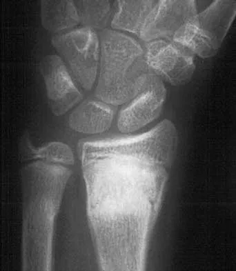
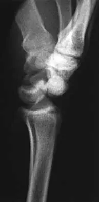
A 25-year-old man reports wrist pain following a motorcycle accident. Examination reveals minimal swelling, slightly limited active range of motion, and point tenderness in the snuff box region. AP and oblique radiographs are shown in Figures 40a and 40b. Management should consist of


Explanation

The radiographs reveal a scaphoid fracture with displacement and comminution and an unstable fracture pattern. Treatment should consist of open reduction and internal fixation. In displaced scaphoid fractures and fractures with unstable fracture patterns, closed reduction is ineffective and is likely to lead to nonunion. Limited intercarpal fusion and proximal row carpectomy are used to correct a variety of traumatic and posttraumatic problems of the wrist. Amadio PC, Taleisnik J: Fractures of the carpal bone, in Green DP, Hotchkiss RN, Pederson WC (eds): Green's Operative Hand Surgery, ed 4. Philadelphia, PA, 1999, pp 809-823. Rettig ME, Kozin SH, Cooney WP: Open reduction and internal fixation of acute displaced scaphoid waist fractures. J Hand Surg Am 2001;26:271-276. Cooney WP, Dobyns JH, Linscheid RL: Fractures of the scaphoid: A rational approach to management. Clin Orthop 1980;149:90-97.


Question 7

A 42-year-old woman reports that she has low back pain and had a transient loss of consciousness after falling off a horse. She denies having neck pain but notes that she was involved in a motor vehicle accident 2 years ago and had neck pain at that time. Examination reveals full range of motion of the neck and no localized tenderness. The neurologic examination is normal. A lateral radiograph of the cervical spine is obtained. Figures 41a and 41b show CT and MRI scans. What is the most likely diagnosis?


Explanation

The examination findings do not correlate with an acute injury (full range of cervical motion and the absence of pain). Radiographically, the fracture appears old based on the smooth contour of the fracture fragments and the absence of soft-tissue swelling. Flexion-extension radiographs can be obtained to determine potential instability; if present, stabilization and fusion should be considered. Schatzker J, Rorabeck CH, Waddell JP: Non-union of the odontoid process: An experimental investigation. Clin Orthop 1975;108:127-137.


Question 8

What neurologic structure is most at risk when performing intramedullary screw fixation of a fifth metatarsal base fracture?


Explanation

The sural nerve and its terminal branches course through the lateral hindfoot and midfoot area and are directly at risk in surgeries involving the peroneal tendon complex and the fifth metatarsal. The first branch of the lateral plantar nerve originates in the tarsal tunnel region and courses across the plantar heel area to innervate the abductor digiti minimi; it is not at direct risk with fifth metatarsal surgery. The saphenous, superficial peroneal, and deep peroneal nerves are not at risk anatomically with a lateral midfoot incision. Donley BG, McCollum MJ, Murphy GA, Richardson EG: Risk of sural nerve injury with intramedullary screw fixation of fifth metatarsal fractures: A cadaver study. Foot Ankle Int 1999;20:182-184.

Question 9

A 25-year-old man sustained an L1 compression fracture in a fall from his roof. He is neurologically intact and has no other injuries. Radiographs reveal a 25% loss of height anteriorly and 5 degrees of kyphosis at the fracture site. A CT scan reveals no compromise of the posterior column. Management should consist of


Explanation

The patient has a stable fracture that can be initially treated with bed rest, followed by bracing and quick mobilization. The outcome is good and surgery is not required. These fractures can be treated nonsurgically if there is less than 50% compression, 15 degrees of angulation, and intact posterior structures. Cantor JB, Lebwohl NH, Garvey T, Eismont FJ: Nonoperative management of stable thoracolumbar burst fractures with early ambulation and bracing. Spine 1993;18:971-976.

Question 10

A 35-year-old man sustained a 10% compression fracture of the C5 vertebra in a diving accident. Radiographs show good alignment, and examination reveals no neurologic compromise. An MRI scan reveals no significant soft-tissue disruption posteriorly. Management should consist of


Explanation

The patient has a stable flexion-compression injury of the cervical spine. The fracture occurs as a result of compression failure of the vertebral body. If the force continues, a tension failure of the posterior structures occurs, leading to potential dislocation. Immobilization in a rigid cervical orthosis will allow this fracture to heal. Clark CR (ed): The Cervical Spine, ed 3. Philadelphia, PA, Lippincott-Raven, 1998, pp 457-464.

Question 11

Figures 42a and 42b shows the radiographs of a 20-year-old man who sustained a hyperextension injury to his little finger. Multiple attempts at closed reduction have been unsuccessful. Management should now consist of


Explanation

The radiographs show a complex dislocation of the little finger metacarpophalangeal joint. This is characterized by obvious dislocation on the AP and lateral views and a type of bayonet apposition best visualized on the lateral view. Irreducibility of this injury is caused by displacement of the volar plate that has been traumatically avulsed from its origin on the metacarpal, with subsequent displacement into the metacarpophalangeal joint. This abnormal position of the volar plate causes irreducibility that can be corrected only by open reduction. This can be effected either by dorsal or palmar approaches. Stern PJ: Fractures of the metacarpals and phalanges, in Green DP, Hotchkiss RN, Pederson WC (eds): Green's Operative Hand Surgery, ed 4. Philadelphia, PA, 1999, pp 711-771. Becton JL, Christian JD Jr, Goodwin HN, Jackson JG III: A simplified technique for treating the complex dislocation of the index metacarpophalangeal joint. J Bone Joint Surg Am 1975;57:698-700.


Question 12

A 34-year-old man sustains an extra-articular fracture of the proximal phalanx of his right index finger in a fall. Examination reveals that the fracture is closed and oblique in orientation. Closed reduction and splinting fail to maintain the reduction. Management should now consist of


Explanation

The patient has an unstable oblique fracture of the proximal phalanx that is easily reducible but unstable; therefore, the treatment of choice is closed reduction and percutaneous pin fixation, followed by casting. Closed reduction and percutaneous pin fixation offers a better functional result than open reduction and plate fixation. Repeat closed reduction and buddy taping is inadequate because of the inherently unstable fracture pattern. Buddy taping will allow the dislocation to recur. The other options represent more aggressive surgical techniques than are necessary to treat this fracture. Stern PJ: Fractures of the metacarpals and phalanges, in Green DP, Hotchkiss RN, Pederson WC (eds): Green's Operative Hand Surgery, ed 4. Philadelphia, PA, 1999, pp 711-771.

Question 13

Figures 43a and 43b show the AP and lateral radiographs of the radius and ulna of a 9-year-old patient. The fracture is manipulated and placed in a long arm cast with the elbow flexed to 90 degrees and the forearm to neutral rotation. Figures 43c and 43d show the alignment of the fracture after the manipulation. What is the next most appropriate step in management?


Explanation

By placing the forearm at neutral rotation, as shown in Figures 43c and 43d, the distal fragment has become malrotated by 90 degrees. This is evident by the fact that the bicipital tuberosity is rotated 90 degrees to the radial styloid. Normally, it should be directly opposite (180 degrees) to the radial styloid. The correct alignment was present in the original radiographs shown in Figures 43a and 43b. Another clue to the malrotation in the postreduction radiographs is the difference in the diameters of the opposing radial shafts. To correct this rotational malalignment, the distal fragment needs to be remanipulated into supination so that it is correctly aligned with the supinated proximal radius. Evans EM: Fractures of the radius and ulna. J Bone Joint Surg Br 1951;33:548-561.


Question 14

Which of the following findings is an indication for adjunctive use of high-dose steroids?


Explanation

According to NASCIS III, the high-dose steroid protocol involves infusion of 30 mg/kg methylprednisolone followed by 5.4 mg/kg/h for 24 hours if the patient has sustained a spinal cord injury within the last 3 hours. The drip is continued for 48 hours if administration is started between 3 and 8 hours of the onset of neurologic deficit. No benefit has been conclusively demonstrated with steroids administered beginning 8 hours or longer after injury. Steroid use is not indicated for nerve root deficits, brachial plexus deficits, or gunshot wounds. Kellam JF, Fischer TJ, Tornetta P III, Bosse MJ, Harris MB (eds): Orthopaedic Knowledge Update: Trauma 2. Rosemont, IL, American Academy of Orthopaedic Surgeons, 2000, pp 319-328.

Question 15

A 22-year-old man sustained a stable pelvic fracture, bilateral femur fractures, and a left closed humeral shaft fracture in a motor vehicle accident. Examination 24 hours after injury reveals that the patient is confused and has shortness of breath. A clinical photograph of his conjunctiva is shown in Figure 44. He has a temperature of 101 degrees F (38.3 degrees C) and a pulse rate of 120/min. Laboratory studies show a hemoglobin level of 8 g/dL, a platelet count of 50,000/mm3, and a PaO2 of 57 mm Hg on 2L of oxygen. What is the most likely diagnosis?


Explanation

The major criteria for the diagnosis of fat embolism syndrome include hypoxemia (PaO2 of less than 60 mm Hg), central nervous system depression, and a petechial rash that is most often located in the axillae, conjunctivae, and palate. The rash is often transient. Tachycardia, pyrexia, anemia, thrombocytopenia, and the presence of fat in the urine are all considered minor criteria. To establish the diagnosis of fat embolism syndrome, one major and four minor signs should be present. Pulmonary embolism, which is the major differential diagnosis, usually is not associated with conjunctival petechia or thrombocytopenia.


Question 16

Figure 45 shows the current radiograph of an 11-year-old girl who sustained a simple nondisplaced fracture of the distal radius 4 weeks ago. Management at the time of injury consisted of application of a short arm cast but no manipulation. What is the major concern at this time?


Explanation

The fracture pattern represents a Peterson type I physeal injury, which is a comminuted metaphyseal fracture in which the fracture lines extend up to the physis. Because there is no displacement of the physis and the fracture lines do not cross the physis, there may be a tendency to dismiss this injury as a simple metaphyseal fracture with no significant sequelae. A small percentage of patients (3% in Peterson's series) experience growth arrest. In this patient, a disabling ulnar plus deformity, defined as increased ulnar length in relationship to the distal radius, developed. Peterson HA: Physeal fractures: Part 2. Two previously unclassified types. J Pediatr Orthop 1994;14:431-438.


Question 17

Which of the following is considered the best measure of the adequacy of resuscitation in the first 6 hours after injury?


Explanation

The end point of resuscitation is adequate tissue perfusion and oxygenation. Blood lactate is the end point of anaerobic metabolism. The level of blood lactate reflects global hypoperfusion and is directly proportional to oxygen debt. Two separate prospective studies have verified a significant difference in mortality when blood lactate was used as a measure of resuscitation when compared to traditional parameters (mean arterial pressure, urine output, central venous pressure, and heart rate). Base deficit is a direct measure of metabolic acidosis and an indirect measure of blood lactate levels. It correlates well with organ dysfunction, mortality, and adequacy of resuscitation. It is easy to measure, can be obtained rapidly, and is an excellent assessment of the adequacy of resuscitation. Porter JM, Ivatury RR: In search of the optimal end points of resuscitation in trauma patients: A review. J Trauma 1998;44:908-914.

Question 18

A 26-year-old man sustains a displaced bimalleolar fracture by sliding into second base while playing baseball. Following initial closed reduction and splinting of the fracture, moderate swelling is noted. What is the safest time to perform surgery?


Explanation

Following any closed fracture, the most important determinant for the timing of surgery is the condition of the soft tissues and especially the skin. The best determinant of appropriate soft-tissue condition is the presence of wrinkling of the skin (wrinkle sign) at the site of the incision. A wrinkle sign is present when all the interstitial edema has left the skin; this may take up to 14 to 21 days of elevation. Any abrasion must be epithelialized so that there are no bacteria left at the site. To date, no other method of soft-tissue viability measurement has been shown to be of any clinical benefit. Stover MD, Kellam JF: Articular fractures: Principles, in Ruedi TP, Murphy WM (eds): AO Principles of Fracture Management. Stuttgart, Thieme, 2000, pp 105-119. Hahn DM, Colton CL, Malleolar fractures, in Ruedi TP, Murphy WM (eds): AO Principles of Fracture Management. Stuttgart, Thieme, 2000, pp 559-581.

Question 19

A 28-year-old woman sustained an injury to her dominant right arm after falling off her porch. Examination reveals a deformity at the elbow. She is neurovascularly intact. Figures 46a and 46b show the radiographs obtained before closed reduction, and postreduction radiographs are shown in Figure 46c and 46d. What is the most likely early complication?


Explanation

The patient has a complex fracture-dislocation of the elbow. The radial head is fractured, and there is a displaced coronoid fracture. These associated fractures indicate that the elbow is at high risk for recurrent instability after initial treatment. To prevent this complication, surgical treatment will most likely be required and will consist of some or all of the following: radial head open reduction and internal fixation or replacement, coronoid open reduction and internal fixation, medial and lateral ligament repairs, and even articulated external fixation. This patient was treated with open reduction and internal fixation of the radial head, and the elbow redislocated postoperatively. Ring D, Jupiter JB: Reconstruction of posttraumatic elbow instability. Clin Orthop 2000;370:44-56. O'Driscoll SW: Classification and evaluation of recurrent instability of the elbow. Clin Orthop 2000;370:34-43.


Question 20

What is the most likely long-term sequela of the injury shown in Figures 47a and 47b?


Explanation

The imaging studies show a comminuted lateral talar process fracture. This injury is often missed on plain radiographs; therefore, CT provides the best method of diagnostic evaluation. The most likely long-term sequela of this injury is subtalar joint arthrosis. Although this injury involves the fibular gutter region, progression to true ankle arthritis is unlikely. There does not appear to be any association with this injury and chronic mechanical instability of the ankle or disruption of the superior peroneal retinaculum and subsequent peroneal tendon instability. Entrapment of the flexor hallucis longus tendon may occur with fractures of the sustentaculum tali but not with injuries of the lateral talar process. Surgical management includes open reduction and internal fixation versus excision; the goal is preservation of the large articular surface fragments. In this patient, there is significant comminution and early fragment excision may be the best option for acute treatment. Tucker DJ, Feder JM, Boylan JP: Fractures of the lateral process of the talus: Two case reports and a comprehensive literature review. Foot Ankle Int 1998;19:641-646.


Question 21

A 16-year-old high school football player has diffuse pain with attempted digital flexion after injuring the ring finger of the dominant hand 1 week ago. Examination reveals that he is unable to flex the distal interphalangeal joint. Management should consist of


Explanation

The patient has an avulsion of the flexor digitorum profundus. Treatment should include surgical exploration and tendon reinsertion. This is not an avulsion of the flexor digitorum superficialis because the patient's deficiency is the inability to flex the distal interphalangeal joint, not the proximal interphalangeal joint. Surgical release of the anterior interosseous nerve is not indicated because the flexor digitorum profundus of the ring finger is innervated by the ulnar nerve. A median nerve contusion causes wrist pain and/or numbness and tingling in the median nerve distribution. Strickland JW: Flexor tendons: Acute injuries, in Green DP, Hotchkiss RN, Pederson WC (eds): Green's Operative Hand Surgery, ed 4. Philadelphia, PA, 1999, pp 1851-1897.

Question 22

A 25-year-old construction worker lands on his outstretched hand in a fall. The position of his wrist at the time of impact causes a force that leads to hyperextension, ulnar deviation, and intercarpal supination. Radiographs are shown in Figures 48a and 48b. What type of injury pattern is shown?


Explanation

The patient has a transscaphoid dorsal perilunate dislocation. The radiographs clearly define a dorsal dislocation of the capitolunate joint, and the scaphoid fracture component is easily visible on the AP view. A scaphoid fracture alone is an unlikely diagnosis because of the midcarpal dislocation component. The radiocarpal joint is not dislocated because the lunate is sitting in the lunate fossa of the radius. Isolated radiocarpal dislocations are not associated with a midcarpal disruption. While a midcarpal dislocation is a component of a dorsal perilunate dislocation, this diagnosis does not address the scaphoid fracture. A volar lunate dislocation is not seen because the lunate is reduced in the lunate fossa of the distal radius. Volar lunate dislocations are in the spectrum of injury of perilunate dislocations and fracture-dislocations; however, the radiographs show a transscaphoid dorsal perilunate dislocation. Mayfield JK, Johnson RP, Kilcoyne RK: Carpal dislocations: Pathomechanics and progressive perilunar instability. J Hand Surg Am 1980;5:226-241.


Question 23

A 25-year-old construction worker lands on his outstretched hand in a fall. The position of his wrist at the time of impact causes a force that leads to hyperextension, ulnar deviation, and intercarpal supination. Radiographs are shown in Figures 48a and 48b. Management should consist of


Explanation

Open reduction and internal fixation is the treatment of choice for accurate reduction of the disrupted intercarpal ligaments. In addition, the displaced scaphoid fracture will require open reduction and internal fixation and possible bone grafting. Closed reduction and long arm casting will not allow accurate reduction of the dislocated intracarpal intervals, and it is unlikely to allow accurate reduction of the scaphoid. The maneuver required to effect closed reduction of a displaced scaphoid fracture will most likely cause the scaphoid lunate interval to displace. Closed reduction with percutaneous pin fixation or with an external fixator is unable to effect anatomic reduction of the injury. Proximal row carpectomy is used as a salvage procedure for a variety of degenerative and posttraumatic problems of the wrist. Kozin SH: Perilunate injuries: Diagnosis and treatment. J Am Acad Orthop Surg 1998;6:114-120. Herzberg G, Comtet JJ, Linscheid RL, Amadio PC, Cooney WP, Stalder J: Perilunate dislocations and fracture-dislocations: A multicenter study. J Hand Surg Am 1993;18:768-779.


Question 24

A 17-year-old boy who fell on a pitchfork in a barn 1 day ago now has a painful, swollen forearm. Examination reveals erythema, exquisite tenderness, and crepitus to palpation of the forearm. He has a pulse rate of 110/min and a blood pressure of 80/60 mm Hg. Radiographs show subcutaneous air and no fractures. Gram stain of wound drainage reveals a gram-positive bacillus. The next most appropriate step in management should consist of


Explanation

The successful treatment of necrotizing soft-tissue infections such as clostridial myonecrosis depends on prompt recognition and aggressive surgical debridement of all involved muscle, fascia, and soft tissue, resecting to a clearly normal healthy, viable margin. The effective antibiotic regimen for clostridial infection is high-dose penicillin; however, necrotizing infections are frequently polymicrobial so initially broad-spectrum antibiotics are indicated. Hyperbaric oxygen therapy may be used as an adjunct to surgical treatment but is insufficient as a primary therapy. Prolonged application of tourniquets and wound closure should be avoided. Pellegrini VD, Evarts CM: Complications, in Rockwood CA Jr, Green DP (eds): Fractures in Adults, ed 3. Philadelphia, PA, JB Lippincott, 1991, pp 365-370. Gerding DN, Peterson LR: Infections caused by anaerobic bacteria, in Shulman ST, Phair JP, Peterson LR, Warren JR (eds): Infectious Diseases, ed 5. Philadelphia, PA, WB Saunders, 1997, pp 416-417.

Question 25

In the management of an open tibia fracture, what factor is considered most important in preventing deep infection?


Explanation

The most important aspect of management of any open fracture, and in particular the tibia, is the degree and the completeness of the debridement of the soft tissue and most importantly, the muscle. The ultimate function is determined by the amount of muscle left, as well as the ability to heal. The amount of necrotic muscle left in the wound also determines the predisposition to infection. The method of fixation, the size of the wound, and the amount of contamination are controlled by the surgeon or the injury and have little to do with the long-term outcome. Initial wound cultures have little predictive value. Clifford P: Open fractures, in Ruedi TP, Murphy WM (eds): AO Principles of Fracture Management. Stuttgart, Thieme, 2000, pp 617-638.

Question 26

A 40-year-old male presents after a high-energy motor vehicle collision with a closed Schatzker IV tibial plateau fracture. Which of the following associated injuries must be most highly suspected and systematically ruled out?


Explanation

Schatzker IV (medial plateau) fractures often result from a high-energy varus force, functioning similarly to a knee dislocation. Consequently, there is a high association with popliteal artery and common peroneal nerve injuries that must be carefully ruled out.

Question 27

In the treatment of a complex pilon fracture, the surgeon identifies a large anterolateral articular fragment. Which of the following ligaments provides the primary soft-tissue attachment to this specific fragment?


Explanation

The anterolateral articular fragment in a pilon fracture is often referred to as the Chaput fragment. It serves as the main tibial attachment site for the anterior inferior tibiofibular ligament (AITFL).

Question 28

A 25-year-old polytrauma patient sustains bilateral femoral shaft fractures and severe pulmonary contusions. Initial labs reveal a pH of 7.21, base excess of -8, and core temperature of 34.5°C. What is the most appropriate initial management of the femoral fractures?


Explanation

This patient is in extremis based on acidosis (pH < 7.24) and hypothermia, meeting the criteria for Damage Control Orthopedics (DCO). The safest initial management is rapid temporary stabilization with spanning external fixators to minimize physiological hit.

Question 29

A surgeon is planning a posteromedial approach to address a displaced posteromedial shear fragment in a bicondylar tibial plateau fracture. Which surgical interval is typically utilized for this approach?


Explanation

The standard posteromedial approach to the proximal tibia exploits the interval between the pes anserinus anteriorly and the medial head of the gastrocnemius posteriorly. This safely exposes the posteromedial cortex for anti-glide plating.

Question 30

When utilizing an anterolateral approach for open reduction and internal fixation of a distal tibia pilon fracture, which neurologic structure is at greatest risk of iatrogenic injury?


Explanation

The superficial peroneal nerve routinely crosses the operative field from lateral to medial over the distal third of the leg. It must be carefully identified and protected during the anterolateral approach to the distal tibia.

Question 31

Following a high-energy Schatzker VI tibial plateau fracture treated with a spanning external fixator, the patient develops increasing leg pain out of proportion to the injury. Which of the following is the most sensitive early clinical finding for compartment syndrome?


Explanation

Pain out of proportion to the injury and pain with passive stretch of the involved compartment's muscles are the earliest and most sensitive clinical indicators of acute compartment syndrome. Loss of pulses and pallor are late, unreliable signs.

Question 32

A 45-year-old male sustains a closed, high-energy tibial pilon fracture initially managed with a spanning external fixator. What is the most reliable clinical indicator that the soft tissue envelope is ready for definitive open reduction and internal fixation?


Explanation

The "wrinkle sign" demonstrates that the initial massive soft tissue swelling has subsided, allowing the skin to wrinkle with joint motion or palpation. This typically occurs 10 to 21 days post-injury and indicates it is safe to proceed with surgical incisions.

Question 33

During the initial resuscitation phase of a severely injured polytrauma patient, which of the following metabolic parameters is the most reliable prognostic indicator of adequate global tissue perfusion and resuscitation?


Explanation

Clearance of serum lactate and normalization of base deficit are the most reliable markers that oxygen debt has been repaid and global tissue perfusion is restored. Standard vital signs can often normalize before adequate resuscitation is achieved.

Question 34

A 55-year-old female undergoes ORIF for a Schatzker II tibial plateau fracture. The depressed articular segment is elevated and supported with bone graft. Which fixation construct is most biomechanically critical to prevent secondary subsidence of the elevated articular fragment?


Explanation

Subchondral rafting screws provide the crucial structural support directly beneath the elevated articular fragments. This acts as a mechanical floor, preventing secondary subsidence into the metaphyseal void during early rehabilitation.

Question 35

A patient treated nonoperatively for a displaced pilon fracture develops a symptomatic varus malunion. Which of the following joint reactive force alterations is most likely to accelerate post-traumatic ankle arthrosis in this patient?


Explanation

A varus malunion shifts the mechanical axis of weight-bearing medially. This substantially increases contact pressures over the medial talar dome, leading to accelerated medial compartment post-traumatic arthritis.

Question 36

A 28-year-old male polytrauma patient with bilateral femur fractures and a pelvic ring injury suddenly develops acute confusion, dyspnea, and a petechial rash over his axillae on hospital day 2. Which of the following is the most effective initial management?


Explanation

This patient exhibits the classic triad of Fat Embolism Syndrome (FES): hypoxemia, neurologic abnormalities, and petechial rash. The mainstay of treatment is rigorous supportive care, primarily focusing on maintaining adequate oxygenation and ventilation.

Question 37

When performing a direct posterolateral approach for a complex tibial plateau fracture, an osteotomy of the fibular head or neck is occasionally required for adequate visualization. Which of the following structures must be carefully identified and protected during this osteotomy?


Explanation

The common peroneal nerve wraps directly around the fibular neck from posterior to anterior. It is at extremely high risk of injury during a posterolateral approach, especially if a fibular osteotomy is performed.

Question 38

Which of the following radiographic patterns is most characteristic of a low-energy, rotational pilon fracture rather than a high-energy axial load pilon fracture?


Explanation

Low-energy rotational mechanisms typically produce spiral fracture patterns of the distal tibia that extend into the plafond, with minimal comminution or articular impaction. High-energy injuries usually present with significant comminution and articular "die-punch" depression.

Question 39

During the initial ATLS survey of a hemodynamically unstable polytrauma patient with an AP compression pelvic ring injury, a pelvic binder is indicated. To be maximally effective, the binder must be centered over which of the following anatomic landmarks?


Explanation

Proper placement of a pelvic binder is centered directly over the greater trochanters. This allows the trochanters to act as levers, effectively internally rotating the hemipelvises and reducing the pelvic volume to tamponade bleeding.

Question 40

A patient with a Schatzker VI bicondylar tibial plateau fracture is treated with dual orthogonal plating through a single extensile anterior midline incision. What is the most significant risk associated with this surgical strategy?


Explanation

Accessing both the medial and lateral plateau through a single anterior midline incision requires massive subcutaneous tissue stripping. This severely disrupts the blood supply to the skin flaps, resulting in an unacceptably high rate of wound necrosis and deep infection.

Question 41

In a highly comminuted pilon fracture, the posterior articular fragment (Volkmann fragment) is typically displaced due to the pull of which attached structure?


Explanation

The Volkmann fragment corresponds to the posterior malleolus or the posterior aspect of the tibial plafond. It typically displaces with the fibula because of the strong, intact attachment of the posterior inferior tibiofibular ligament (PITFL).

Question 42

In managing a bleeding polytrauma patient, the trauma team must rapidly intervene to prevent the "lethal triad" of trauma. Which three clinical entities comprise this triad?


Explanation

The lethal triad of trauma consists of hypothermia, acidosis, and coagulopathy. These three derangements create a vicious cycle that exacerbates bleeding and represents a severe failure of physiologic compensation.

Question 43

Which specific type of tibial plateau fracture is most strongly associated with an injury to the medial collateral ligament (MCL) or a lateral meniscal tear?


Explanation

Schatzker II (lateral split-depression) fractures are typically caused by a valgus load with axial compression. This mechanism frequently results in a concurrent lateral meniscal tear or stretching/rupture of the medial collateral ligament (MCL).

Question 44

Despite achieving anatomic articular reduction and stable fixation of a high-energy pilon fracture, the patient has a high risk of developing post-traumatic ankle arthrosis. What is the primary pathophysiologic reason for this outcome?


Explanation

The massive energy transfer during an axial load injury causes immediate, irreversible mechanical damage and subsequent apoptosis of the articular chondrocytes. This leads to early post-traumatic arthritis regardless of how anatomically the joint surface is surgically restored.

Question 45

When placing an emergency spanning external fixator across the knee for a severely comminuted tibial plateau fracture in a polytrauma patient, where should the femoral half-pins be optimally placed to minimize complications?


Explanation

Femoral pins for a knee-spanning external fixator should be placed anterolaterally or directly laterally into the femur. This avoids impaling the rectus femoris tendon, which can cause severe knee stiffness, and avoids the medial neurovascular structures.

Question 46

A 45-year-old male sustains a high-energy Schatzker VI tibial plateau fracture. CT imaging demonstrates a displaced posteromedial shear fragment. You elect to utilize a posteromedial approach for buttress plating. Which of the following represents the correct internervous or intermuscular plane for this approach?


Explanation

The posteromedial approach to the tibial plateau utilizes the interval between the medial head of the gastrocnemius (tibial nerve) and the pes anserinus (femoral and tibial nerves). Retracting the medial gastrocnemius laterally protects the neurovascular bundle.

Question 47

A 35-year-old male sustains a high-energy Schatzker VI tibial plateau fracture. During the initial emergency department evaluation, which clinical finding is the most reliable early indicator of acute compartment syndrome?


Explanation

Pain out of proportion to the injury, especially elicited by passive stretch of the involved muscles, is the earliest and most sensitive clinical sign of acute compartment syndrome. Pulselessness and paralysis are late findings indicating irreversible nerve and muscle ischemia.

Question 48

In a polytraumatized patient with bilateral femur fractures, which of the following metabolic markers is the most accurate indicator of adequate tissue resuscitation to safely proceed with Early Total Care (ETC)?


Explanation

Serum lactate (< 2.5 mmol/L) and base deficit are the most reliable indicators of global tissue perfusion. Normalization of these markers indicates adequate resuscitation in polytrauma patients, guiding the safe transition from Damage Control Orthopedics to Early Total Care.

Question 49

A 42-year-old female presents with a closed, severe tibial pilon fracture (AO/OTA 43-C3) with massive soft tissue swelling and fracture blisters. A spanning external fixator is placed. When is the optimal time to proceed with definitive open reduction and internal fixation (ORIF)?


Explanation

Definitive ORIF of pilon fractures should be delayed until the soft tissue envelope has adequately recovered to minimize wound complications. This is clinically indicated by the presence of the "wrinkle sign" and the re-epithelialization of fracture blisters, typically taking 10-21 days.

Question 50

A 28-year-old male sustains a Schatzker IV medial tibial plateau fracture. A CT scan reveals a displaced posteromedial coronal shear fragment. Which surgical approach is most appropriate for direct visualization and buttress plating of this specific fragment?


Explanation

A posteromedial approach allows direct visualization and application of an anti-glide or buttress plate on the posterior aspect of the medial tibial condyle. This mechanically counteracts the typical apex distal and posterior displacement of a posteromedial shear fragment.

Question 51

Which of the following inflammatory cytokines is considered the best early predictor for the severity of systemic inflammatory response syndrome (SIRS) and the subsequent development of acute respiratory distress syndrome (ARDS) in polytrauma patients?


Explanation

Interleukin-6 (IL-6) is a key pro-inflammatory cytokine. Its serum levels peak early after major trauma and strongly correlate with the severity of the injury, the magnitude of the SIRS response, and the risk of multiorgan failure.

Question 52

When utilizing an anterolateral surgical approach for the definitive fixation of a tibial pilon fracture, which neurovascular structure is at the highest risk of iatrogenic injury during the superficial dissection?


Explanation

The superficial peroneal nerve crosses the surgical field from medial to lateral in the anterolateral approach to the distal tibia and ankle. It must be carefully identified and protected during the superficial dissection to avoid neuroma formation or sensory deficits.

Question 53

A patient with a Schatzker II (split-depression) lateral tibial plateau fracture undergoes an MRI prior to surgery. Which of the following soft tissue injuries is most frequently associated with this specific fracture pattern?


Explanation

Lateral meniscal tears are highly associated with Schatzker II lateral tibial plateau fractures. The meniscus is frequently injured, trapped, or driven into the metaphyseal defect as the lateral femoral condyle impacts and depresses the articular surface.

Question 54

According to the principles of Damage Control Orthopedics (DCO), which of the following scenarios is an absolute indication for temporary external fixation of a femoral shaft fracture instead of primary intramedullary nailing?


Explanation

DCO is mandated for patients in extremis or those exhibiting the 'lethal triad' of coagulopathy, hypothermia, and acidosis. Primary intramedullary nailing in this setting risks a severe "second hit" phenomenon, worsening the systemic inflammatory response.

Question 55

In the staged treatment of a highly comminuted, intra-articular tibial pilon fracture (OTA 43-C3) with an associated distal fibula fracture, what is the primary rationale for plating the fibula first?


Explanation

Plating the fibula restores the length, rotation, and alignment of the lateral column of the leg. Through the intact anterior and posterior syndesmotic ligaments, this assists in the indirect reduction of the anterolateral (Chaput) and posterolateral (Volkmann) tibial fragments.

Question 56

When using a laterally applied pre-contoured locking plate for a bicondylar tibial plateau fracture (Schatzker VI), what is the primary biomechanical advantage of the locking screws in the proximal segment?


Explanation

Locking screws thread directly into the plate, creating a fixed-angle construct. In metaphyseal bone, this construct strongly resists cantilever bending and prevents varus collapse of the medial plateau when relying on a single lateral plate.

Question 57

A 30-year-old intubated and sedated polytrauma patient has a normal, high-quality multidetector CT scan of the cervical spine. According to current Eastern Association for the Surgery of Trauma (EAST) guidelines, what is the most appropriate next step regarding cervical spine clearance?


Explanation

Current EAST guidelines state that a high-quality, normal multidetector CT scan is highly sensitive for detecting clinically significant cervical spine injuries. In obtunded patients, the cervical collar can be safely removed based on the negative CT scan alone.

Question 58

A 50-year-old male sustains a pilon fracture characterized by significant articular comminution and metaphyseal impaction, without massive diaphyseal extension. According to the Ruedi-Allgower classification, what grade is this injury?


Explanation

The Ruedi-Allgower classification categorizes pilon fractures into three types based on articular displacement and comminution. Type III fractures involve significant articular comminution and metaphyseal impaction, representing the most severe grade in this system.

Question 59

A patient presents with a hyperextension-varus injury to the knee, resulting in a small anteromedial tibial plateau fracture and an avulsion fracture of the fibular head. Which concomitant ligamentous injury must be highly suspected?


Explanation

A hyperextension-varus force characteristically results in a posterolateral corner (PLC) injury, often manifesting radiographically as a fibular head avulsion (arcuate sign). The anteromedial tibial plateau fracture occurs from impaction during the varus stress.

Question 60

Following open reduction and internal fixation of a severe tibial pilon fracture, what is the most common angular deformity that alters ankle biomechanics and accelerates post-traumatic arthrosis?


Explanation

Valgus malunion is the most common deformity observed following pilon fractures. It typically occurs due to inadequate surgical restoration of the comminuted medial column or subsequent late medial collapse.

Question 61

In a hemodynamically unstable polytrauma patient with an anteroposterior compression (APC) pelvic ring injury, what is the primary mechanism by which a pelvic binder improves hemodynamics?


Explanation

The primary source of hemorrhage in pelvic ring injuries is the presacral venous plexus and exposed cancellous bone. A pelvic binder mechanically reduces the pelvic volume, facilitating the tamponade of this low-pressure venous bleeding.

Question 62

A 33-year-old male sustains a severe bicondylar tibial plateau fracture. Following provisional gross reduction and splinting, his dorsalis pedis pulse is weakly palpable, and his Ankle-Brachial Index (ABI) is calculated at 0.8. What is the most appropriate next step?


Explanation

An Ankle-Brachial Index (ABI) of less than 0.9 in the setting of high-energy lower extremity trauma indicates a high suspicion for vascular injury. The appropriate next step is a CT angiogram to accurately define the arterial anatomy and injury.

Question 63

A 24-year-old male with bilateral femur fractures is admitted to the ICU. On post-injury day 2, he develops confusion, tachypnea, and a petechial rash over his axillae. What is the most effective intervention for preventing the development of this syndrome?


Explanation

The clinical triad of hypoxemia, neurological abnormalities, and petechial rash describes Fat Embolism Syndrome (FES). Early immobilization and definitive surgical fixation of long bone fractures (within 24 hours) is the most proven method to reduce the incidence of FES.

Question 64

When applying a delta-frame spanning external fixator for a severe pilon fracture, what is the optimal placement technique for the transfixing calcaneal pin to minimize the risk of neurovascular injury?


Explanation

Transfixing calcaneal pins should be placed from medial to lateral to protect the posterior tibial neurovascular bundle, which can be injured if the pin exits medially. The safe zone is typically 2-3 cm posterior and inferior to the medial malleolus.

Question 65

A 42-year-old polytrauma patient with a severe closed head injury (GCS 7) and a closed diaphyseal femur fracture is evaluated in the trauma bay. He is hemodynamically stable, but his intracranial pressure (ICP) remains elevated despite medical management. What is the most appropriate management of his femur fracture?


Explanation

In a patient with a severe traumatic brain injury and elevated ICP, damage control orthopedics (DCO) with external fixation avoids the secondary hits of prolonged surgery and reaming, which can further elevate ICP.

Question 66

A 35-year-old man sustains a severe, high-energy distal tibia fracture extending into the ankle joint (pilon fracture) with significant soft tissue swelling and fracture blisters. What is the standard protocol for initial management?


Explanation

The standard of care for high-energy pilon fractures with severe soft tissue compromise is a staged protocol utilizing an initial spanning external fixator until soft tissues recover (usually 10-21 days), followed by definitive fixation.

Question 67

A 28-year-old woman sustains a Schatzker IV tibial plateau fracture. Which of the following mechanisms and associated injuries is most characteristic of this fracture pattern?


Explanation

Schatzker IV fractures (medial plateau) result from a high-energy varus and axial loading mechanism. They are frequently associated with knee subluxation or dislocation, carrying a high risk of popliteal artery and peroneal nerve injuries.

Question 68

During an anterolateral approach to the distal tibia for a pilon fracture, the skin incision is made in line with the fourth ray. Which nerve is at greatest risk of injury during the superficial dissection?


Explanation

The superficial peroneal nerve is at significant risk during the superficial dissection of the anterolateral approach to the distal tibia and must be carefully identified and protected.

Question 69

A polytrauma patient presents with bilateral femur fractures and a pelvic ring injury. Initial labs show a serum lactate of 5.5 mmol/L and base excess of -8. After initial resuscitation in the ICU, at what threshold is it considered physiologically safe to proceed with early total care (ETC) rather than damage control orthopedics (DCO)?


Explanation

A serum lactate of < 2.5 mmol/L and an improving base excess indicate adequate tissue perfusion and clearance of shock, favoring a safe transition to early total care in borderline polytrauma patients.

Question 70

In the setting of a complex posterior pilon fracture, a posterolateral surgical approach is planned. Which of the following anatomic intervals is typically utilized to access the posterolateral fragment (Volkmann's fragment)?


Explanation

The posterolateral approach to the distal tibia utilizes the interval between the flexor hallucis longus (FHL) medially and the peroneal tendons laterally to safely access the posterolateral Volkmann fragment.

Question 71

A 40-year-old man falls from a height and sustains a highly comminuted Schatzker VI tibial plateau fracture. He undergoes a two-incision dual plating technique. To minimize the risk of wound complications and skin necrosis, what is the minimum recommended width of the skin bridge between the anterolateral and posteromedial incisions?


Explanation

When performing dual incisions for complex tibial plateau fractures, a minimum skin bridge of 7 cm is highly recommended to preserve adequate vascularity and minimize the risk of wound necrosis.

Question 72

You are managing a 55-year-old man with a displaced Schatzker II tibial plateau fracture. Preoperative MRI indicates a peripheral tear of the anterior horn of the lateral meniscus that is trapped within the fracture site. Which of the following is the most appropriate management?


Explanation

A submeniscal arthrotomy allows clear visualization of the joint surface and accurate reduction of the fracture. The entrapped meniscus should be elevated, preserved, and repaired to prevent early post-traumatic arthritis.

Question 73

A 25-year-old male sustains a high-energy polytrauma including bilateral pulmonary contusions and a femur fracture. On day 2, he develops a petechial rash, hypoxemia, and confusion. Which of the following pathophysiological mechanisms primarily drives this specific syndrome?


Explanation

Fat embolism syndrome is characterized by the classic triad of hypoxemia, neurologic abnormalities, and a petechial rash. It is primarily driven by the systemic release of marrow fat from long bone fractures and the subsequent severe inflammatory cascade.

Question 74

During the application of a knee-spanning external fixator for a highly comminuted proximal tibia fracture, pins are placed in the distal femur. To avoid intra-articular placement of the femoral pins, how far proximal to the knee joint line should the pins be placed?


Explanation

The suprapatellar pouch reflects proximally about 4-6 cm from the knee joint line. To avoid intra-articular pin placement and the catastrophic complication of septic arthritis, femoral pins must be placed at least 6-8 cm proximal to the joint.

Question 75

In the management of a high-energy pilon fracture, axial CT imaging reveals a large anterolateral articular fragment still attached to the anterior inferior tibiofibular ligament (AITFL). This specific fracture fragment is classically known as the:


Explanation

The Chaput fragment is the anterolateral corner of the distal tibia, which serves as the strong tibial attachment site for the anterior inferior tibiofibular ligament (AITFL).

Question 76

A polytraumatized patient is classified as "borderline" based on initial physiological parameters. According to the concepts of Damage Control Orthopedics (DCO), which of the following intraoperative developments is an absolute indication to abandon early total care and immediately proceed with DCO?


Explanation

The acute development of intraoperative coagulopathy, worsening acidosis, or hypothermia (the lethal triad) in a borderline patient is an absolute indication to abort definitive fracture fixation and switch immediately to a damage control approach.

Question 77

A 24-year-old male presents with a Schatzker I tibial plateau fracture. Which of the following patient profiles and mechanisms most closely matches the typical presentation for this specific fracture pattern?


Explanation

Schatzker I fractures represent a pure wedge split of the lateral plateau. They typically occur in younger patients with strong cancellous bone that splits rather than depresses under an axial or valgus load.

Question 78

When planning definitive open reduction and internal fixation for a complex pilon fracture (AO/OTA 43-C), the sequence of reconstruction is critical. Which of the following represents the classic and most widely accepted sequential approach?


Explanation

The classic sequence for pilon fracture reconstruction is to first restore fibular length (if fractured), then reconstruct the tibial articular surface, attach the reconstructed articular block to the tibial shaft, and finally bone graft any resulting metaphyseal voids.

Question 79

A 32-year-old polytrauma patient with a severe chest injury and bilateral femur fractures is initially treated with bilateral damage control external fixators. When converting the femoral external fixators to intramedullary nails, which of the following strategies best minimizes the risk of deep infection?


Explanation

To minimize deep infection risk when converting from an external fixator to an intramedullary nail, especially if pin sites are inflamed or the fixator has been on for more than 2 weeks, a staged protocol utilizing a "pin-site holiday" to allow tracts to heal is strongly recommended.

Question 80

A 45-year-old male sustains a high-energy Schatzker IV tibial plateau fracture with a significant posteromedial shear fragment. Which of the following describes the most appropriate surgical approach and interval for fixing this specific fragment?


Explanation

The posteromedial approach is ideal for direct visualization and buttress plating of a posteromedial shear fragment. The classic surgical interval is between the medial head of the gastrocnemius and the pes anserinus.

Question 81

A 35-year-old construction worker falls from a height, sustaining a highly comminuted, closed distal tibia pilon fracture. On presentation, the ankle is severely swollen with fracture blisters over the medial and lateral malleoli. What is the most appropriate initial management?


Explanation

High-energy pilon fractures with severe soft tissue compromise should be managed with staged treatment. A joint-spanning external fixator stabilizes the fracture and allows for soft tissue recovery before definitive internal fixation.

Question 82

In a polytrauma patient with bilateral femoral shaft fractures and a severe pulmonary contusion, which of the following laboratory parameters is the most reliable indicator of adequate global tissue perfusion and the end-point of resuscitation prior to early total care (ETC)?


Explanation

Serum lactate and base deficit are the most reliable indicators of global tissue perfusion. Normalization of serum lactate (< 2.0 mmol/L) suggests adequate resuscitation, favoring early definitive fixation.

Question 83

A 45-year-old man sustains a high-energy bicondylar tibial plateau fracture following a motor vehicle collision. CT imaging demonstrates a large, displaced posteromedial coronal shear fragment. Which of the following describes the most appropriate surgical approach and fixation strategy for managing this specific fragment?


Explanation

A displaced posteromedial shear fragment in a bicondylar tibial plateau fracture cannot be adequately completely reduced or stabilized with lateral locking screws alone. It requires a dedicated posteromedial approach and placement of a posterior anti-glide (buttress) plate to successfully counteract the vertical shear forces.

None

Clinic OS
Medically Verified Content by
Prof. Clinic OS
Consultant Orthopedic & Spine Surgeon
Chapter Index