العربية
Part of the Master Guide

Orthopedic Surgery Board Prep: Set 403 (100 MCQs on Fractures & Casting)

AAOS Pediatric Orthopedics MCQs (Set 1): Fractures & Deformities | OITE & ABOS Review

23 Apr 2026 62 min read 98 Views
Pediatrics 2007 MCQs - Part 1

Key Takeaway

This high-yield question set (Set 1) for the AAOS, ABOS, and OITE exams focuses on core topics in Pediatric Orthopedics. Questions cover diagnosis and management of common pediatric fractures, developmental dysplasia of the hip (DDH), scoliosis, clubfoot, and other essential musculoskeletal conditions for board preparation.

AAOS Pediatric Orthopedics MCQs (Set 1): Fractures & Deformities | OITE & ABOS Review

Comprehensive 100-Question Exam


00:00

Start Quiz

Question 1

A pediatric orthopaedic surgeon refers a child to a neurologist. The neurologist's office requests the office records of the pediatric orthopaedic surgeon. To maintain Health Insurance Portability and Accountability Act (HIPAA) compliance, what must the surgeon obtain from the parent(s) prior to sending records?


Explanation

The privacy rules do not require an individual's written authorization for certain permitted or required uses and disclosures of the medical records. Patient or parental authorization is not required for disclosures for certain purposes related to treatment, payment, or health care operations. Specifically, HIPAA does not require a covered entity to obtain patient authorization for many of the health care industry's most fundamental activities such as providing care. Carroll R: Risk Management Handbook for Health Care Organizations, ed 4. Hoboken, NJ, Jossey-Bass, 2003, p 1142.

Question 2

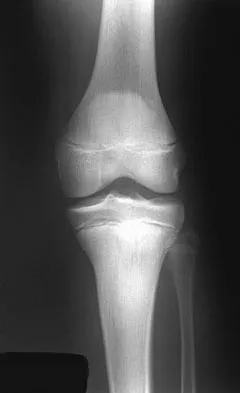
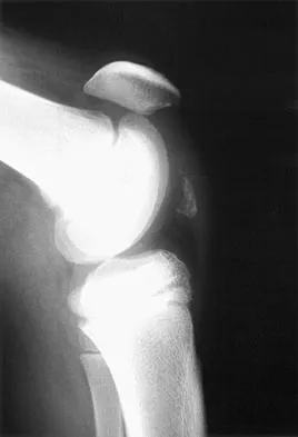
A 13-year-old boy injured his knee playing basketball and is now unable to bear weight. Examination reveals tenderness and swelling at the proximal anterior tibia, with a normal neurologic examination. AP and lateral radiographs are shown in Figures 1a and 1b. Management should consist of


Explanation

The patient has a displaced intra-articular tibial tuberosity fracture; therefore, the treatment of choice is open reduction and internal fixation. Periosteum is often interposed between the fracture fragments and prevents satisfactory closed reduction. Fortunately, most patients with this injury are close to skeletal maturity and therefore, growth arrest and recurvatum are unusual. Nondisplaced fractures can be treated with a cast, but displaced fractures are best treated with open reduction and internal fixation. Intra-articular fractures can disrupt the joint surface and are sometimes associated with a meniscal tear; therefore, arthroscopy may be needed at the time of open reduction and internal fixation. McKoy BE, Stanitski CL: Acute tibial tubercle avulsion fractures. Orthop Clin North Am 2003;34:397-403.


Question 3

A 12-year-old boy sustained a grade III open tibial fracture 1 week ago and underwent multiple debridements and fracture fixation. He now has a soft-tissue defect that measures 6 cm x 6 cm, with an area of exposed bone and muscle on the distal medial leg that is a few centimeters proximal to the ankle. Management of the soft-tissue defect should now consist of


Explanation

The soft-tissue defect is in a very difficult position - the distal tibia. The defect is too distal for a gastrocnemius flap, and the exposed bone precludes an immediate skin graft. A free flap and skin graft would be required for closure. VAC is very effective in soft-tissue defects such as this one. Healthy granulation tissues form quickly. VAC can be the definitive treatment, or it can be used before skin grafting. Wet-to-dry dressings could promote granulation, but the process is hastened substantially by VAC. Amputation is not a consideration because there are no signs of infection or fracture healing problems at this time. Mooney JF III, Argenta LC, Marks MW, et al: Treatment of soft tissue defects in pediatric patients using the V.A.C. system. Clin Orthop 2000;376:26-31.


Question 4

A 6-year-old child sustained a closed nondisplaced proximal tibial metaphyseal fracture 1 year ago. She was treated with a long leg cast with a varus mold, and the fracture healed uneventfully. She now has a 15-degree valgus deformity. What is the next step in management?


Explanation

The tibia has grown into valgus secondary to the proximal fracture. This occurs in about one half of these injuries, and maximal deformity occurs at 18 months postinjury. The deformity gradually improves over several years, with minimal residual deformity. Therefore, treatment at this age is unnecessary as there is a high rate of recurrence and complications regardless of technique. The valgus deformity is not a result of physeal injury or growth arrest. Medial proximal tibial hemiepiphysiodesis is an excellent method of correcting the residual deformity but is best reserved until close to the end of growth. Brougham DI, Nicol RO: Valgus deformity after proximal tibial fractures in children. J Bone Joint Surg Br 1987;69:482. McCarthy JJ, Kim DH, Eilert RE: Posttraumatic genu valgum: Operative versus nonoperative treatment. J Pediatr Orthop 1998;18:518-521.


Question 5

To control most spontaneous bleeding into the knee in children with hemophilia, factor VIII must be replaced to what percentage of normal?


Explanation

The knee is the most common location of spontaneous bleeding in children with hemophilia. Treatment generally requires replacement to 40% to 50% of normal. For surgery, the replacement should be to 100%. The plasma level generally rises 2% for every unit (per kg body weight) of factor VIII administered. Rodriquez-Merchan EC: Management of the orthopaedic complications of hemophilia. J Bone Joint Surg Br 1998;80:191-196.


Question 6

A 6-year-old girl is referred for the elbow injury seen in Figure 2. What is the most appropriate treatment?


Explanation

The patient has a displaced lateral condyle fracture; therefore, simple immobilization for 3 to 8 weeks is likely to result in malunion or nonunion. Closed reduction of such injuries is rarely successful. The fracture is unstable, so fixation is required after open reduction. Because the fixation must cross the physis, smooth pins are indicated for the skeletally immature elbow. Open reduction with fixation has been shown to reduce the risk of delayed union and malunion. Beaty JH, Kasser JR: The elbow: Physeal fractures, apophyseal injuries of the distal humerus, avascular necrosis of the trochlea, and T-condylar fractures, in Beaty JH, Kasser JR (eds): Fractures in Children, ed 5. Philadelphia, PA, Lippincott Williams & Wilkins, 2001, pp 625-703. Rutherford A: Fractures of the lateral humeral condyle in children. J Bone Joint Surg Am 1985;67:851-856.


Question 7

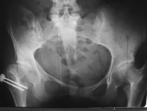
A patient who underwent closed reduction of the hips as an infant now reports pain. An abduction internal rotation view shows an incongruous joint. Based on the findings shown in Figure 3, what is the most appropriate type of pelvic osteotomy for the right hip?


Explanation

Pelvic osteotomies that redirect hyaline cartilage over the femoral head offer the potential for long-term preservation of the hip; however, salvage procedures such as the Chiari osteotomy are indicated in patients without a concentrically reducible hip. Ito and associates reported that moderate dysplasia and moderate subluxation without complete obliteration of the joint space and a preoperative center-edge angle of at least minus 10 degrees are desirable selection criteria. Ohashi H, Hirohashi K, Yamano Y: Factors influencing the outcome of Chiari pelvic osteotomy: A long-term follow-up. J Bone Joint Surg Br 2000;82:517-525.


Question 8

An 18-year-old girl with quadriplegic cerebral palsy underwent posterior spinal fusion from T2 to the pelvis 3 weeks ago. She now has a low-grade fever and mild midline erythema in a 1-cm area from which there is slight clear yellowish drainage. What is the next most appropriate step in management?


Explanation

The presence of drainage 3 weeks after surgery is a sign of wound infection. This infection most likely involves deep tissues until proven otherwise. Oral or IV antibiotics, in the absence of debridement, are not sufficient. Removal of the hardware would lead to rapid progression of the scoliosis in a spine that has been surgically destabilized by removal of the facet joints. The appropriate treatment is debridement with wound culture, IV antibiotics, and retention of hardware. The wound should be closed over drains. Theiss SM, Lonstein JE, Winter RB: Wound infections in reconstructive spine surgery. Orthop Clin North Am 1996;27:105-110.


Question 9

A 13-year-old girl is referred for a painful progressive valgus deformity of the right knee. Examination reveals an antalgic gait with an obvious valgus deformity. The right distal femur has a palpable, tender mass with erythema and warmth. Figures 4a and 4b show a clinical photograph and a radiograph. Management should consist of


Explanation

The radiograph shows a pathologic fracture through a destructive lesion of the distal femur metaphysis with osteolytic and osteoblastic features. The lateral cortex is destroyed, and there is periosteal new bone formation. These findings are consistent with malignancy, most likely an osteogenic sarcoma. Patients with suspected malignant tumors are best managed by surgeons with specific expertise in orthopaedic oncology. The biopsy of a malignant lesion should be deferred to the surgeon who is capable of definitive management of the patient. Enneking W: Principles of musculoskeletal oncologic surgery, in Evarts C (ed): Surgery of the Musculoskeletal System. New York, NY, Churchill Livingston, 1990.


Question 10

An 18-month-old boy with obstetric brachial plexus palsy is being evaluated for limited right shoulder motion. Physical therapy for the past 6 months has failed to result in improvement of the contracture. Which of the following studies is necessary prior to any shoulder reconstruction?


Explanation

The child sustained a brachial plexus injury at birth, and internal rotation/adduction contractures frequently develop at the shoulder. Initial treatment should consist of physical therapy to increase the range of motion. If this fails, as in this patient, MRI is used to evaluate the glenohumeral joint. Commonly, there is joint deformity with increased retroversion of the glenoid and even posterior shoulder subluxation. If the deformity is mild, an anterior release, coupled with teres major and latissimus transfers, is very effective. If the deformity is severe and the shoulder is unreconstructable, then humeral derotation osteotomy is the procedure of choice. MRI of the brain, a radiograph of the elbow, and aspiration of the shoulder would not be helpful. Waters PM: Update on management of pediatric brachial plexus palsy. J Pediatr Orthop B 2005;14:233-244. Waters PM, Bae DS: Effect of tendon transfers and extra-articular soft-tissue balancing on glenohumeral development in brachial plexus birth palsy. J Bone Joint Surg Am 2005;87:320-325.


Question 11

Where is the underlying defect in a rhizomelic dwarf with the findings shown in Figure 5?


Explanation

The radiograph shows the typical findings of achondroplasia. The defect is in fibroblast growth factor receptor 3. The pedicles narrow distally in the lumbar spine. The pelvis is low and broad with narrow sciatic notches and ping-pong paddle-shaped iliac wings. This is often called a champagne glass pelvis. Type I collagen abnormalities are typically found in osteogenesis imperfecta, and type II collagen defects are found in spondyloepiphyseal dysplasia and Kneist syndrome. COMP is defective in multiple epiphyseal dysplasia. Sulfate transport defects are seen in diastrophic dysplasia. Johnson TR, Steinbach LS: Essentials of Musculoskeletal Imaging. Rosemont, IL, American Academy of Orthopaedic Surgeons, 2004, pp 809-812.


Question 12

A 2-year-old boy has complete absence of the sacrum and lower lumbar spine. What is the most likely long-term outcome if no spinal pelvic stabilization is performed?


Explanation

Without stabilization, progressive kyphosis will develop between the spine and pelvis. The kyphosis progresses to the point that the child must use his or her hands to support the trunk, and therefore is unable to use his or her hands for other activities. Neck extension contracture does not usually develop. Neurologic deficit, including sexual dysfunction, is generally present at birth and static. Tachdjian MO: The spine: Congenital absence of the sacrum and lumbosacral vertebrae (lumbosacral agenesis), in Wickland EH Jr (ed): Pediatric Orthopaedics, ed 2. Philadelphia, PA, WB Saunders, 1990, vol 3, p 2228.


Question 13

Which of the following findings is most prognostic for the ability of a young child with cerebral palsy to walk?


Explanation

Several studies have shown that sitting ability by age 2 years is highly prognostic of walking. Molnar and Gordon reported that children not sitting independently by age 2 years had a poor prognosis for walking. Wu and associates reported that children sitting without support by age 2 years had an odds ratio of 26:1 of walking compared with those unable to sit. This was far higher than the odds ratios for cerebral palsy location, motor dysfunction, crawling, creeping, scooting, or rolling. Molnar GE, Gordon SU: Cerebral palsy: Predictive value of selected clinical signs for early prognostication of motor function. Arch Phys Med Rehabil 1976;57:153-158.


Question 14

A 2-year-old girl has had a 2-day history of fever and refuses to move her left shoulder following varicella. Laboratory studies show an erythrocyte sedimentation rate of 75 mm/h and a peripheral WBC count of 18,000/mm3. What is the most common organism in this scenario?


Explanation

The most common bacterial etiologic agent following varicella is group A beta-hemolytic streptococcus. The other organisms are much less common. Staphylococcus aureus is the most common bone infection organism. Staphylococcus epidermidis is increasingly a bone infection organism. Group B streptococcus occurs more commonly in newborns. Kingella kingae is a common joint pathogen but is not as common following varicella. Schreck P, Schreck P, Bradley J, et al: Musculoskeletal complications of varicella. J Bone Joint Surg Am 1996;78:1713-1719.


Question 15

Which of the following is considered the best method to measure limb-length discrepancy in a patient with a knee flexion contracture?


Explanation

The most effective way to measure a limb-length discrepancy in a patient with a knee flexion contracture is a lateral CT scanogram. All the other methods listed provide inaccurate results with a knee flexion contracture because the measurements are made in the coronal plane. Aaron A, Weinstein D, Thickman D, et al: Comparison of orthoroentgenography and computed tomography in the measurement of limb-length discrepancy. J Bone Joint Surg Am 1992;74:897-902.


Question 16

A 5-year-old boy sustained an elbow injury. Examination in the emergency department reveals that he is unable to flex the interphalangeal joint of his thumb and the distal interphalangeal joint of his index finger. The radial pulse is palpable at the wrist, and sensation is normal throughout the hand. Radiographs are shown in Figures 6a and 6b. In addition to reduction and pinning of the fracture, initial treatment should include


Explanation

The findings are consistent with a neurapraxia of the anterior interosseous branch of the median nerve. This is the most common nerve palsy seen with supracondylar humerus fractures, followed closely by radial nerve palsy. Nearly all cases of neurapraxia following supracondylar humerus fractures resolve spontaneously, and therefore, further diagnostic studies and surgery are not indicated. Cramer KE, Green NE, Devito DP: Incidence of anterior interosseous nerve palsy in supracondylar humerus fractures in children. J Pediatr Orthop 1993;13:502-505.


Question 17

An 11-year-old basketball player reports that he felt a painful pop in the left knee when he stumbled while running. He is unable to bear weight on the extremity and cannot actively extend the knee against gravity. Examination reveals a large knee effusion. A lateral radiograph is shown in Figure 7. Management should consist of


Explanation

The radiograph shows an avulsion fracture, or "sleeve fracture," of the distal pole of the patella. The distal fragment is much larger than it appears on the radiograph because it largely consists of cartilage; therefore, excision of the fragment is contraindicated. The treatment of choice is open reduction and tension band fixation to correct patella alta and restore the extensor mechanism. Maguire JK, Canale ST: Fractures of the patella in children and adolescents. J Pediatr Orthop 1993;13:567-571.


Question 18

Figures 8a and 8b show the clinical photograph and radiograph of a 4-month-old infant who has a left foot deformity. Examination reveals that the foot deformity is an isolated entity, and the infant has no known neuromuscular conditions or genetic syndromes. Which of the following studies will best confirm the diagnosis?


Explanation

The clinical photograph shows a rocker-bottom deformity, and the lateral radiograph suggests a congenital vertical talus deformity. A lateral radiograph of the foot in maximum plantar flexion is needed to demonstrate the fixed position of the deformity with malalignment of the talar-metatarsal axis. A fixed dislocation of the navicular on the talus differentiates a congenital vertical talus from the oblique talus with talonavicular subluxation. Kumar SJ, Cowell HR, Ramsey PL: Vertical and oblique talus. Instr Course Lect 1982;31:235-251. Kodros SA, Dias LS: Single-stage correction of congenital vertical talus. J Pediatr Orthop 1999;19:42-48.


Question 19

An 8-year-old girl was treated for a Salter-Harris type I fracture of the right distal femur 2 years ago. Examination reveals symmetric knee flexion, extension, and frontal alignment compared to the contralateral knee. She has 1-cm of shortening of the right femur. History reveals that she has always been in the 50th percentile for height, and her skeletal age matches her chronologic age. Radiographs are shown in Figure 9. What is the expected consequence at maturity?


Explanation

The child has a near complete central physeal arrest of the distal femur and worsening limb-length discrepancy will develop. She is growing at the average rate for the population. The distal femoral physis grows at a rate of roughly 9 mm per year. Girls finish their growth at approximately age 14 years. Thus, at maturity the left leg will be 6.4 cm longer than the right. An angular deformity has not developed at this point and her arrest is central; therefore, angular deformity is unlikely to develop in any plane. Little DG, Nigo L, Aiona MD: Deficiencies of current methods for the timing of epiphysiodesis. J Pediatr Orthop 1996;16:173-179.


Question 20

Examination of an obese 3-year-old girl reveals 30 degrees of unilateral genu varum. A radiograph of the involved leg with the patella forward is shown in Figure 10. Management should consist of


Explanation

The clinical scenario describes infantile tibia vara (Blount's disease). The radiograph shows severe deformity with the characteristic Langenskiold stage 3 changes of the medial proximal tibial metaphysis that distinguish it from physiologic bowing. The preferred treatment is proximal tibiofibular osteotomy with acute correction into slight valgus to unload the damaged area of the physis. This method provides the best results in patients younger than age 4 years. Continued observation would result in progressive deformity. Bracing is most effective in younger children with less severe deformity. Lateral proximal tibial hemiepiphysiodesis relies on growth of the injured medial physis for correction and would result in severe tibial shortening in this young child. Complete epiphysiodesis also produces severe shortening and requires multiple lengthening procedures. Johnston CE II: Infantile tibia vara. Clin Orthop 1990;255:13-23.


Question 21

What is the most important consideration in the preoperative evaluation of a child with polyarticular or systemic juvenile rheumatoid arthritis (JRA)?


Explanation

The cervical spine may be involved in a child with polyarticular or systemic JRA; fusion or instability can occur. Radiographic assessment of the cervical spine should include lateral flexion-extension views. The potential exists for spinal cord injury during intubation or positioning in the presence of an unstable cervical spine. Limitations of the TMJ and micrognathia may affect ease of intubation and administration of anesthesia via a mask. If the TMJ and jaw are involved, some patients may have dental findings such as dental caries and even abscesses which can affect surgery. Some children, particularly those with systemic arthritis, may be taking corticosteroids long-term and may need stress dosing with complex surgeries. Although it is important to routinely check for uveitis and iritis in children with JRA, this usually is not needed preoperatively. Uveitis and iritis are less likely in a child with systemic JRA. Cassity JT, Petty RE (eds): Textbook of Pediatric Rheumatology, ed 5. Philadelphia, PA, WB Saunders, 2005. Ilowite N: Current treatment of juvenile rheumatoid arthritis. Pediatrics 2002;109:109-115. Ruddy S, Harris ED, Sledge CB (eds): Kelley's Textbook of Rheumatology, ed 6. Philadelphia, PA, WB Saunders, 2001.


Question 22

A 15-year-old boy has a mass at the knee. Radiographs show an aggressive tumor involving the proximal tibia, and biopsy findings reveal a high-grade osteosarcoma. Staging studies show that the tumor impinges on the neurovascular bundle. The tumor enlarges during preoperative chemotherapy. Management should now consist of


Explanation

Limb salvage procedures have become the usual treatment for even high-grade osteosarcomas. However, tumors associated with pathologic fracture, tumors encasing the neurovascular bundle, and tumors that enlarged during preoperative therapy and are adjacent to the neurovascular bundle require amputation.


Question 23

Figure 11 shows the radiograph of a 2-year-old child with marked genu varum and tibial bowing. Based on these findings, what is the best initial course of action?


Explanation

The radiograph shows multiple wide physes, consistent with a diagnosis of rickets. A low serum phosphorous level and an elevated alkaline phosphatase level are the hallmarks in diagnosing familial hypophosphatemic Vitamin D-resistant rickets. Serum calcium is usually normal or low normal. This disease is inherited as an X-linked dominant trait and usually presents at age 18 to 24 months. The disease results from a poorly defined problem with renal phosphate transport in which normal dietary intake of vitamin D is insufficient to achieve normal bone mineralization. Renal tubular dysfunction is associated with urinary phosphate wasting. Treatment involves oral phosphate supplementation, which can cause hypocalcemia and secondary hyperparathyroidism. To prevent associated problems, high doses of Vitamin D are administered. While obtaining a scanogram may be clinically indicated in an associated limb-length discrepancy, and subsequent corrective surgery may be indicated, either of these choices would not be the first course of action. An orthosis may slow the progression of genu varum in this disorder but is less important than establishing the correct diagnosis to begin pharmacologic treatment. This amount of varum and tibial bowing far exceeds the normal limits of physiologic genu varum. Skeletal dysplasias usually are not associated with abnormal laboratory values. Herring JA: Metabolic and endocrine bone diseases, in Tachdjian's Pediatric Orthopaedics, ed 3. New York, NY, WB Saunders, 2002, pp 1685-1743.


Question 24

Figure 12 shows the radiograph of a 15-year-old boy with cerebral palsy who has pain at the first metatarsophalangeal joints. He is a community ambulator. Management consisting of accommodative shoes has failed to provide relief. What is the treatment of choice?


Explanation

While other surgeries have provided some success, first metatarsophalangeal joint arthrodesis has the highest overall success rate compared to other surgeries in ambulatory and nonambulatory children with cerebral palsy. The recurrence rate is unacceptably high with the other procedures listed above. In contrast, neurologically normal children are amenable to osteotomies and soft-tissue procedures. Davids JR, Mason TA, Danko A, et al: Surgical management of hallux valgus deformity in children with cerebral palsy. J Pediatr Orthop 2001;21:89-94.


Question 25

What risk factor is most associated with progression of idiopathic scoliosis to a curve requiring surgery?


Explanation

The magnitude of the curve at the time of the peak height velocity is the most prognostic sign in relationship to surgery. More than 70% of curves that measure more than 30 degrees at this time are likely to reach surgical range. Little DG, Song KM, Katz D, et al: Relationship of peak height velocity to other maturity indicators in idiopathic scoliosis in girls. J Bone Joint Surg Am 2000;82:685-693.


Question 26

A 6-year-old boy falls from monkey bars and sustains a displaced extension-type supracondylar humerus fracture. On presentation, his hand is warm and pink, but the radial pulse is not palpable. After closed reduction and percutaneous pinning, the hand remains warm and pink, and capillary refill is less than 2 seconds, but the pulse is still absent on Doppler. What is the most appropriate next step in management?


Explanation

In a pediatric supracondylar fracture with a "pulseless, pink" hand post-reduction, the extremity is well-perfused via collateral circulation. The standard of care is close clinical observation rather than immediate surgical exploration or angiography.

Question 27

A 45-year-old man presents with progressive numbness in his ring and small fingers and weakness in hand grip. He reports a history of an elbow fracture as a young child that was treated in a cast. Radiographs reveal a severe cubitus valgus deformity. Which of the following pediatric injuries is most likely responsible for his current presentation?


Explanation

Nonunion of a lateral condyle fracture leads to a progressive cubitus valgus deformity. Over time, this causes stretch on the ulnar nerve, presenting decades later as tardy ulnar nerve palsy.

Question 28

A 6-week-old infant is being treated with a Pavlik harness for developmental dysplasia of the hip (DDH). During the follow-up visit, the parents report the infant is no longer actively extending the knee on the treated side. What is the most appropriate management?


Explanation

Decreased active knee extension indicates a femoral nerve palsy, a known complication of hyperflexion in a Pavlik harness. Management consists of loosening the anterior straps or temporarily discontinuing the harness until nerve function returns.

Question 29

When correcting a classic congenital idiopathic clubfoot using the Ponseti method, what is the correct sequence of deformity correction?


Explanation

The Ponseti method corrects deformities in a specific sequence remembered by the acronym CAVE: Cavus (elevating the first ray), Adductus, Varus, and finally Equinus (usually requiring a percutaneous Achilles tenotomy).

Question 30

A 13-year-old girl sustains an ankle injury while skateboarding. Radiographs show a Salter-Harris III fracture of the anterolateral distal tibia. Which of the following ligaments imparts the avulsion force responsible for this specific fracture pattern?


Explanation

A juvenile Tillaux fracture is a Salter-Harris III fracture of the anterolateral distal tibial epiphysis. It is caused by an avulsion force from the anterior inferior tibiofibular ligament (AITFL) during an external rotation injury.

Question 31

A 12-year-old boy with a BMI of 32 presents with a left-sided stable slipped capital femoral epiphysis (SCFE). Which of the following is considered an absolute indication for prophylactic in situ pinning of the asymptomatic contralateral right hip?


Explanation

Prophylactic pinning of the contralateral hip in SCFE is highly recommended in patients with endocrine disorders (e.g., hypothyroidism, renal osteodystrophy) or prior radiation therapy due to the high risk of bilateral involvement.

Question 32

A 14-year-old boy sustains an ankle fracture. CT imaging confirms a classic triplane fracture. This injury consists of which of the following fracture patterns?


Explanation

A classic triplane ankle fracture features a sagittal fracture line through the epiphysis, an axial fracture through the physis, and a coronal fracture line extending through the posterior metaphysis.

Question 33

A 10-year-old girl falls on her outstretched hand and sustains an elbow dislocation. After closed reduction, radiographs show an intra-articular fragment. Which of the following physical exam findings is most likely associated with this incarcerated fracture fragment?


Explanation

Medial epicondyle fractures are highly associated with elbow dislocations. An incarcerated medial epicondyle can impinge or injure the ulnar nerve, leading to decreased sensation over the volar small finger and weakness in ulnar-innervated intrinsic hand muscles.

Question 34

A 6-year-old boy presents with a "snapping" and painful lateral left knee. MRI demonstrates a discoid lateral meniscus. During arthroscopy, the meniscus is noted to be hypermobile due to a lack of posterior coronary ligament attachments, with its only posterior attachment being the meniscofemoral ligament. Which variant of discoid meniscus does this represent?


Explanation

The Wrisberg variant of a discoid meniscus lacks the normal posterior capsular attachments (coronary ligaments). Its only posterior attachment is the meniscofemoral ligament of Wrisberg, leading to hypermobility and a snapping sensation.

Question 35

A 9-month-old infant is brought to the emergency department with a swollen right thigh. Radiographs demonstrate a spiral fracture of the midshaft femur. The parents report the child tripped and fell while pulling to stand. Which of the following is the most appropriate next step in management?


Explanation

Femur fractures in non-ambulatory infants (under 1 year of age) are highly suspicious for non-accidental trauma. Admission, consultation with child protective services, and a full skeletal survey are mandatory.

Question 36

In a 7-year-old child diagnosed with Legg-Calvé-Perthes disease, which of the following radiographic classifications is most prognostic for long-term hip joint congruency and outcome?


Explanation

The Herring Lateral Pillar classification, assessed during the fragmentation phase, is the most accurate prognostic indicator for long-term outcomes in Legg-Calvé-Perthes disease based on the height of the lateral pillar of the femoral head.

Question 37

A 12-year-old girl presents with adolescent idiopathic scoliosis (AIS). She has a right thoracic curve of 35 degrees. Her Risser stage is 0, and she is pre-menarchal. What is her approximate risk of curve progression to greater than 50 degrees?


Explanation

Patients with large curves (greater than 30-35 degrees) before skeletal maturity (Risser 0, pre-menarchal) have a very high risk of progression, often cited as approaching or exceeding 90%. Bracing or surgery is indicated depending on exact curve magnitude and progression.

Question 38

A 13-year-old boy presents with vague midfoot pain and frequent ankle sprains. Examination reveals rigid pes planus and limited subtalar motion. Radiographs show a "C-sign" on the lateral view. What is the most likely diagnosis?


Explanation

The "C-sign" on a lateral radiograph is a classic finding representing the continuous bony outline of the medial talar dome and the sustentaculum tali, highly indicative of a talocalcaneal coalition.

Question 39

A 3-year-old girl presents with progressive bilateral genu varum and lateral thrust during gait. Radiographs show a sharp varus angulation at the proximal tibial metaphysis with breaking of the medial cortex. Which radiographic measurement is most useful to distinguish infantile Blount disease from physiologic bowing?


Explanation

The metaphyseal-diaphyseal angle (Drennan angle) is used to differentiate physiologic bowing from infantile Blount disease. An angle greater than 16 degrees is highly predictive of Blount disease.

Question 40

A 13-year-old boy is brought to the ED after sudden onset of severe groin pain following a minor slip. He is unable to bear weight on the affected limb. Radiographs confirm a severe slipped capital femoral epiphysis. What is the most significant complication associated with this specific injury pattern?


Explanation

The inability to bear weight defines an "unstable" SCFE. Unstable SCFE has a significantly higher rate of avascular necrosis (AVN) of the femoral head (up to 47%) compared to stable SCFE.

Question 41

A 15-year-old male gymnast complains of chronic lower back pain exacerbated by extension. Radiographs show a grade II isthmic spondylolisthesis at L5-S1. He has failed 6 months of physical therapy and bracing, and his hamstring tightness is worsening. What is the recommended surgical management?


Explanation

For a symptomatic low-grade (Grade I or II) isthmic spondylolisthesis failing conservative management, an in situ posterolateral fusion of L5-S1 (with or without instrumentation) is the standard surgical treatment. Pars repair is generally reserved for isolated spondylolysis without slip or very early slips in younger patients.

Question 42

A 6-year-old boy falls from monkey bars and presents with a Gartland type III extension-type supracondylar humerus fracture. Examination reveals an inability to flex the interphalangeal joint of the thumb and the distal interphalangeal joint of the index finger. Which nerve is most likely injured?


Explanation

The anterior interosseous nerve (AIN) is the most commonly injured nerve in extension-type supracondylar humerus fractures. It is assessed by asking the patient to make an "OK" sign, which requires FPL and FDP function.

Question 43

A 5-year-old child sustains a displaced lateral condyle fracture of the humerus that is missed and subsequently goes on to nonunion. Years later, which of the following is the most likely late clinical complication?


Explanation

Nonunion of a lateral condyle fracture typically results in a progressive cubitus valgus deformity. Over time, the valgus stretching of the medial elbow structures can lead to a tardy ulnar nerve palsy.

Question 44

A 3-year-old girl sustains an isolated, closed spiral fracture of the middle third of the femoral shaft with 1.5 cm of shortening. What is the most appropriate definitive management?


Explanation

Early spica casting is the standard of care for isolated femoral shaft fractures in children aged 6 months to 5 years with acceptable shortening (< 2 cm). Flexible nailing is typically reserved for older children (ages 5-11).

Question 45

A 2-year-old boy presents with bilateral genu varum. To differentiate between physiologic bowing and infantile Blount disease, a standing AP radiograph is obtained. A metaphyseal-diaphyseal (Drennan) angle greater than which of the following values is most predictive of progression to Blount disease?


Explanation

A metaphyseal-diaphyseal (Drennan) angle greater than 16 degrees has a high positive predictive value for progression to infantile Blount disease, whereas angles less than 10 degrees typically resolve as physiologic bowing.

Question 46

Prophylactic pinning of the contralateral, asymptomatic hip is most strongly indicated in which of the following patients presenting with a unilateral slipped capital femoral epiphysis (SCFE)?


Explanation

Prophylactic pinning of the contralateral hip is indicated in patients with metabolic or endocrine disorders (e.g., renal osteodystrophy, hypothyroidism) due to an extremely high incidence of bilateral involvement.

Question 47

Which of the following pediatric physeal fractures carries the highest risk of premature physeal closure and subsequent growth arrest?


Explanation

Distal femur physeal fractures carry the highest risk of premature physeal closure and growth arrest, approaching 50% in some series, largely due to the undulating topography of the physis.

Question 48

When utilizing the Ponseti method for the treatment of idiopathic clubfoot, what is the correct sequential order of deformity correction?


Explanation

The Ponseti method corrects deformities sequentially using the CAVE acronym: Cavus (by elevating the first ray), Adductus, Varus, and finally Equinus (which often requires a percutaneous Achilles tenotomy).

Question 49

A 6-week-old infant is being treated with a Pavlik harness for developmental dysplasia of the hip (DDH). During follow-up, the anterior straps are found to be adjusted such that the hips are held in 130 degrees of flexion. The infant is noted to have decreased active knee extension. What is the most likely complication?


Explanation

Hyperflexion of the hip in a Pavlik harness (>120 degrees) can compress the femoral nerve against the inguinal ligament, causing a femoral nerve palsy characterized by decreased active knee extension.

Question 50

An 11-year-old boy sustains a proximal humerus fracture with 60% translation. Management consists of a sling and observation due to the tremendous remodeling potential of this region. Approximately what percentage of longitudinal growth of the entire humerus is contributed by the proximal humeral physis?


Explanation

The proximal humerus physis contributes approximately 80% to the longitudinal growth of the humerus. This significant growth contribution explains the massive remodeling potential in children, allowing nonoperative treatment for highly displaced fractures.

Question 51

A 13-year-old girl sustains an ankle injury resulting in a juvenile Tillaux fracture. This fracture pattern is directly related to the normal closure sequence of the distal tibial physis. Which of the following describes the correct chronological sequence of distal tibial physeal closure?


Explanation

The distal tibial physis closes in a predictable pattern: central, then anteromedial, then posteromedial, and finally lateral. The late open lateral physis predisposes adolescents to Tillaux fractures (avulsion by the AITFL).

Question 52

A 14-year-old boy presents after an external rotation injury to his ankle. Radiographs demonstrate a triplane fracture. How does this fracture typically appear on standard anteroposterior (AP) and lateral ankle radiographs?


Explanation

A triplane fracture typically appears as a Salter-Harris III fracture on the anteroposterior (AP) radiograph and a Salter-Harris II fracture on the lateral radiograph. It represents a true Salter-Harris IV injury occurring in three planes.

Question 53

A 6-year-old boy falls from the monkey bars and sustains an extension-type Gartland III supracondylar humerus fracture. On presentation, his hand is pink but pulseless. He is unable to flex the interphalangeal joint of his thumb and the distal interphalangeal joint of his index finger. Which of the following nerves is most likely injured?


Explanation

The anterior interosseous nerve (AIN) is the most commonly injured nerve in extension-type supracondylar humerus fractures. Injury results in the inability to make an 'A-OK' sign due to weakness of the flexor pollicis longus and flexor digitorum profundus to the index finger.

Question 54

A 5-year-old child sustains a displaced lateral condyle fracture of the distal humerus.

Following appropriate treatment with open reduction and internal fixation with pins, which of the following is the most common long-term complication?


Explanation

Lateral condylar overgrowth (spurring) is the most common complication following both operative and nonoperative treatment of lateral condyle fractures. Nonunion is a severe complication but is less common when appropriate operative reduction and fixation are achieved.

Question 55

A 9-year-old boy weighing 110 lbs (50 kg) sustains a severely comminuted, length-unstable midshaft femoral fracture in a motor vehicle collision. Which of the following is the most appropriate definitive management?


Explanation

For pediatric patients older than 5 years and weighing more than 45 kg (100 lbs), titanium elastic nails have a high failure rate, especially in length-unstable patterns. Submuscular plating or a rigid lateral-entry intramedullary nail are the treatments of choice.

Question 56

A 12-year-old boy with a BMI in the 98th percentile is diagnosed with a unilateral stable slipped capital femoral epiphysis (SCFE) and treated with in situ pinning. Prophylactic pinning of the contralateral asymptomatic hip is most strongly indicated in which of the following clinical scenarios?


Explanation

Prophylactic pinning of the contralateral hip in SCFE is strongly recommended for patients with endocrine disorders (e.g., hypothyroidism, renal osteodystrophy) or prior radiation, as they have a very high risk of bilateral involvement.

Question 57

When utilizing the Ponseti method for the treatment of idiopathic clubfoot, the sequence of deformity correction is critical. Which of the following represents the correct sequential order of correction?


Explanation

The Ponseti method follows the CAVE sequence: Cavus is corrected first by elevating the first ray, followed by Adductus and Varus corrected simultaneously, and Equinus is corrected last, often requiring a percutaneous Achilles tenotomy.

Question 58

A 4-year-old boy sustains a nondisplaced proximal tibial metaphyseal fracture that is treated successfully in a long leg cast for 4 weeks. One year later, the parents bring him to the clinic concerned about a deformity in the injured leg. What is the most likely deformity and its anticipated natural history?


Explanation

Cozen's phenomenon is a valgus overgrowth deformity that occurs after proximal tibial metaphyseal fractures in children. It typically peaks at 12 to 18 months post-injury and generally remodels spontaneously over 2 to 3 years.

Question 59

A 13-year-old gymnast sustains a fall and presents with an elbow dislocation.

Following closed reduction, radiographs reveal an associated medial epicondyle fracture. Which of the following is an absolute indication for operative fixation of the medial epicondyle?


Explanation

An incarcerated medial epicondyle fragment within the ulnohumeral joint that cannot be extracted by closed means is an absolute indication for open reduction and internal fixation. Ulnar nerve symptoms and displacement >5-10 mm are relative indications.

Question 60

An 8-month-old infant with developmental dysplasia of the hip (DDH) undergoes a closed reduction and spica casting. To minimize the risk of iatrogenic avascular necrosis (AVN), the hip must be positioned within the 'safe zone' of Ramsey. Which of the following best describes this optimal position?


Explanation

The safe zone of Ramsey defines the position where the hip is reduced without excessive capsular tension. Immobilization in extreme abduction forces the femoral head against the acetabulum, severely increasing the risk of AVN. The ideal position is ~90-100 degrees of flexion and mild to moderate abduction.

Question 61

A 14-year-old boy sustains a twisting injury to his ankle while playing soccer. Radiographs reveal a Salter-Harris III fracture of the anterolateral aspect of the distal tibia epiphysis. Avulsion of this fragment is caused by tension from which of the following structures?


Explanation

A Tillaux fracture is a Salter-Harris III fracture of the anterolateral distal tibial epiphysis. It is caused by an avulsion force from the anterior inferior tibiofibular ligament (AITFL) during external rotation of the foot.

Question 62

A 13-year-old boy sustains a midshaft both-bone forearm fracture. What is the maximum acceptable angular deformity in the sagittal plane to consider nonoperative management with a cast in this age group?


Explanation

In children older than 10 years, remodeling potential is significantly decreased. The acceptable angulation for a midshaft both-bone forearm fracture in this age group is typically less than 10 degrees.

Question 63

A 2-year-old girl is evaluated for bilateral severe bowing of the legs. Radiographs demonstrate a metaphyseal-diaphyseal (MD) angle of 18 degrees bilaterally. What is the most appropriate initial management?


Explanation

An MD angle greater than 16 degrees in a child aged 2 to 3 years is highly suggestive of infantile Blount's disease. The standard initial management for early-stage infantile Blount's disease is KAFO bracing.

Question 64

A 13-year-old boy presents with a rigid flatfoot, recurrent ankle sprains, and deep hindfoot pain. Radiographs demonstrate a continuous 'C-sign' on the lateral view. This radiographic finding is pathognomonic for which of the following conditions?


Explanation

The 'C-sign' is a continuous C-shaped arc formed by the medial outline of the talar dome and the posterior outline of the sustentaculum tali. It is highly indicative of a talocalcaneal coalition.

Question 65

A 4-year-old boy with a history of recurrent low-energy fractures presents to the clinic. Examination reveals blue sclerae and mild joint hyperlaxity. A genetic defect affecting which of the following is the most likely cause of his condition?


Explanation

Osteogenesis imperfecta is a genetic disorder of connective tissue characterized by bone fragility, blue sclerae, and dentinogenesis imperfecta. It is caused by mutations in the COL1A1 or COL1A2 genes, which encode Type I collagen.

Question 66

When evaluating a 7-year-old child with Legg-Calvé-Perthes disease, which of the following radiographic findings during the fragmentation stage is associated with the worst long-term prognosis?


Explanation

The Herring Lateral Pillar classification during the fragmentation stage is highly prognostic. A Lateral Pillar Group C classification (where the lateral pillar is <50% of its original height) is associated with the worst outcomes, often resulting in an aspherical, incongruent hip.

Question 67

A 6-year-old child sustains an isolated fracture of the proximal third of the ulna with an associated anterior dislocation of the radial head. According to the Bado classification, what type of Monteggia lesion is this?


Explanation

The Bado classification categorizes Monteggia fractures based on the direction of the radial head dislocation. Type I is anterior, Type II is posterior, Type III is lateral, and Type IV involves fractures of both the radius and ulna with an anterior radial head dislocation.

Question 68

A 10-year-old boy sustains a Salter-Harris II fracture of the distal femur. What is the approximate reported incidence of premature physeal closure (growth arrest) associated with this specific injury pattern at this location?


Explanation

Distal femur physeal fractures have an exceptionally high rate of premature physeal closure, historically reported to be up to 40% to 50%, even in low-grade (Salter-Harris I and II) injuries, due to the undulating nature of the distal femoral physis.

Question 69

A 15-year-old track athlete experiences a sudden 'pop' and intense pain in his right groin while sprinting. Radiographs reveal an avulsion fracture of the anterior inferior iliac spine (AIIS). Which of the following muscles is responsible for this avulsion?


Explanation

The rectus femoris has its direct head origin on the anterior inferior iliac spine (AIIS) and is responsible for avulsion fractures at this site. The sartorius avulses the ASIS, and the hamstrings avulse the ischial tuberosity.

Question 70

A 12-year-old premenarchal female with Risser stage 0 is diagnosed with adolescent idiopathic scoliosis (AIS). She has a right thoracic curve. Which of the following scenarios is the most appropriate indication to initiate treatment with a Thoracolumbosacral Orthosis (TLSO)?


Explanation

Bracing in AIS is indicated for growing children (Risser 0-2) with curves between 25 and 44 degrees that have documented progression (>5 degrees) or those presenting initially with curves of 30 to 39 degrees. Curves >45-50 degrees generally require surgery.

Question 71

A 6-year-old boy presents with a completely displaced supracondylar humerus fracture. Upon examination, his hand is warm and pink, but the radial pulse is not palpable. What is the most appropriate initial management?


Explanation

The initial management of a pink, pulseless hand associated with a supracondylar humerus fracture is urgent closed reduction and percutaneous pinning. Vascular status often returns to normal once the fracture is anatomically reduced and pinned.

Question 72

A 5-year-old girl sustains a lateral condyle fracture of the humerus. Radiographs show 4 mm of displacement. What is the most common complication if this injury is treated with cast immobilization alone?


Explanation

Lateral condyle fractures displaced >2 mm are highly prone to nonunion if treated non-operatively due to the pull of the common extensor origin and bathing of the fracture in synovial fluid. Operative fixation is indicated for displacement >2 mm.

Question 73

A 13-year-old girl presents with an ankle injury after an external rotation mechanism. Radiographs demonstrate a Salter-Harris III fracture of the anterolateral distal tibial epiphysis. What ligament is directly responsible for the avulsion of this fracture fragment?


Explanation

A Tillaux fracture is a Salter-Harris III fracture of the anterolateral distal tibia. It is an avulsion injury caused by the strong pull of the anterior inferior tibiofibular ligament (AITFL) as the medial distal tibial physis closes before the lateral side.

Question 74

A 12-year-old obese boy presents with severe groin pain and an inability to bear weight after a minor fall. Radiographs show a slipped capital femoral epiphysis (SCFE). According to the Loder classification, what specific clinical finding defines this slip as "unstable"?


Explanation

The Loder classification functionally defines an unstable slipped capital femoral epiphysis (SCFE) as the inability to ambulate with or without crutches. Unstable SCFE has a significantly higher rate of avascular necrosis (up to 47%) compared to stable SCFE.

Question 75

A 3-year-old boy sustains a completely displaced, isolated midshaft femur fracture after a fall from a low playground structure. What is the most appropriate definitive management?


Explanation

For children aged 6 months to 5 years with an isolated femoral shaft fracture and less than 2 cm of shortening, early spica casting is the standard of care. Flexible intramedullary nails are typically reserved for older children, generally aged 5 to 11 years.

Question 76

In the Ponseti method of idiopathic clubfoot casting, what is the correct sequence of deformity correction?


Explanation

The Ponseti method corrects clubfoot deformities in the specific order of the acronym CAVE: Cavus, Adductus, Varus, and finally Equinus. The cavus is corrected first by elevating the first ray to align the forefoot with the hindfoot.

Question 77

A 2-year-old girl is evaluated for severe bilateral bowing of her legs. Radiographs demonstrate an abrupt angulation and breaking of the medial proximal tibial metaphysis with a metaphyseal-diaphyseal angle of 18 degrees. What is the most appropriate initial management?


Explanation

In a child under 3 years with infantile Blount disease and a metaphyseal-diaphyseal angle > 16 degrees, the initial treatment is a trial of bracing with KAFOs. Surgical intervention is indicated if bracing fails or the child presents at an older age.

Question 78

A 9-year-old boy falls on an outstretched hand and sustains a radial neck fracture. Which of the following is considered the upper limit of acceptable angulation for non-operative management in this age group?


Explanation

In older children (around 10 years of age), up to 30 degrees of angulation is acceptable for radial neck fractures. In younger children, up to 45 degrees may remodel adequately, but 30 degrees is a standard threshold for intervention in a 9-year-old.

Question 79

A 6-week-old female infant is diagnosed with a dislocated left hip that is reducible on the Ortolani maneuver. A Pavlik harness is prescribed. Excessive flexion of the hips in the harness increases the risk of which complication?


Explanation

Excessive flexion (>120 degrees) in a Pavlik harness can compress the femoral nerve against the inguinal ligament, leading to femoral nerve palsy. Excessive forced abduction increases the risk of avascular necrosis of the femoral head.

Question 80

A 6-year-old boy sustains a midshaft both-bone forearm fracture. Which of the following fracture parameters has the LEAST potential for spontaneous remodeling?


Explanation

In pediatric forearm fractures, rotational malalignment does not remodel significantly and must be corrected during reduction. Angulation in the plane of joint motion and bayonet apposition remodel very well in children under the age of 10.

Question 81

A 14-year-old boy sustains a triplane fracture of the distal tibia. Which of the following correctly describes the Salter-Harris classification of the fracture lines typically seen on AP and lateral radiographs, respectively?


Explanation

A classic triplane fracture appears as a Salter-Harris III on the AP radiograph (due to the vertical fracture through the epiphysis) and a Salter-Harris II on the lateral radiograph (due to the fracture extending through the posterior metaphysis).

Question 82

A 12-year-old boy presents with an acute elbow dislocation. After closed reduction, radiographs reveal a medial epicondyle fracture fragment incarcerated within the joint. Which nerve is most at risk of injury or entrapment in this scenario?


Explanation

The ulnar nerve courses posterior to the medial epicondyle in the cubital tunnel. It is the most commonly injured nerve in medial epicondyle fractures, especially when the fracture fragment becomes incarcerated within the ulnohumeral joint.

Question 83

A 15-year-old male sprinter feels a sudden pop in his pelvis during a race. Radiographs demonstrate an avulsion fracture of the anterior superior iliac spine (ASIS). Which muscle is responsible for this specific avulsion?


Explanation

The sartorius muscle originates from the anterior superior iliac spine (ASIS) and can cause an avulsion fracture during forceful hip flexion or sprinting. In contrast, the rectus femoris originates from the anterior inferior iliac spine (AIIS).

Question 84

A 14-year-old gymnast presents with persistent low back pain exacerbated by extension. Imaging reveals an acute, unilateral pars interarticularis stress fracture at L5 without spondylolisthesis. What is the most appropriate initial treatment?


Explanation

The initial treatment for an acute spondylolysis (pars stress fracture) without slip is conservative. This typically consists of activity restriction and bracing (such as a TLSO or Boston brace) to limit extension and allow for bony healing.

Question 85

A 2-year-old girl presents with her arm held closely to her side in slight flexion and pronation. Her father reports pulling her by the arm to prevent a fall. Radiographs are normal. What anatomical structure is subluxated?


Explanation

Nursemaid's elbow is a radial head subluxation caused by axial traction on a pronated and extended forearm. The annular ligament slips over the radial head and becomes interposed between the radius and the capitellum.

Question 86

A 6-year-old boy falls from monkey bars and sustains a Gartland type III supracondylar humerus fracture. On arrival, his hand is pink but the radial pulse is absent. The fracture undergoes immediate closed reduction and percutaneous pinning. Following fixation, the hand remains pink and well-perfused, but the radial pulse remains absent by Doppler. What is the most appropriate next step in management?


Explanation

A pink, pulseless hand following reduction and pinning of a supracondylar fracture indicates adequate collateral circulation. The standard of care is close observation for 24 to 48 hours rather than immediate vascular exploration.

Question 87

A 15-year-old boy presents with progressive numbness and tingling in his ring and small fingers. He sustained an elbow fracture at age 4 that was treated nonoperatively. Examination reveals weakness of intrinsic hand muscles. What elbow deformity is most likely present and responsible for his current symptoms?


Explanation

The patient has a tardy ulnar nerve palsy, a classic complication of a nonunion of a pediatric lateral condyle fracture. This nonunion results in a progressive cubitus valgus deformity, stretching the ulnar nerve over time.

Question 88

A 3-week-old infant with idiopathic clubfoot is undergoing serial casting using the Ponseti method. The deformity is corrected in a specific sequence (CAVES). Which aspect of the deformity is corrected last, frequently necessitating a minor surgical procedure?


Explanation

In the Ponseti method, the sequence of correction is Cavus, Adductus, Varus, and finally Equinus. Equinus correction often requires a percutaneous Achilles tenotomy as the final step before the application of the definitive cast.

Question 89

A 14-year-old boy sustains an ankle injury while playing soccer. Radiographs demonstrate a Salter-Harris type III fracture of the anterolateral distal tibia epiphysis. Which of the following is the most likely mechanism of injury for this specific fracture pattern?


Explanation

A juvenile Tillaux fracture is an avulsion of the anterolateral distal tibial epiphysis caused by the anterior inferior tibiofibular ligament (AITFL). It occurs via an external rotation mechanism during the transitional period of physeal closure.

Question 90

A 13-year-old girl is diagnosed with a triplane fracture of the distal tibia following a fall. Which combination of Salter-Harris fracture patterns is classically observed on the standard anteroposterior (AP) and lateral radiographs of the ankle, respectively?


Explanation

A triplane fracture typically appears as a Salter-Harris type III fracture on the AP radiograph (vertical fracture line through the epiphysis) and a Salter-Harris type II fracture on the lateral radiograph (posterior metaphyseal Thurston-Holland fragment).

Question 91

A 12-year-old obese boy presents to the emergency department with severe acute hip pain and inability to bear weight. Radiographs confirm a slipped capital femoral epiphysis (SCFE). Because he cannot bear weight, even with crutches, this is classified as an unstable SCFE. Which of the following complications is significantly higher in this patient compared to a stable SCFE?


Explanation

An unstable SCFE is defined clinically by the inability to bear weight. This instability implies a disruption of the epiphyseal vascular supply, carrying a much higher risk of avascular necrosis (up to 47%) compared to stable slips.

Question 92

A 3-month-old girl with developmental dysplasia of the hip (DDH) is treated with a Pavlik harness. At her follow-up visit, the parents report that she is no longer actively extending her left knee. Which improper adjustment of the Pavlik harness is the most likely cause of this finding?


Explanation

Femoral nerve palsy is a known complication of Pavlik harness treatment caused by excessive hip flexion (typically >120 degrees). This occurs when the anterior straps are pulled too tightly, compressing the nerve against the pubis.

Question 93

A 4-year-old boy sustains an isolated, closed diaphyseal fracture of the right femur with 1.5 cm of shortening. He has no other injuries. What is the most appropriate definitive management for this patient?


Explanation

For children aged 1 to 5 years with an isolated femur fracture and acceptable shortening (< 2-3 cm), early spica casting remains the gold standard of treatment. Flexible nails are generally preferred for children older than 5.

Question 94

An 8-year-old girl is evaluated for a leg length discrepancy 2 years after a distal femur fracture. MRI demonstrates a central physeal bar occupying 25% of the cross-sectional area of the distal femoral physis. The remaining physis is open and healthy. What is the most appropriate surgical management?


Explanation

Physeal bar resection is indicated if the bar involves less than 50% of the physis and the child has at least 2 years or 2 cm of growth remaining. Interposition of fat or cranioplast prevents bar reformation.

Question 95

An 11-year-old boy sustains a posterolateral elbow dislocation and an associated fracture. Following closed reduction of the joint in the emergency department, post-reduction radiographs reveal that the medial epicondyle fragment is incarcerated within the ulnohumeral joint. What is the most appropriate management?


Explanation

Incarceration of the medial epicondyle fragment in the joint following reduction of an elbow dislocation is an absolute indication for operative intervention (ORIF) to extract and fix the fragment and restore joint congruity.

Question 96

A 7-year-old boy is diagnosed with Legg-Calvé-Perthes disease. Radiographs demonstrate that the lateral pillar of the femoral head maintains 60% of its normal height. According to the Herring Lateral Pillar Classification, which group does this represent, and what is the general prognosis?


Explanation

Group B of the Herring classification is defined by >50% maintenance of the lateral pillar height. It carries a better prognosis than Group C (<50%), though children older than 8 years in Group B benefit significantly from surgical containment.

Question 97

A 6-year-old boy with a history of recurrent fractures, blue sclerae, and dentinogenesis imperfecta is treated with intravenous pamidronate. What is the primary mechanism of action of this medication in the treatment of Osteogenesis Imperfecta?


Explanation

Pamidronate is a bisphosphonate that inhibits osteoclast activity and induces osteoclast apoptosis. This reduces bone turnover and improves bone mineral density in patients with osteogenesis imperfecta.

Question 98

A 6-year-old girl falls onto an outstretched arm. Radiographs demonstrate an anterior bowing plastic deformation of the ulnar diaphysis combined with an anterior dislocation of the radial head. This injury pattern corresponds to which type of Bado classification?


Explanation

A Bado Type I Monteggia lesion features an anterior dislocation of the radial head with an anteriorly angulated ulnar shaft fracture (or plastic deformation). It is the most common Monteggia variant in children.

Question 99

A 2.5-year-old child presents with worsening bilateral genu varum. Standing radiographs reveal medial metaphyseal beaking of the proximal tibia and a metaphyseal-diaphyseal angle of 18 degrees. What is the most appropriate initial management for this stage of infantile Blount's disease?


Explanation

Infantile Blount's disease in a child under 3 to 4 years old with a metaphyseal-diaphyseal angle > 16 degrees should initially be treated with bracing (KAFOs). Surgery is considered if bracing fails or if the child is older than 4.

Question 100

A 14-year-old elite gymnast complains of 6 weeks of worsening low back pain exacerbated by lumbar extension. Neurologic examination is normal. Standard AP, lateral, and oblique lumbar radiographs are unremarkable. What is the most appropriate next imaging modality to evaluate for an acute pars interarticularis stress reaction?


Explanation

MRI of the lumbar spine is the imaging modality of choice to detect acute spondylolysis (pars stress reaction) via bone marrow edema. It offers high sensitivity without exposing the pediatric patient to ionizing radiation.

None

Clinic OS
Medically Verified Content by
Prof. Clinic OS
Consultant Orthopedic & Spine Surgeon
Chapter Index