العربية
Part of the Master Guide

Orthopedic Surgery Board Prep: Set 403 (100 MCQs on Fractures & Casting)

Orthopedic Trauma MCQs (Part 3): Upper & Lower Extremity Fractures | AAOS & ABOS 2026 Review

23 Apr 2026 64 min read 115 Views
Figure for Trauma 2009 MCQs - Part 3 - Question 51

Key Takeaway

This high-yield Orthopedic Trauma MCQ set (Part 3) is tailored for AAOS, ABOS, and OITE exams. It focuses on critical topics in upper and lower extremity trauma, including evaluation of complex fractures, management strategies for articular injuries, and recognition of common post-traumatic complications, enhancing board review preparation.

Orthopedic Trauma MCQs (Part 3): Upper & Lower Extremity Fractures | AAOS & ABOS 2026 Review

Comprehensive 100-Question Exam


00:00

Start Quiz

Question 1

A 42-year-old college professor reports persistent pain at the hypothenar eminence 9 months after falling from his bicycle. Initial radiographs were reportedly normal. Use of a wrist splint for the last 2 months has failed to provide relief. A radiograph obtained by his primary care physician prior to referral is seen in Figure 31. What is the most appropriate treatment?


Explanation

The oblique radiograph of the wrist reveals a displaced fracture of the pisiform that usually occurs following a direct blow or sudden contraction of the flexor carpi ulnaris tendon. Persistent pain secondary to pisotriquetral incongruity and degenerative arthritis warrants pisiform excision, which does not compromise wrist flexion strength. Pisiform fractures are usually missed on routine radiographic views. An oblique or carpal tunnel view can be helpful in visualizing the pisotriquetral joint.

Question 2

Figures 32a and 32b show the radiographs of a 13-year-old right hand-dominant boy who sustained a closed Salter-Harris type II fracture of the proximal humerus during a hockey game. The shoulder has significant swelling, but is neurovascularly intact. What treatment offers the best chance of reestablishing normal shoulder motion?


Explanation

32b The patient has a significantly angulated proximal humerus fracture with a high degree of varus angulation, and rotational malalignment is likely. Failure to correct the varus angulation will result in permanent loss of shoulder abduction because the patient's age limits bony remodeling. These fractures are inherently unstable due to the inability to control the proximal fracture alignment. Shoulder spica casts have a high rate of redisplacement after treatment. Adequate open or closed reduction and pin fixation in the operating room optimizes alignment and all but eliminates the chance of redisplacement. Dobbs MB, Luhmann SJ, Gordon JE, et al: Severely displaced proximal humerus epiphyseal fractures. J Pediatr Orthop 2003;23:208-215. Vaccaro AR (ed): Orthopaedic Knowledge Update 8. Rosemont, IL, American Academy of Orthopaedic Surgeons, 2005, p 701.

Question 3

What letter in Figure 33 marks the correct starting point for a transiliac pelvic screw?


Explanation

Iliosacral screws cannot always be placed safely due to variations in pelvic anatomy. Transiliac bars are an alternative method for fixation but are placed using an open technique. A screw can be placed percutaneously through both iliac wings posterior to the posterior border of the sacrum. The starting point is visualized using a lateral C arm shot and is located on the posterior iliac crest at about the level of the S1 body where the crest has its largest area posterior to the sacrum. This area is labeled A in the figure, B represents the sacral canal, C is S1, D is the area cephalad to the iliocortical density, and E is the anterior border of the sacrum. The radiograph demonstrates a well-placed sacroiliac screw. Moed BR, Fissel BA, Jasey G: Percutaneous transiliac pelvic fracture fixation: Cadaver feasibility study and preliminary clinical results. J Trauma 2007;62:357-364.

Question 4

A 57-year-old man involved in a motor vehicle accident sustains an injury to his right shoulder. A spot AP radiograph is shown in Figure 34. What is the next most appropriate step in the orthopaedic management of this patient?


Explanation

The next step in the management of this injury is completion of the shoulder trauma series. An axillary radiograph, which can be quickly performed in the emergency department, must be obtained to accurately assess the humeral head relationship to the glenoid. If difficulty is encountered, a "Velpeau" axillary may be substituted. If that fails to elucidate the status of the glenohumeral joint, a CT scan should be obtained.

Question 5

An 8-year-old girl injures her elbow playing soccer. After attempted reduction in the emergency department, radiographs of the elbow are shown in Figures 35a through 35c. What is the next most appropriate step in treatment?


Explanation

35b 35c Ninety percent of injuries to the proximal radius in children are radial neck fractures, and 50% of these fractures are through the metaphyseal bone. The remaining 50% are Salter-Harris type I or II fractures. These radiographs show a fracture of the radial head and subluxation of the radius anteriorly. Most congenital radial head dislocations are posterior lateral. Nonsurgical treatment modalities are unlikely to be successful due to the wide displacement of the fracture fragments, as well as dislocation of the radial head. Leung AG, Peterson HA: Fractures of the proximal radial head and neck in children with emphasis on those that involve the articular cartilage. J Pediatr Orthop 2000;20:7-14. Hashemi-Nejad A, Goddard NJ: Radial head fractures. Br J Hosp Med 1994;51:223-226.

Question 6

A 30-year-old woman injured the ring finger of her nondominant hand while playing baseball 5 weeks ago. She now reports pain and limited motion of the proximal interphalangeal (PIP) joint. A lateral fluoroscopy image is shown in Figure 36. Treatment of the PIP joint should consist of


Explanation

The patient has a neglected PIP joint fracture-dislocation with comminution involving more than 40% of the volar articular surface of the middle phalanx. Volar plate arthroplasty has been advocated for the treatment of acute unstable and chronic dorsal fracture-dislocations. The volar plate is incised laterally and released from the collateral ligaments. The volar fragments of the middle phalanx are removed and a trough is created for advancement of the volar plate, which is secured with sutures secured on the dorsum of the middle phalanx beneath the extensor mechanism. Dionysian E, Eaton RG: The long-term outcome of volar plate arthroplasty of the proximal interphalangeal joint. J Hand Surg Am 2000;25:429-437. Eaton RG, Malerich MM: Volar plate arthroplasty of the proximal interphalangeal joint: A review of ten years' experience. J Hand Surg Am 1980;5:260-268.

Question 7

A 19-year-old woman fell onto her nondominant hand 6 weeks ago. Radiographs are shown in Figures 37a and 37b. A decision has been made to treat this fracture surgically. What is the best approach to treat this fracture?


Explanation

37b Displaced fractures of the scaphoid are best treated with compression screw fixation. Proximal third fractures (as in this patient) are optimally approached via a dorsal approach to ensure proper reduction and compression. Fractures of the scaphoid waist can be approached either by a volar or a dorsal approach. Kirschner wire fixation is limited to proximal pole fractures that are too small to accommodate the trailing head of a compression screw. Retting ME, Raskin KB: Retrograde compression screw fixation of acute proximal pole fractures. J Hand Surg Am 1999;24:1206-1210.

Question 8

Which of the following findings best describes the acetabular fracture shown in Figure 38?


Explanation

The CT scan shows a posterior wall fracture with impaction of the articular surface and a free fragment within the joint. Proper treatment of this injury requires not only reduction and fixation of the posterior wall fragment but also removal of the free fragment and elevation of the depressed articular segment. Letournel E, Judet R: Fractures of the Acetabulum, ed 2. Berlin, Germany, Springer Verlag, 1993.

Question 9

In the setting of a proximal tibial plateau fracture and its repair, which of the following materials is an isotropic material?


Explanation

An isotropic material is one that has similar mechanical properties regardless of the orientation of the material. Examples of isotropic materials include metals, plastics, and methacrylate. Most biologic tissues are anisotropic, meaning their mechanical properties alter depending on the materials' orientation to the applied stress.

Question 10

A 28-year-old female firefighter fell from the top of a three-story building in the line of duty. She sustained a displaced pelvic fracture with more than 5 mm displacement. Compared to normal healthy controls, these patients have a higher incidence of


Explanation

Pelvic trauma in women has been shown to increase the risk of sexual dysfunction and dyspareunia. Additionally, caesarean section childbirth is almost universal following pelvic trauma regardless of whether anterior pelvic hardware is present or not. Copeland CE, Bosse MJ, McCarthy ML et al: Effect of trauma and pelvic fracture on female genitourinary, sexual, and reproductive function. J Orthop Trauma 1997;11:73-81.

Question 11

A 30-year-old man falls off a 7-foot ladder and sustains the injury seen in the radiograph and the CT scan shown in Figures 39a and 39b. Medical history is negative. Management of this injury should include which of the following?


Explanation

39b A Sanders type 2 intra-articular calcaneus fracture in a young healthy nonsmoker is best treated with open reduction and internal fixation. Whereas nonsurgical management is an option, Buckley and associates have shown that these fractures have a better outcome with surgical care. Percutaneous fixation is reserved for tongue-type fractures and subtalar arthrodesis is used in some type 4 fractures. External fixation has not been shown to be advantageous in closed fractures. Buckley R, Tough S, McCormack R, et al: Operative compared with nonoperative treatment of displaced intra-articular calcaneal fractures: A prospective, randomized, controlled multicenter trial. J Bone Joint Surg Am 2002;84:1733-1744. Sanders R: Displaced intraarticular fractures of the calcaneus. J Bone Joint Surg Am 2000;82:225-250.

Question 12

A 24-year-old woman fell from a horse and landed on her outstretched right arm. Radiographs reveal an elbow dislocation with a type II coronoid fracture and a nonreconstructable comminuted radial head fracture. What is the most appropriate management?


Explanation

The combination of an elbow dislocation and a fracture of the radial head and coronoid is known as a terrible triad injury. To restore elbow stability, each injury must be addressed. The nonreconstructable radial head fracture requires implant arthroplasty. Open reduction and internal fixation of the coronoid is also necessary as is repair of the lateral collateral ligament complex which is usually avulsed from the lateral epicondyle region. Ring D, Quintero J, Jupiter JB: Open reduction and internal fixation of fractures of the radial head. J Bone Joint Surg Am 2002;84:1811-1815. Ring D, Jupiter JB, Zilberfarb J: Posterior dislocation of the elbow with fractures of the radial head and coronoid. J Bone Joint Surg Am 2002;84:547-551.

Question 13

A 30-year-old man is brought to the emergency department after a motor vehicle accident. He has a closed midshaft femoral fracture and an intra-abdominal injury. He is currently in the operating room and the exploration of his abdomen has been completed. His initial blood pressure was 70/30 mm Hg and is now 90/50 mm Hg after 4 liters of fluid and 2 units of blood. His initial serum lactate was 3.0 mmol/L (normal < 2.5), 1 hour postinjury it was 3.5 mmol/L, and it is now 5 mmol/L. His core temperature is 93 degrees F (34 degrees C). What is the most appropriate management for the femoral shaft fracture at this point?


Explanation

The patient has several indications that he is not ready for definitive fixation of the femoral shaft fracture at this point. He is cold with a core temperature of 93 degrees F, and hypothermia of less than 95 degrees F (35 degrees C) has been shown to be associated with an increased mortality rate in trauma patients. The patient has also not been resuscitated based on his increasing lactate levels and although controversial, it has been shown that temporary external fixation leads to a lower incidence of multiple organ failure and acute respiratory distress syndrome. Shafi S, Elliot AC, Gentilello L: Is hypothermia simply a marker of shock and injury severity or an independent risk factor for mortality in trauma patients? Analysis of a large national trauma registry. J Trauma 2005;59:1081-1085. Pape HC, Hildebrand F, Pertschy S, et al: Changes in the management of femoral shaft fractures in polytrauma patients: From early total care to damage control orthopedic surgery. J Trauma 2002;53:452-461.

Question 14

A 45-year-old male karate instructor sustained the injury shown in Figures 40a through 40c while practicing karate. The decision to proceed with surgery depends on which of the following factors?


Explanation

40b 40c The most important criteria in determining the need for surgery following a nondisplaced or minimally displaced tibial plateau fracture is knee stability to varus/valgus stress. Soft-tissue injury noted on MRI may be addressed at a later time following fracture healing. This fracture pattern is amenable to nonsurgical management. Decisions regarding surgical intervention may be made up to 2 weeks after injury.

Question 15

A 100-lb 9-year-old boy has a closed midshaft transverse femoral fracture. The oblique fracture is shortened by 3 cm with a 10-degree varus angulation. Surgical management consists of intramedullary, retrograde flexible titanium nailing. To optimize fracture stability, the surgeon should


Explanation

The technique of intramedullary nailing with titanium elastic nails is based on the concept of balanced forces across the fracture site with two equally sized nails. Implantation of the largest sized nails possible, with two equally sized nails, maximizes the stiffness at the fracture site, thereby optimizing fracture alignment and stability. Impacting the nails into the medullary canal can impact the fixation by minimizing distal purchase of the nail at the cortical insertion site. Closed reduction commonly permits bony reduction and passage of the nails; open reduction is reserved for inability to align the fracture. Luhmann SJ, Schootman M, Schoenecker PL, et al: Complications of titanium elastic nails for pediatric femur fractures. J Pediatr Orthop 2003;23:443-447. Lascombes P, Haumont T, Journeau P: Use and abuse of flexible intramedullary nailing in children and adolescents. J Pediatr Orthop 2006;26:827-834.

Question 16

A 16-year-old girl was involved in a motorcycle accident that resulted in a significant right tibial fracture with soft-tissue loss over the distal 4 cm of the anterior medial tibia. The patient has had two irrigations and debridements and recently had an intramedullary nail placed for the skeletal injury. Vacuum-assisted closure (VAC) has been used to cover the defect since the injury. The risk of infection developing in the tibia is


Explanation

The risk of infection in a 3B open tibia fracture is most directly related to the timing of the soft-tissue coverage and less related to the size or location of the wound. The wound VAC does not lower or raise the risk of infection in open fractures. It does appear to increase the window of time to obtain coverage without increasing the risk of infection. Additionally, the wound VAC may decrease the probability of needing free tissue coverage. Intramedullary nailing has not been shown to lower the risk of infection in 3B fractures. Godina M: Early microsurgical reconstruction of complex trauma of the extremities. Plast Reconstr Surg 1986;78:285-292. Dedmond BT, Kortesis B, Punger K, et al: The use of negative-pressure wound therapy (NPWT) in the temporary treatment of soft-tissue injuries associated with high-energy open tibia shaft fractures. J Orthop Trauma 2007;21:11-17.

Question 17

A 12-year-old boy falls from a bicycle. A radiograph of his injured shoulder is shown in Figure 41. What is the optimal method of treatment?


Explanation

The radiograph reveals a distal clavicle fracture. In children, a periosteal sleeve will remain attached to the intact coracoclavicular ligament, and as such, remodeling can be expected. Therefore, nonsurgical management with a sling is preferred. Surgical treatment is not necessary, and a shoulder spica cast offers no advantage over a simple sling.

Question 18

The major benefit of irrigation with a castile soap solution over irrigation with bacitracin solution for the treatment of the open fracture shown in Figure 42 can be seen in which of the following outcomes?


Explanation

The mainstay of early treatment of open fractures includes irrigation and debridement. Prior to the development of antibiotics, this was traditionally accomplished with some form of detergent irrigation. Antibiotic irrigation has been in favor more recently but has mixed scientific results related to its use. Results of at least one major study show the use of a nonsterile liquid soap additive (castile soap) is at least as effective as the use of bacitracin with regards to the rate of postoperative infection and fracture healing, and shows a significant decrease in problems with soft-tissue healing.

Question 19

A 22-year-old cheerleader who fell from the top of a pyramid now reports anterior and posterior pelvic pain. A radiograph and CT scans are shown in Figures 43a through 43c. What is the best treatment for this injury?


Explanation

43b 43c Symphyseal widening of greater than 2.5 cm and less than 5 cm denotes an AP II injury and a rotationally unstable pelvis. An AP II pelvic ring injury is best treated with anterior open reduction and internal fixation. Nonsurgical management is reserved for AP I injuries. Pelvic binders are used only acutely and should not be used for definitive management. Iliosacral screws usually are not necessary in the acute management of AP II injuries. Matta JM: Indications for anterior fixation of pelvic fractures. Clin Orthop Relat Res 1996;329:88-96. Templeman DC, Schmidt AH, Sems AS, et al: Diastasis of the symphysis pubis: Open reduction internal fixation, in Wiss D (ed): Masters Techniques in Orthopaedic Surgery-Fractures, ed 2. Philadelphia, PA, Lippincott Williams and Wilkins, 2006, pp 639-648.

Question 20

What vessel is marked with an asterisk in Figure 44?


Explanation

The superior gluteal artery is a branch of the posterior division of the internal iliac artery and exits the pelvis through the greater sciatic notch. It can be injured as a result of a pelvic ring fracture or acetabular fracture that has a fracture of the posterior column. Agur AM, Dalley AF (eds): Grant's Atlas of Anatomy, ed 12. Philadelphia, PA, Lippincott Williams and Wilkins, 2008.

Question 21

Figures 45a and 45b show the radiographs of a 14-year-old boy who sustained a distal radius fracture while playing hockey. After 1 year the patient is asymptomatic. Follow-up and comparison radiographs and an MRI scan are shown in Figures 45c and 45d. What is the next most appropriate step in management?


Explanation

45b 45c 45d The patient sustained a growth plate fracture of the distal radius and ulna. Although treated with closed reduction and casting, the follow-up radiographs demonstrate shortening of the radius in comparison to the ulna, and the MRI scan confirms thinning of the distal radius growth plate and bony bars consistent with a growth arrest. At this time, the discrepancy in length is too minor to consider lengthening of the radius; in addition, excision of a physeal bar with minimal growth potential is not likely to restore the gross discrepancy. Ulnar styloid fractures are rarely symptomatic and do not require treatment in the asymptomatic patient. Closure of the distal ulna growth plate will prevent further discrepancy between the radius and ulna. Vanheest A: Wrist deformities after fracture. Hand Clin 2006;22:113-120.

Question 22

A 13-year-old girl sustained an isolated midshaft left femoral fracture in a motor vehicle accident. The fracture was treated with a rigid, antegrade intramedullary nail placed through the piriformis fossa. The fracture healed uneventfully, as shown in Figure 46a; however, at 12 months postoperatively she now reports left hip pain. A current AP radiograph and MRI scan are shown in Figures 46b and 46c. What complication occurred in this patient?


Explanation

46b 46c The development of femoral head ischemic necrosis is the iatrogenically created complication in this skeletally immature patient. Placement of a rigid, antegrade intramedullary nail through the piriformis fossa is likely to damage the vascular supply to the femoral head as the vessels ascend the femoral neck on the way to the femoral head. The MRI scan reveals ischemic necrosis with early collapse of the femoral head. The joint space is preserved on the MRI scan, ruling out chondrolysis. Letts M, Jarvis J, Lawton L, et al: Complications of rigid intramedullary rodding of femoral shaft fractures in children. J Trauma 2002;52:504-516. Buckley SL: Current trends in the treatment of femoral shaft fractures in children and adolescents. Clin Orthop Relat Res 1997;338:60-73.

Question 23

A 30-year-old man caught his dominant little finger on the straps of his windsurfing board 10 days ago. He reports swelling about the distal phalanx and has difficulty completely extending the distal interphalangeal joint. A radiograph is shown in Figure 47. What is the most appropriate treatment for this injury?


Explanation

The radiograph reveals a "bony mallet injury." As the distal phalanx is not volarly subluxated, extension splinting, similar to a classic mallet injury without bony involvement, is appropriate. If there is volar subluxation associated with a large bony fragment, surgical intervention is appropriate. Baratz ME, Schmidt CC, Hughes TB: Extensor tendon injuries, in Green DP, Hotchkiss RN, Pederson WC, et al (eds): Green's Operative Hand Surgery, ed 5. Philadelphia, PA, Elsevier, 2005, p 192.

Question 24

A 40-year-old man sustains a fracture-dislocation of C4-5. Examination reveals no motor or sensory function below the C5 level. All extremities are areflexic. The bulbocavernosus reflex is absent. The prognosis for this patient's neurologic recovery can be best determined by


Explanation

The patient has spinal shock. Steroid administration and MRI are appropriate therapeutic and diagnostic procedures. Myelography with CT is of little value unless there is an unusual skeletal variant. Spinal cord-evoked potentials have no value. The best method to determine the patient's neurologic recovery is repeated physical examinations over the first 48 to 72 hours. Spivak JM, Connolly PF (eds): Orthopaedic Knowledge Update: Spine 3. Rosemont, IL, American Academy of Orthopaedic Surgeons, 2006, pp 183-184.

Question 25

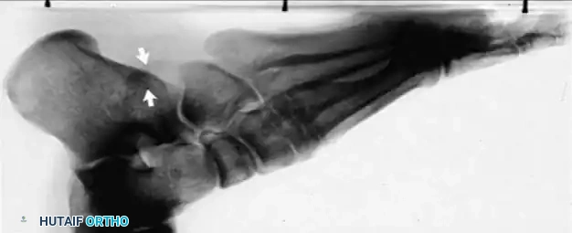
Figures 48a and 48b show the radiographs of a 26-year-old woman who fell down two steps and twisted her foot and ankle. What is the most appropriate treatment for this injury?


Explanation

48b The patient has a zone 1 base of the fifth metatarsal fracture (Pseudojones) that represents a less serious injury compared to zone 2 and 3 fractures with regard to healing potential. Treatment is symptomatic and casting is not necessary. These fractures are well treated with a hard-soled shoe for comfort and weight bearing as tolerated. Surgical intervention is not warranted. Vorlat P, Achtergael W, Haentjens P: Predictors of outcome of non-displaced fractures of the base of the fifth metatarsal. Int Orthop 2007;31:5-10. Wiener BD, Linder JF, Giattini JF: Treatment of fractures of the fifth metatarsal: A prospective study. Foot Ankle Int 1997;18:267-269.

Question 26

A 32-year-old male presents with a high-energy knee injury. Radiographs reveal a posteromedial tibial plateau fracture. Which surgical approach and fixation strategy is most appropriate?


Explanation

Posteromedial tibial plateau fractures typically require a posteromedial approach with an anti-glide or buttress plate to counteract shear forces. Anterolateral plating alone will not capture the posteromedial fragment adequately.

Question 27

A 25-year-old female sustains a closed distal humerus fracture involving the capitellum and lateral trochlea extending into the lateral column. Which classification best describes this injury?


Explanation

The Bryan-Morrey Type IV fracture involves a shear fracture of the capitellum that extends medially to include most of the trochlea. This is also known as a Hahn-Steinthal fracture with lateral trochlear extension.

Question 28

A 28-year-old male sustains a vertically oriented femoral neck fracture (Pauwels Type III). What is the primary biomechanical advantage of adding a fully threaded positional screw or a medial buttress plate to sliding hip screw fixation?


Explanation

Pauwels Type III fractures are highly unstable due to vertical shear forces. Adding a fully threaded positional screw or medial buttress plate mitigates shear stress and prevents varus collapse.

Question 29

A 40-year-old male falls on an outstretched hand, sustaining a volar Barton's fracture of the distal radius. What is the pathognomonic feature of this fracture?


Explanation

A volar Barton's fracture is an intra-articular fracture of the volar margin of the distal radius with associated volar subluxation of the carpus along with the fracture fragment.

Question 30

In a Sanders Type II calcaneus fracture, what anatomical structure is primarily evaluated on the coronal CT scan to determine the classification?


Explanation

The Sanders classification is based on the number and location of fracture lines through the posterior facet of the subtalar joint on coronal CT images. Type II consists of a two-part fracture of the posterior facet.

Question 31

During open reduction and internal fixation of a severe Pilon fracture, the Chaput fragment is identified. Which ligament attaches to this fragment?


Explanation

The Chaput fragment is the anterolateral fragment of the distal tibia. The anterior inferior tibiofibular ligament (AITFL) attaches the Chaput fragment to the Wagstaffe fragment of the fibula.

Question 32

A 35-year-old male undergoes nonoperative treatment for a talar neck fracture. At 8 weeks, a subchondral radiolucent band is seen in the talar dome on an AP mortise radiograph. What does this finding indicate?


Explanation

Hawkins sign is a subchondral radiolucent band seen in the talar dome, indicating subchondral osteopenia. This demonstrates active bone resorption, which requires an intact vascular supply, thereby ruling out avascular necrosis.

Question 33

A 22-year-old athlete sustains a proximal pole scaphoid fracture. What is the primary reason for the high rate of avascular necrosis in this specific fracture pattern?


Explanation

The major blood supply to the scaphoid enters distally and flows retrogradely to the proximal pole. Fractures at the proximal pole disrupt this retrograde flow, predisposing the fragment to ischemia and avascular necrosis.

Question 34

A 45-year-old male sustained a distal femur fracture in a motor vehicle collision. The CT scan reveals a coronal plane fracture of the lateral femoral condyle. What is the eponymous name for this fracture?


Explanation

A Hoffa fracture is a coronal plane fracture of the femoral condyle, most commonly involving the lateral condyle. It typically requires anterior-to-posterior interfragmentary screw fixation for stability.

Question 35

A 24-year-old male with a closed tibial shaft fracture develops disproportionate leg pain. Intracompartmental pressures are measured. Which delta pressure measurement indicates a need for emergent fasciotomy?


Explanation

Acute compartment syndrome is diagnosed when the delta pressure (diastolic blood pressure minus intracompartmental pressure) is less than 30 mmHg. When this threshold is met, emergent four-compartment fasciotomy is required.

Question 36

Following standard tension band wiring of a transverse olecranon fracture, what is the most common complication necessitating reoperation?


Explanation

The most common complication after tension band wiring of an olecranon fracture is prominent and symptomatic hardware (such as K-wires backing out), which frequently necessitates secondary hardware removal.

Question 37

In the management of midshaft clavicle fractures, which of the following is considered an absolute indication for immediate open reduction and internal fixation?


Explanation

Absolute indications for operative management of clavicle fractures include open fractures, neurovascular compromise, and severe skin tenting that threatens skin integrity. Displacement and shortening are relative indications.

Question 38

A 65-year-old female sustains a 3-part proximal humerus fracture. According to Hertel's criteria, which structural feature is the most important predictor of humeral head ischemia?


Explanation

Hertel's criteria for predicting humeral head ischemia include a metaphyseal head extension < 8mm, medial hinge disruption > 2mm, and basicervical fracture patterns. Complete disruption of the medial hinge is a major predictor of avascular necrosis.

Question 39

A 30-year-old male has an unstable ankle syndesmosis after fixation of a Weber C fibula fracture. Which of the following is true regarding suture button fixation compared to rigid syndesmotic screws?


Explanation

Suture button constructs provide dynamic stabilization, allowing physiologic motion while maintaining reduction. They generally allow earlier weight-bearing and eliminate the need for routine hardware removal compared to syndesmotic screws.

Question 40

A 42-year-old female sustains a displaced transverse patella fracture and is treated with anterior tension band wiring. What biomechanical principle does this fixation construct rely upon?


Explanation

The anterior tension band construct converts anterior tension forces created by the extensor mechanism during knee flexion into compressive forces at the articular surface, promoting stability and healing.

Question 41

A 78-year-old male with a highly comminuted, reverse obliquity intertrochanteric femur fracture is treated with a cephalomedullary nail. Why is a sliding hip screw (DHS) contraindicated in this fracture pattern?


Explanation

In reverse obliquity and lateral wall-deficient intertrochanteric fractures, the femoral shaft tends to displace medially. A sliding hip screw relies on an intact lateral wall; without it, excessive sliding leads to medialization and construct failure.

Question 42

A 34-year-old construction worker falls from a ladder and sustains a severely comminuted, open (Gustilo type IIIA) tibial pilon fracture. Initial management includes formal debridement and application of a delta-frame spanning external fixator. What is the most reliable clinical indicator that the patient is ready for definitive open reduction and internal fixation (ORIF)?


Explanation

The 'wrinkle sign' indicates that soft tissue swelling has subsided adequately, significantly reducing the risk of wound dehiscence and deep infection following extensile surgical approaches for pilon fractures.

Question 43

A 65-year-old female presents with an inability to extend her thumb at the interphalangeal joint 6 weeks after sustaining a nondisplaced distal radius fracture treated with cast immobilization. She reports a sudden, painless loss of motion while grasping a jar. What is the most likely etiology of this complication?


Explanation

Extensor pollicis longus (EPL) tendon rupture is a well-known complication of nondisplaced distal radius fractures. It occurs secondary to vascular watershed ischemia and increased pressure within the intact third dorsal compartment.

Question 44

A 32-year-old male sustains a proximal-third extra-articular tibial shaft fracture. He undergoes intramedullary nailing via an infrapatellar approach. Postoperatively, what is the most common malalignment deformity expected with this specific fracture pattern and surgical approach?


Explanation

Proximal-third tibial shaft fractures treated with standard infrapatellar intramedullary nailing are highly prone to apex anterior (recurvatum) and valgus malalignment. This is due to the unopposed pull of the patellar tendon on the proximal fragment and the anatomy of the tibial metaphysis.

Question 45

A 28-year-old unrestrained driver is involved in a high-speed motor vehicle collision and sustains a highly vertical (Pauwels Type III) femoral neck fracture. Which of the following internal fixation constructs provides the greatest biomechanical stability to resist the predominant shear forces?


Explanation

Pauwels Type III fractures are highly vertical and subjected to intense shear and varus deforming forces. A sliding hip screw supplemented with a derotation screw provides the most biomechanically stable construct to resist these specific forces in young adults.

Question 46

A 45-year-old male presents with a coronal plane fracture of the lateral femoral condyle (Hoffa fracture) following a motorcycle accident. Which of the following anatomic structures acts as the primary deforming force on the fractured fragment?


Explanation

A Hoffa fracture is a coronal plane fracture of the femoral condyle, most often the lateral condyle. The primary deforming force on this fragment is the lateral head of the gastrocnemius, which pulls the fragment posteriorly and inferiorly.

Question 47

A 22-year-old athlete sustains a proximal pole scaphoid fracture. The treating orthopedic surgeon counsels the patient on the high risk of nonunion and avascular necrosis. This risk is primarily due to the blood supply originating from which of the following vessels?


Explanation

The scaphoid receives 70-80% of its blood supply from the dorsal carpal branch of the radial artery. The blood vessels enter the scaphoid at the distal pole and flow in a retrograde fashion, placing proximal pole fractures at high risk for avascular necrosis.

Question 48

An 8-week postoperative radiograph of a 35-year-old male who underwent ORIF for a talar neck fracture demonstrates a subchondral radiolucent band extending across the talar dome. What does this radiographic finding indicate?


Explanation

The presence of a subchondral radiolucency in the talar dome at 6 to 8 weeks post-injury is known as the Hawkins sign. It represents subchondral atrophy from disuse in the setting of intact vascularity, effectively ruling out avascular necrosis.

Question 49

A surgeon utilizes an extensile lateral approach for open reduction and internal fixation of a joint-depressed calcaneus fracture. During the elevation of the full-thickness flap, which of the following nerves is at greatest risk of injury near the proximal vertical limb of the incision?


Explanation

The sural nerve is at significant risk during the extensile lateral approach to the calcaneus, particularly where it crosses the superior aspect of the proximal vertical limb of the incision. Careful, subperiosteal flap elevation is required to protect it.

Question 50

A 29-year-old male sustains a closed, spiral fracture of the distal third of the humeral shaft (Holstein-Lewis fracture) and exhibits an immediate inability to extend his wrist and fingers. Closed reduction is performed, and acceptable alignment is achieved; however, the nerve palsy persists. What is the most appropriate next step in management?


Explanation

Radial nerve palsies associated with closed humeral shaft fractures are overwhelmingly neurapraxias. The standard of care is clinical observation for 3 months; if no spontaneous recovery is noted by then, an EMG and potential nerve exploration are indicated.

Question 51

A 40-year-old female sustains a comminuted subtrochanteric femur fracture. During closed reduction attempts, the proximal fragment exhibits a characteristic deformity. What are the primary deforming forces acting on this proximal fragment?


Explanation

In a subtrochanteric femur fracture, the proximal fragment is typically flexed by the iliopsoas, abducted by the gluteus medius and minimus, and externally rotated by the short external rotators.

Question 52

A 24-year-old collegiate football player sustains a purely ligamentous Lisfranc injury. An MRI confirms complete disruption of the Lisfranc ligament complex with multi-directional instability, but no fractures are noted. Based on recent literature, what is the best operative treatment for optimizing long-term function?


Explanation

For purely ligamentous Lisfranc injuries, primary arthrodesis of the first, second, and third tarsometatarsal joints has been shown to yield better functional outcomes and lower reoperation rates compared to traditional ORIF.

Question 53

A 48-year-old male sustains a high-energy pilon fracture. Initial management consists of a spanning external fixator. He develops significant soft tissue swelling and clear fracture blisters. When is it most appropriate to proceed with definitive open reduction and internal fixation?


Explanation

Definitive ORIF of high-energy pilon fractures must be delayed until the soft tissue envelope has adequately recovered. This is indicated by the resolution of swelling (a positive skin 'wrinkle sign') and the complete re-epithelialization of fracture blisters.

Question 54

A 6-year-old child presents with a Bado Type I Monteggia fracture-dislocation (anterior dislocation of the radial head with an anteriorly angulated ulnar fracture). What is the optimal closed reduction maneuver?


Explanation

Bado Type I Monteggia fractures are characterized by anterior bowing of the ulna and anterior radial head dislocation. The standard closed reduction technique involves traction, full supination, and flexing the elbow past 90 degrees to relax the biceps and stabilize the radiocapitellar joint.

Question 55

A 55-year-old female sustains a Dubberley Type 3B capitellum-trochlea fracture, characterized by a highly comminuted articular fragment involving the posterior condyle with complete loss of soft tissue attachment. Which surgical approach provides the best exposure for internal fixation?


Explanation

Complex capitellar-trochlear fractures with posterior comminution (Dubberley Type 3) require extensive visualization of the articular surface and the posterior column. A posterior approach utilizing an olecranon osteotomy provides the optimal exposure for rigid fixation.

Question 56

Following open reduction and internal fixation of a bimalleolar ankle fracture with syndesmotic instability, the surgeon obtains intraoperative fluoroscopy to assess the syndesmotic reduction. Which of the following parameters is the most reliable radiographic indicator of a normally reduced syndesmosis on a mortise view?


Explanation

The tibiofibular clear space is the most reliable plain radiographic parameter for evaluating syndesmotic integrity. It is measured 1 cm proximal to the joint line, and a clear space of less than 5 mm on both AP and mortise views indicates a normal relationship.

Question 57

A 25-year-old male sustains a completely displaced midshaft clavicle fracture with a Z-deformity and 2.5 cm of shortening. The surgeon recommends operative fixation over nonoperative management. Based on the most robust current evidence, what is the primary clinical advantage of surgery in this specific scenario?


Explanation

In highly displaced midshaft clavicle fractures (especially those with >2 cm shortening and complete displacement), operative fixation significantly decreases the rate of nonunion and symptomatic malunion compared to nonoperative management.

Question 58

A 38-year-old male develops acute compartment syndrome of the lower leg following a tibial plateau fracture. The surgeon performs a dual-incision, four-compartment fasciotomy. Which compartment is historically the most frequently inadequately released or missed during this procedure?


Explanation

The deep posterior compartment is the most frequently missed or incompletely released compartment during a standard fasciotomy of the leg. This often occurs because the surgeon fails to adequately detach the soleus bridge from the posteromedial tibia.

Question 59

A 75-year-old female presents with a periprosthetic distal femur fracture (Lewis and Rorabeck Type II) above a stable, well-fixed posterior-stabilized total knee arthroplasty component. Which of the following is the most appropriate definitive management?


Explanation

A Lewis and Rorabeck Type II periprosthetic distal femur fracture involves a fracture around a well-fixed, stable component. The standard of care is surgical fixation with either a lateral locking plate or a retrograde intramedullary nail (if the femoral component box allows).

Question 60

A 45-year-old man sustains a closed isolated scapular body fracture after an all-terrain vehicle accident. Radiographs demonstrate a displaced scapular body fracture with 10 mm of medialization and 15 degrees of angular deformity. The glenoid is not involved. What is the most appropriate management?


Explanation

Sling immobilization and early range of motion is the standard of care for most extra-articular scapular body fractures. Operative intervention is typically reserved for extreme displacement, such as medialization greater than 25 mm or angulation exceeding 45 degrees.

Question 61

A 78-year-old woman with a history of severe osteoporosis sustains a 4-part proximal humerus fracture with significant medial calcar comminution and varus angulation. To optimize her functional outcome and minimize complications, what is the most appropriate surgical treatment?


Explanation

Reverse total shoulder arthroplasty is indicated for elderly patients with complex 4-part proximal humerus fractures, especially in the setting of poor bone quality or tuberosity comminution. It provides more reliable functional outcomes and pain relief compared to hemiarthroplasty or ORIF in this demographic.

Question 62

A 25-year-old man sustains a closed spiral fracture of the distal third of the humeral shaft (Holstein-Lewis fracture). Upon initial emergency department evaluation, he is unable to actively extend his wrist or fingers. What is the most appropriate initial management?


Explanation

Primary radial nerve palsy in the setting of a closed humeral shaft fracture is generally managed expectantly with functional bracing and observation. The vast majority of these nerve injuries are neuropraxias that will spontaneously recover within 3 to 4 months.

Question 63

A 75-year-old woman with a history of rheumatoid arthritis sustains an intra-articular, bicolumnar distal humerus fracture (OTA/AO 13-C3) with severe comminution and osteopenia. What treatment option provides the best chance for early functional recovery and reliable pain relief?


Explanation

Total elbow arthroplasty is the preferred treatment for highly comminuted, intra-articular distal humerus fractures in low-demand, elderly patients with poor bone quality or pre-existing inflammatory arthritis. It allows for immediate range of motion and has reliable outcomes compared to the high failure rate of ORIF in this population.

Question 64

A 32-year-old man sustains a Galeazzi fracture-dislocation. Following anatomic open reduction and internal fixation of the radial shaft, intraoperative evaluation reveals that the distal radioulnar joint (DRUJ) remains unstable in supination. What is the most appropriate next step in management?


Explanation

If the DRUJ remains unstable after anatomic fixation of the radius in a Galeazzi fracture, it should be transfixed with a K-wire in the position of maximum stability (typically supination). The pins are generally left in place for 4 to 6 weeks.

Question 65

Eight weeks following nonoperative management of a nondisplaced distal radius fracture in a short arm cast, a 60-year-old woman reports the sudden inability to actively extend her thumb interphalangeal joint. What is the most likely etiology of her new deficit?


Explanation

Extensor pollicis longus (EPL) tendon rupture is a well-known complication of nondisplaced distal radius fractures. It occurs secondary to mechanical attrition or local ischemia within the unreleased third extensor compartment.

Question 66

A 28-year-old man is involved in a motorcycle collision and sustains a displaced, vertically oriented (Pauwels type III) femoral neck fracture. To minimize the risk of mechanical failure and nonunion, which of the following is the most biomechanically stable fixation construct?


Explanation

In young adults with high shear angle (Pauwels III) femoral neck fractures, a fixed-angle construct such as a sliding hip screw (DHS) with an adjunctive derotational screw provides superior biomechanical stability. This construct resists vertical shear forces better than parallel cancellous screws.

Question 67

A 72-year-old woman sustains an intertrochanteric femur fracture. Radiographs show the fracture line exiting the lateral cortex below the vastus ridge, indicating an incompetent lateral wall. Which of the following fixation implants is biomechanically optimal for this specific fracture pattern?


Explanation

Intertrochanteric fractures with an incompetent lateral wall (reverse obliquity or lateral wall blowout) have a high rate of excessive medialization and failure if treated with a sliding hip screw. A cephalomedullary nail provides an intramedullary buttress that prevents excessive sliding and lateral wall collapse.

Question 68

A 35-year-old man presents with a high-energy Schatzker VI tibial plateau fracture. Which of the following physical examination findings is the earliest and most reliable clinical indicator of acute compartment syndrome?


Explanation

Pain out of proportion to the apparent injury, which is classically exacerbated by passive stretch of the muscles in the involved compartment, is the most sensitive and earliest clinical sign of acute compartment syndrome. Pulselessness and paralysis are late, often irreversible signs.

Question 69

A 42-year-old man undergoes intramedullary nailing of a proximal third tibial shaft fracture. Which of the following intraoperative techniques is most effective in preventing the common apex anterior (procurvatum) and valgus deformity associated with this injury?


Explanation

Proximal third tibial fractures frequently malalign into procurvatum and valgus during traditional hyperflexed nailing due to the unresisted pull of the patellar tendon. A semi-extended (suprapatellar) approach relaxes the extensor mechanism, facilitating anatomic reduction and proper nail trajectory.

Question 70

A 48-year-old construction worker sustains a severe, comminuted tibial pilon fracture with massive soft tissue swelling and hemorrhagic fracture blisters around the ankle. What is the most appropriate initial management strategy?


Explanation

High-energy pilon fractures with severe soft tissue compromise should be managed with a staged protocol. Initial management consists of a spanning external fixator and elevation until the soft tissues recover (the "wrinkle sign" appears), typically followed by definitive ORIF 10 to 21 days later.

Question 71

A 29-year-old snowboarder is diagnosed with a Hawkins type III fracture of the talar neck. What specific pattern of displacement defines a Hawkins type III injury?


Explanation

In the Hawkins classification for talar neck fractures, Type III is defined as a displaced talar neck fracture with dislocation of both the subtalar and tibiotalar joints. This injury carries a very high risk of avascular necrosis (AVN) of the talar body.

Question 72

A 22-year-old collegiate athlete sustains a hyperplantarflexion injury to his midfoot. Weight-bearing radiographs demonstrate a 3 mm diastasis between the medial cuneiform and the base of the second metatarsal. What is the most appropriate definitive management?


Explanation

Lisfranc injuries exhibiting instability, defined as greater than 2 mm of diastasis on weight-bearing radiographs, require operative stabilization. This is achieved via open reduction and internal fixation (ORIF) or primary arthrodesis, depending on the extent of ligamentous disruption and articular damage.

Question 73

A 35-year-old man sustains a high-energy distal femur fracture. CT imaging reveals a displaced coronal plane fracture of the lateral femoral condyle. What is the most biomechanically stable method of internal fixation for this specific articular fragment?


Explanation

A coronal fracture of the femoral condyle is known as a Hoffa fracture. It requires orthogonal fixation with AP or PA interfragmentary lag screws to adequately neutralize the shear forces of the knee.

Question 74

A 40-year-old woman is undergoing open reduction and internal fixation of a bicondylar tibial plateau fracture with a large posteromedial shear fragment. Which anatomic interval is utilized for the classic posteromedial approach to the knee?


Explanation

The classic posteromedial approach to the tibial plateau uses the interval between the medial head of the gastrocnemius (retracted laterally) and the pes anserinus tendons (retracted anteriorly/medially). This protects the neurovascular bundle while providing direct access to the posteromedial fragment.

Question 75

A 25-year-old man sustains a Hawkins Type II talar neck fracture. At his 6-week follow-up radiograph, a subchondral radiolucent band is observed in the talar dome. What does this radiographic finding indicate?


Explanation

This finding is the Hawkins sign, which represents subchondral atrophy secondary to hyperemia. Its presence indicates intact vascularity to the talar body and makes the development of avascular necrosis highly unlikely.

Question 76

A 28-year-old man sustains a closed midshaft clavicle fracture. Non-operative management is initially chosen. Which of the following initial radiographic findings is the most reliable predictor of subsequent nonunion?


Explanation

The most reliable predictors of nonunion in midshaft clavicle fractures treated non-operatively are completely displaced fractures (no cortical contact) with shortening greater than 2 cm.

Question 77

During surgical approach for internal fixation of a severe proximal humerus fracture, the surgeon attempts to preserve the primary blood supply to the humeral head. Which vessel supplies the majority of the blood to the humeral head?


Explanation

Historically, the anterior circumflex humeral artery (via the arcuate branch) was thought to be the primary supply. However, recent quantitative studies have proven that the posterior circumflex humeral artery provides the dominant blood supply to the humeral head.

Question 78

A 30-year-old man sustains a Bado Type I Monteggia fracture-dislocation. The ulnar shaft fracture is anatomically reduced and plated, but the radial head remains dislocated anteriorly. What is the most common anatomic structure blocking the reduction of the radial head?


Explanation

In Monteggia fracture-dislocations, anatomic fixation of the ulna typically reduces the radial head. If the radial head remains unreduced, the most common structure interposed and blocking reduction is the annular ligament.

Question 79

A 55-year-old woman undergoes volar plate fixation for a distal radius fracture. Six months later, she suddenly loses the ability to actively flex the interphalangeal joint of her thumb. What is the most likely cause of this complication?


Explanation

Prominence of a volar plate distal to the watershed line of the distal radius can cause attritional wear and subsequent rupture of the flexor pollicis longus (FPL) tendon. This presents as a loss of active thumb IP joint flexion.

Question 80

A 22-year-old man sustains a vertically oriented (Pauwels Type III) femoral neck fracture. Which of the following fixation constructs provides the most biomechanical stability against the high shear forces seen in this fracture pattern?


Explanation

Pauwels Type III fractures are highly unstable due to vertical shear forces. A fixed-angle device like a sliding hip screw combined with a derotational screw provides superior biomechanical stability compared to multiple cannulated screws.

Question 81

In a complete subtrochanteric femur fracture, the proximal fracture fragment is characteristically displaced by strong muscular forces. What is the typical position of the proximal fragment, and which muscles are responsible?


Explanation

The proximal fragment in a subtrochanteric fracture is classically pulled into flexion (iliopsoas), abduction (gluteus medius/minimus), and external rotation (short external rotators).

Question 82

A 45-year-old man presents with a "terrible triad" injury of the elbow following a fall. What is the widely accepted standard sequence for surgical reconstruction of this injury?


Explanation

The standard surgical algorithm for a terrible triad injury works from deep to superficial: repair the coronoid first, then fix or replace the radial head, and finally repair the lateral ulnar collateral ligament (LUCL). The MCL is only addressed if the elbow remains grossly unstable after lateral-sided repair.

Question 83

A 35-year-old man sustains a severe closed tibial pilon fracture with massive soft tissue swelling. What is the most appropriate initial management strategy to minimize the risk of wound complications?


Explanation

High-energy pilon fractures are fraught with soft tissue complications. The standard of care is a staged protocol: initial spanning external fixation with fibular fixation (if needed), followed by delayed definitive ORIF once the soft tissue envelope has healed (indicated by the presence of skin wrinkles).

Question 84

A surgeon utilizes the standard extensile lateral approach for open reduction and internal fixation of a displaced intra-articular calcaneus fracture. Which nerve is at greatest risk of iatrogenic injury during the creation of the full-thickness soft tissue flap?


Explanation

The sural nerve crosses the lateral border of the hindfoot and is at high risk of injury or entrapment during the extensile lateral approach to the calcaneus.

Question 85

A 20-year-old football player sustains a midfoot injury. Weight-bearing radiographs show widening of the interval between the first and second metatarsal bases. The primary stabilizing ligament of this articulation (the Lisfranc ligament) connects which two osseous structures?


Explanation

The Lisfranc ligament is an intra-articular interosseous ligament that connects the lateral aspect of the medial cuneiform to the medial base of the second metatarsal. It is critical for the stability of the midfoot arch.

Question 86

A 24-year-old man sustains a fracture of the proximal pole of the scaphoid. Why is this specific fracture pattern at an exceptionally high risk for developing avascular necrosis (AVN) and nonunion?


Explanation

The scaphoid has a retrograde blood supply. Branches of the radial artery (the dorsal carpal branch) enter the scaphoid near the waist and distal pole, flowing proximally. Fractures of the proximal pole disrupt this supply, leading to high rates of AVN.

Question 87

A 45-year-old woman falls on an outstretched hand and sustains a capitellum fracture that extends medially to involve the majority of the trochlea (Dubberley Type 2). Which surgical approach provides the most optimal visualization for anatomic reduction of this complex articular injury?


Explanation

Fractures involving the capitellum and significant portions of the trochlea require extensive articular exposure. An extensile lateral approach (often elevating the common extensor origin) or an anterior approach allows direct visualization and fixation of the anterior articular shear fragment.

Question 88

A 32-year-old skier sustains a distal third spiral fracture of the tibial shaft. Which concomitant injury is statistically most likely to be present and must be specifically evaluated with dedicated imaging?


Explanation

Distal third spiral tibia fractures are highly associated with occult posterior malleolus fractures. CT scanning is generally recommended to evaluate the posterior malleolus and plan for appropriate internal fixation.

Question 89

A 19-year-old elite college basketball player sustains an acute Zone 2 fracture of the proximal fifth metatarsal (Jones fracture). What is the most appropriate management to ensure the fastest return to play and lowest risk of nonunion?


Explanation

In elite athletes, acute Jones fractures (Zone 2) are typically treated with early intramedullary screw fixation. This provides the most reliable healing and fastest return to competitive sports, avoiding the high nonunion rates seen with conservative care due to the watershed blood supply.

Question 90

A 26-year-old man is brought to the trauma bay with an Injury Severity Score (ISS) of 42, bilateral pulmonary contusions, and a closed right femoral shaft fracture. His initial lactate is 4.5 mmol/L. According to the principles of Damage Control Orthopedics (DCO), what is the most appropriate initial management for his femur fracture?


Explanation

In an unstable polytrauma patient with severe chest injury and metabolic acidosis (elevated lactate), early total care (IM nailing) can exacerbate systemic inflammation (Second Hit phenomenon). Damage Control Orthopedics dictates rapid, provisional stabilization with spanning external fixation.

Question 91

When comparing operative versus non-operative management of acute complete Achilles tendon ruptures using functional rehabilitation protocols, current literature indicates which of the following regarding complication rates?


Explanation

Recent high-quality evidence shows that operative management slightly decreases the risk of re-rupture compared to traditional non-operative care, but carries a significantly higher risk of complications such as infection, sural nerve injury, and wound breakdown.

Question 92

A 68-year-old woman on long-term alendronate therapy presents with thigh pain and sustains a low-energy transverse fracture of the subtrochanteric femur with lateral cortical thickening. Which surgical implant is considered the gold standard for treating this specific type of atypical femur fracture?


Explanation

Atypical femur fractures associated with bisphosphonate use are inherently prone to delayed healing and stress propagation. A full-length cephalomedullary nail is the gold standard as it spans the entire bone and mechanically protects the femur while it heals.

Question 93

A 28-year-old male sustains a high-energy lateral Hoffa fracture (coronal shear fracture of the lateral femoral condyle). If lag screw fixation is chosen, biomechanical studies suggest which of the following screw configurations provides the most stable fixation and highest pullout strength?


Explanation

Biomechanical studies have demonstrated that anterior-to-posterior (AP) directed lag screws provide significantly greater pullout strength and construct stiffness for Hoffa fractures compared to posterior-to-anterior (PA) screws. AP screws engage the denser bone of the posterior condyle more effectively.

Question 94

A 32-year-old male sustains a Hawkins Type III talar neck fracture following a motor vehicle collision. What is the approximate rate of avascular necrosis (AVN) of the talar body expected in this injury pattern?


Explanation

Hawkins Type III talar neck fractures involve displacement of the talar neck with subluxation or dislocation of both the subtalar and tibiotalar joints. This severe injury disrupts all three major blood supplies to the talar body, leading to an AVN rate approaching 70 to 100 percent.

Question 95

A 45-year-old female treated non-operatively for a nondisplaced distal radius fracture presents 6 weeks later unable to actively extend her thumb interphalangeal joint. Tenodesis effect is absent. What is the most appropriate and reliable surgical treatment?


Explanation

Extensor pollicis longus (EPL) tendon rupture is a classic complication of nondisplaced distal radius fractures due to ischemia or attrition in the third dorsal compartment. Because the tendon ends retract and degenerate, primary repair is rarely feasible, making an EIP to EPL transfer the gold standard treatment.

Question 96

A 38-year-old male sustains a bicondylar tibial plateau fracture featuring a large, displaced posteromedial coronal shear fragment. Which surgical approach and fixation strategy is most appropriate for addressing this specific fragment?


Explanation

Posteromedial coronal shear fragments in tibial plateau fractures cannot be adequately reduced or stabilized via standard anterior approaches. A posteromedial approach with an under-contoured buttress plate (creating a spring-like anti-glide effect) provides superior biomechanical resistance to vertical shear forces.

Question 97

A 35-year-old male with a high-energy, highly comminuted distal tibia pilon fracture (OTA/AO 43-C3) is treated with a spanning external fixator. Why might a surgeon purposefully delay open reduction and internal fixation of the associated comminuted fibula fracture during this index procedure?


Explanation

In complex, comminuted pilon fractures, early fixation of the fibula can act as a deforming force if length or rotation is slightly off. Delaying fibular fixation allows the surgeon to accurately reduce the critical tibial articular block without being hindered by an arbitrarily fixed fibula.

Question 98

A 22-year-old male falls onto his shoulder and sustains a completely displaced midshaft clavicle fracture. While many factors influence surgical decision-making, which of the following is considered an absolute indication for acute operative fixation?


Explanation

While severe shortening, 100 percent displacement, and comminution are strong relative indications for operative fixation to prevent symptomatic malunion or nonunion, an open clavicle fracture is an absolute indication for urgent surgical debridement and stabilization.

Question 99

A 78-year-old female sustains an unstable reverse obliquity intertrochanteric femur fracture. Which of the following implants is biomechanically optimal to minimize the risk of lateral wall blowout and fixation failure?


Explanation

Reverse obliquity intertrochanteric fractures are highly unstable because the fracture line parallels the vector of weight-bearing, encouraging medial shaft displacement. A cephalomedullary nail acts as an intramedullary buttress against this displacement, providing vastly superior stability compared to extramedullary sliding hip screws.

Question 100

A 6-year-old boy presents with a Bado Type I Monteggia fracture-dislocation. Closed reduction of the ulna correctly restores length and alignment, but the radial head remains anteriorly dislocated on radiographs. What is the most common anatomical block to the reduction of the radial head in this scenario?


Explanation

In Bado Type I Monteggia injuries where the ulna is anatomically reduced but the radial head remains dislocated, the most common block to reduction is interposition of the annular ligament or joint capsule. Open reduction of the radiocapitellar joint is required to clear the interposed tissue.

None

Clinic OS
Medically Verified Content by
Prof. Clinic OS
Consultant Orthopedic & Spine Surgeon
Chapter Index