العربية
Part of the Master Guide

Orthopedic Surgery Board Prep: Set 403 (100 MCQs on Fractures & Casting)

Trauma Board Review 2009: High-Yield MCQs (Set 2)

27 Apr 2026 81 min read 82 Views
Trauma 2009 MCQs - Part 2

Key Takeaway

This article provides essential research regarding Trauma Board Review 2009: High-Yield MCQs (Set 2). Access high-yield Trauma questions for the 2009 board exam. This module (Set 2) covers critical topics including surgical techniques, pathology, and treatment protocols with verified answers.

Trauma Board Review 2009: High-Yield MCQs (Set 2)

Comprehensive 100-Question Exam


00:00

Start Quiz

Question 1

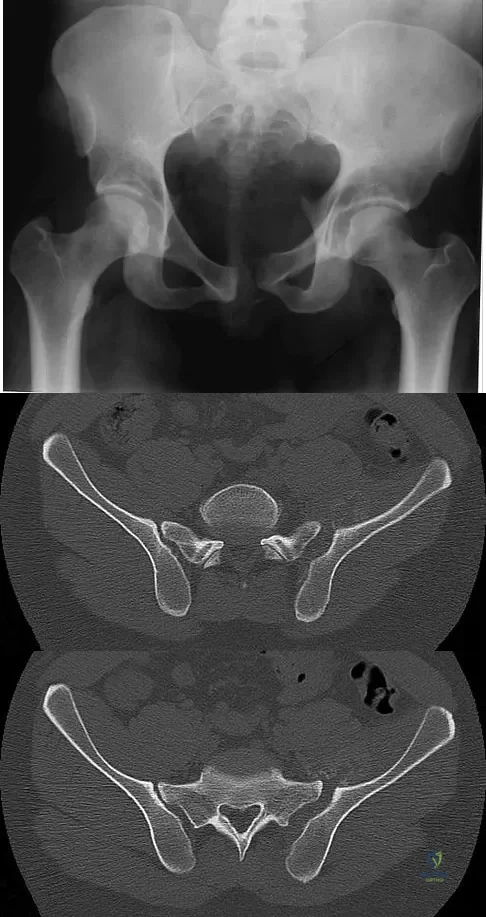
A 20-year-old man is brought to the emergency department after a high-speed motor vehicle accident. His initial blood pressure is 70/40 mm Hg. He is currently receiving intravenous fluids as well as blood. His Focused Assessment with Sonography for Trauma examination did not show any free fluid in his abdomen and his chest radiograph is unremarkable. An AP pelvis radiograph is shown in Figure 15. What is the next most appropriate step in the management of his pelvic injury?

Trauma Board Review 2009: High-Yield MCQs (Set 2) - Figure 1


Explanation

This hypotensive patient has an obvious open book injury of the pelvic ring on the AP pelvis radiograph and further radiographs are not needed prior to the initiation of treatment. Although angiography may be indicated if he does not respond to stabilization of his pelvis and fluid/blood administration, temporary stabilization of the pelvis with a sheet or binder should be performed first because it is simple, quick, and has been shown to be effective. This patient does not need a laparotomy at this point since the FAST examination did not show any free intra-abdominal fluid and his chest radiograph was unremarkable, leaving the most likely source of bleeding the pelvic fracture. Open reduction with internal fixation of a pelvic injury is not indicated in an acutely ill patient. Kreig JC, Mohr M, Ellis TJ, et al: Emergent stabilization of pelvic ring injuries by controlled circumferential compression: A clinical trial. J Trauma 2005;59:659-664. Croce MA, Magnotti LJ, Savage SA, et al: Emergent pelvic fixation in patients with exsanguinating pelvic fractures. J Am Coll Surg 2007;204:935-942.

Question 2

A 7-year-old girl is hit by a motor vehicle and sustains the isolated ipsilateral injuries shown in Figures 16a and 16b. What is the optimal definitive method of treatment?


Explanation

The child has isolated ipsilateral femoral shaft and tibial shaft fractures. Spica cast immobilization is unlikely to accommodate for shortening and alignment in this child with multiple levels of injury. In this instance, efforts should be made to mobilize a least one level of the limb; therefore, treatment should include flexible nailing of the femur and tibia. Rigid reamed nails are not indicated in this young patient secondary to risk of a growth arrest and osteonecrosis of the proximal femur. Poolman RW, Kocher MS, Bhandari M: Pediatric femoral fractures: A systematic review of 2422 cases. J Orthop Trauma 2006;20:648-654. Anglen JO, Choi L: Treatment options in pediatric femoral shaft fractures. J Orthop Trauma 2005;19:724-733.

Question 3

What is the most common cause of errors that harm patients?

Trauma Board Review 2009: High-Yield MCQs (Set 2) - Figure 4


Explanation

The AMA report identified communication breakdown as the most common cause of errors that harm patients. It is extremely important to learn to communicate effectively with your patients. Understanding cultural and language differences helps avoid communication errors. Jimenez R, Lewis VO (eds): Culturally Competent Care Guidebook. Rosemont, IL, American Academy of Orthopaedic Surgeons, 2007. American Medical Association ethical force program report: "Improving communication - Improving care", 2006

Question 4

A 220-lb 20-year-old man was involved in a motor vehicle accident. His work-up reveals that he has multiple long bone fractures as well as a splenic injury that is currently being managed nonsurgically. His initial blood pressure in the trauma bay was 70/30 mm Hg. After receiving 4 liters of fluid and 3 units of packed red blood cells, his blood pressure is currently 110/70, his heart rate is 100, his urine output is 90 mL/h (normal 0.5 to 1 mL/kg/h), and his core temperature is 97.9 degrees F (36.5 degrees C). At this point, the patient's resuscitation can be described as which of the following?

Trauma Board Review 2009: High-Yield MCQs (Set 2) - Figure 5


Explanation

Although the end points of resuscitation are still unclear, what is known is that normalization of the standard hemodynamic parameters (blood pressure, heart rate, and urine output) is not adequate. Up to 85% of patients with normal hemodynamic parameters can still have inadequate tissue oxygenation or uncompensated shock. The initial base deficit, lactate level, or gastric pHi can be used to stratify patients for resuscitation needs, risks of death, and multiple organ failure (level 1 evidence). The time it takes to normalize the base deficit, the lactate level, or gastric pHi, can predict survival (level 2 evidence). Patients who have been in uncompensated shock (abnormal vital signs) should have their resuscitation monitored using data other than vital signs. Tisherman SA, Barie P, Bokhari F, et al: Clinical practice guideline: Endpoints of resuscitation. J Trauma 2004;57:898-912. Moore FA, McKinley BA, Moore EE, et al: Inflammation and the Host Response to Injury, a large-scale collaborative project: Patient-oriented research core--standard operating procedures for clinical care. III. Guidelines for shock resuscitation. J Trauma 2006;61:82-89.

Question 5

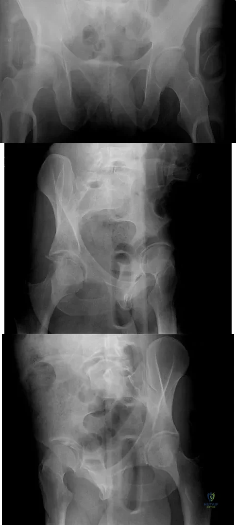
A 30-year-old man who sustained a work-related injury 6 weeks ago reports persistent back and left-sided buttock pain that has been attributed to lumbar transverse process fractures. A pelvic radiograph and CT scans obtained 2 days ago are seen in Figures 17a through 17c. What is the best treatment for his injury?


Explanation

Fortunately, surgical treatment of sub-acute pelvic ring injuries is relatively uncommon as acute management has become more common. Delayed reconstruction of pelvic ring malunion and impending malunion is rare. Nonsurgical management may have a role as long as the hemipelvis does not flex, shorten, and/or externally rotate. The AP pelvic radiograph suggests that all three motions are happening in this patient. These are just a few of the indications to repair the pelvic ring and this is best done with anterior and posterior fixation. Anterior symphyseal plating will help correct most of the deformity. Posterior fixation can and should be added to lessen the forces on the anterior ring reconstruction when repair is performed in a sub-acute or delayed fashion. Posterior fixation can help obtain a more anatomic reduction and helps decrease the risk of anterior hardware failure. Mears DC: Management of pelvic pseudarthroses and pelvic malunion. Orthopade 1996;25:441-448. Matta JM, Dickson KF, Markovich GD: Surgical treatment of pelvic nonunions and malunions. Clin Orthop Relat Res 1996;329:199-206.

Question 6

A 36-year-old woman was injured in a train derailment. She has a significant open depressed skull fracture with active bleeding, a hemopneumothorax, and blood in the left upper quadrant and colic gutter by Focused Assessment with Sonography for Trauma (FAST) examination. Additionally, she has the pelvic injury seen on the CT scans in Figures 18a and 18b. The mortality rate for this patient approaches


Explanation

Mortality following trauma that requires surgical intervention for head, chest, and abdominal injury exceeds 90%. The type of pelvic fracture is a predictor of associated injury, blood requirements, and overall mortality. AP III pelvic fractures require the most blood, and are associated with significant abdominal trauma and shock. Lateral compression pelvic fractures are more associated with head, chest, and occasionally abdominal trauma, and mortality often occurs from associated injuries. Dalal SA, Burgess AR, Siegel JH, et al: Pelvic fracture in multiple trauma: Classification by mechanism is key to pattern of organ injury, resuscitative requirements and outcome. J Trauma 1989;29:981-1000. Eastridge BJ, Burgess AR: Pedestrian pelvic fractures: 5-year experience of a major urban trauma center. J Trauma 1997;42:695-700.

Question 7

A 19-year-old collegiate baseball player injures the ring finger on his dominant hand while sliding headfirst into second base. He reports that he is unable to actively flex or extend the distal interphalangeal joint of the finger. Radiographs are shown in Figures 19a and 19b. What is the anatomic lesion leading to this injury?
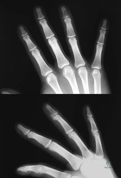


Explanation

The radiographs reveal a bony avulsion of the flexor profundus insertion (Jersey finger). The large bony fragment classifies this as a Leddy type III injury. The bony fragment has retracted to the level of the annular pulley (A4). Leddy JP, Packer JW: Avulsion of the insertion of the profundus tendon insertion in athletes. J Hand Surg 1977;2:66-69.

Question 8

A 72-year-old man was involved in an automobile accident 4 weeks ago. Initially he noted pain about his nondominant left shoulder, which resolved within a few weeks after the accident. He now describes trouble with gripping and carrying items in his left hand. Radiographs are shown in Figures 20a through 20c. His signs and symptoms are the result of injury to which of the following ligaments?


Explanation

The radiographs reveal a gap between the scaphoid and lunate bones, indicative of disruption of the scapholunate ligament complex. The three components of the complex are the dorsal scapholunate ligament, the volar (or palmar) scapholunate ligament, and the proximal fibrocartilaginous membrane, listed in decreasing yield strength. Disruption of the stout dorsal interosseous ligament is required for scapholunate dissociation to occur. Berger RA: The ligaments of the wrist: A current overview of anatomy with considerations of their potential functions. Hand Clin 1997;13:63-82.

Question 9

To avoid an injury to the L5 nerve root when placing an S1 sacroiliac screw, what area of the sacrum should be avoided on the lateral C arm image shown in Figure 21?

Trauma Board Review 2009: High-Yield MCQs (Set 2) - Figure 16


Explanation

Safe placement of a sacroiliac screw depends on excellent imaging of and understanding of pelvic anatomy. There are variations in the anatomy of the upper sacrum. Patients with dysplasia of the sacrum can have "in-out-in" screws placed that exit the ilium, pass anterior to the sacral ala, and injure the L5 nerve root. To make sure that this does not occur, a lateral image of the sacrum is used to ensure that the starting point is in the "safe zone." The starting point needs to be below the iliac cortical density (ICD) which parallels the sacral alar slope. This will prevent placing screws into the recessed ala of patients with a dysplastic sacrum. The triangular area anterior to the ICD is labeled A in the figure, B represents the sacral canal, C is S2, D is the anterior border of the sacrum, and E represents the greater sciatic notches. Routt ML Jr, Simonian PT, Agnew SG, et al: Radiographic recognition of the sacral alar slope for optimal placement of iliosacral screws: A cadaveric and clinical study. J Orthop Trauma 1996;10:171-177.

Question 10

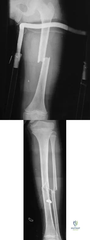
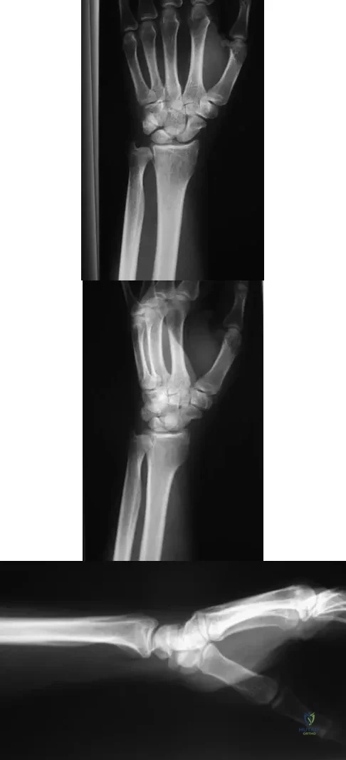
An otherwise healthy 37-year-old man fell off the flatbed of a delivery truck and landed directly on his dominant left hand. Surgical stabilization of a distal radius fracture is performed. An intraoperative radiograph is shown in Figure 22. What is the next most appropriate step in management?

Trauma Board Review 2009: High-Yield MCQs (Set 2) - Figure 17


Explanation

The intraoperative radiograph reveals a scapholunate ligament disruption. Repair of the stout dorsal scapholunate interosseous ligaments is required. Interestingly, the results of scapholunate ligament injuries associated with distal radius fractures appear to be superior to those of isolated ligament injuries. Smith DW, Henry MK: Comprehensive management of soft-tissue injuries associated with distal radius fractures. J ASSH 2002;3:153-164.

Question 11

A 36-year-old woman is placed in a short arm cast for a nondisplaced extra-articular distal radius fracture. Seven weeks later she notes the sudden inability to extend her thumb. What is the most likely cause of her condition?

Trauma Board Review 2009: High-Yield MCQs (Set 2) - Figure 18


Explanation

A recent review of 200 consecutive distal radius fractures noted that the overall incidence of extensor pollicis longus rupture was 3%. The causes are believed to be mechanical irritation, attrition, and vascular impairment. The fracture is usually nondisplaced and the patient notes weeks to months after injury the sudden, painless inability to extend the thumb. Treatment involves extensor indicis proprius tendon transfer or free palmaris longus tendon grafting. Skoff HD: Postfracture extensor pollicis longus tenosynovitis and tendon rupture: A scientific study and personal series. Am J Orthop 2003;32:245-247. Bonatz E, Kramer TD, Masear VR: Rupture of the extensor pollicis longus tendon. Am J Orthop 1996;25:118-122.

Question 12

In Gustilo type III open tibial diaphyseal fractures, which of the following factors is associated with an increased risk of a poor functional outcome?

Trauma Board Review 2009: High-Yield MCQs (Set 2) - Figure 19


Explanation

According to the published outcomes analyses from the Lower Extremity Assessment Project (LEAP) study group of patients prospectively followed for 2 to 7 years, definitive fixation with an intramedullary nail has shown improved outcomes when compared to definitive external fixation. The findings showed that the timing of wound debridement (within 6 hours from injury as compared to within 6 to 24 hours), the timing of soft-tissue coverage (3 days or less from injury as compared to more than 3 days), and the timing of bone grafting after injury (within or after 3 months) did not impact the infection or union rates and had no effect on functional outcome. The LEAP study has shown at 7-year follow-up that patients who are definitively treated with external fixation have a significantly longer time to union, poorer functional outcomes, longer time to achieve full weight bearing, and more time in the hospital.

Question 13

Figures 23a and 23b show the radiographs of a 75-year-old woman who sustained an injury to her nondominant hand. Initial treatment should consist of
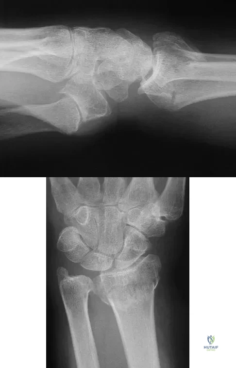


Explanation

Definitive treatment decisions for displaced distal radius fractures in the elderly are based on a number of factors related to the fracture pattern and patient demographics. The first step in any treatment algorithm is a closed reduction and splinting with reassessment of alignment parameters. This is an extra-articular fracture with dorsal angulation. Low-demand elderly patients can be treated well with accepted minor malreduction. Handoll HH, Madhok R: Conservative interventions for treating distal radial fractures in adults. Cochrane Database Syst Rev 2003;2:CD000314.

Question 14

A 43-year-old man sustained a closed, intra-articular pilon fracture. It has now been 1 year since he underwent open reduction and internal fixation. Which of the following statements most accurately describes his perceived outcome?

Trauma Board Review 2009: High-Yield MCQs (Set 2) - Figure 22


Explanation

Marsh and associates retrospectively reviewed 56 tibial plafond fractures and found that the patients perceived improvement in their function and pain for an average of 2.4 years. They demonstrated some limitations in recreational activities but not marked limitations. Patients were unlikely to need a late arthrodesis (13%), and their outcomes did not correlate well with assessments of reduction or arthritis scores.

Question 15

The injury shown in Figure 24 was most likely caused by what mechanism of injury?

Trauma Board Review 2009: High-Yield MCQs (Set 2) - Figure 23


Explanation

The CT cut shows a fracture through the posterior portion of the iliac wing or a crescent fracture. This occurs after a laterally directed force is applied to the anterior part of the involved iliac wing.

Question 16

A 21-year-old college student fell from a balcony and landed on his outstretched right hand. He is seen in the emergency department 4 hours later and reports wrist pain and diffuse hand numbness. The volar forearm compartment is soft and there is no pain with passive finger extension. Radiographs are shown in Figures 25a and 25b. Definitive treatment should consist of
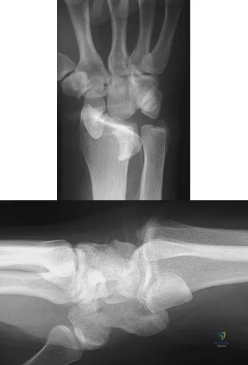


Explanation

A spectrum of perilunate injury patterns exists, with the dorsal trans-scaphoid perilunate fracture-dislocation being the most common. Perilunate injuries are highly unstable complex carpal disruptions that are not amenable to closed treatment. Open reduction and internal fixation is necessary to accurately restore carpal alignment via fracture reduction and fixation and intercarpal ligament repair. Controversy exists regarding the need for dorsal or combined dorsal and volar approaches. Based on the radiographic findings of a volar dislocation of the lunate and the associated median nerve injury, the patient requires open reduction and internal fixation via combined dorsal and volar approaches with a concomitant carpal tunnel release. Herzberg G, Forissier D: Acute dorsal trans-scaphoid perilunate fracture-dislocations: Medium-term results. J Hand Surg Br 2002;27:498-502. Melone CP Jr, Murphy MS, Raskin KB: Perilunate injuries: Repair by dual dorsal and volar approaches. Hand Clin 2000;16:439-448.

Question 17

A 9-year-old boy falls from a scooter and sustains the injury shown in the radiographs in Figure 26. After closed reduction and cast immobilization, what is the most likely complication that can result?

Trauma Board Review 2009: High-Yield MCQs (Set 2) - Figure 26


Explanation

The radiographs show a fracture of the distal radius and ulna physis. The most likely complication is growth arrest of the distal ulna. In contradistinction to physis fractures of the radius (growth arrest incidence of less than 5%), the incidence of growth arrest in the ulna is between 30% and 40%. Entrapment of the EPL tendon and cross union between the two bones is extremely rare. Vanheest A: Wrist deformities after fracture. Hand Clin 2006;22:113-120. Cannata G, De Maio F, Mancini F, et al: Physeal fractures of the distal radius and ulna: Long-term prognosis. J Orthop Trauma 2003;17:172-179. Ray TD, Tessler RH, Dell PC: Traumatic ulnar physeal arrest after distal forearm fractures in children. J Pediatr Orthop 1996;16:195-200.

Question 18

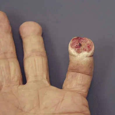
A 69-year-old man sustains a traumatic amputation to the distal phalanx of his little finger while working with power tools. Radiographs are shown in Figures 27a and 27b. The patient was instructed how to perform wet-to-dry dressing changes in the emergency department. Clinical pictures taken in the office are shown in Figures 27c through 27e. What is the most appropriate management of this soft-tissue wound?
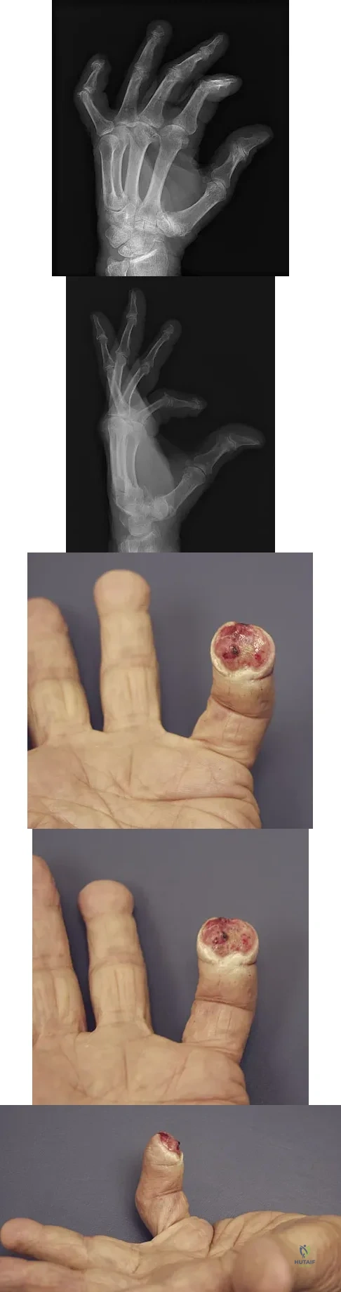


Explanation

The clinical photographs and radiographs reveal a distal phalangeal amputation with soft-tissue coverage over nonexposed bone. This is an ideal circumstance to allow healing by secondary intention with wet-to-dry dressing changes. There are few complications and the aesthetics surpass that of any soft-tissue reconstruction procedure. Volar advancement flaps (Moberg flaps) are limited to small defects about the thumb. A thenar flap will provide good coverage; however, the results are not comparable to simple dressing changes. A V-Y flap is useful when there is more tissue loss dorsally. Jebson PL, Louis DS: Amputations, in Green DP, Hotchkiss RN, Pederson WC, et al (eds): Green's Operative Hand Surgery, ed 5. Philadelphia, PA, Elsevier, 2005, p 1947.

Question 19

A 32-year-old man has an open comminuted humeral shaft fracture. Examination reveals absence of sensation in the first web space and he is unable to fully extend the thumb, fingers, and wrist. What is the recommended treatment following irrigation and debridement of the fracture?

Trauma Board Review 2009: High-Yield MCQs (Set 2) - Figure 32


Explanation

There is a high incidence of partial or complete laceration of the radial nerve with high-energy open fractures of the humeral shaft. The recommended treatment is irrigation and debridement of the fracture followed by open reduction and internal fixation and exploration of the radial nerve. If the nerve is completely lacerated, primary repair may be performed but poor outcomes have been reported. If a large zone of nerve injury is identified, delayed nerve grafting is advocated. Ring D, Chin K, Jupiter JB: Radial nerve palsy associated with high energy humeral shaft fractures. J Hand Surg 2004;29:144-147. Foster RJ, Swiontkowski MR, Bach AW, et al: Radial nerve palsy caused by open humeral shaft fractures. J Hand Surg Am 1993;18:121-124.

Question 20

Which of the following complications is associated with the use of a short cephalomedullary nail for fixation of intertrochanteric hip fractures?

Trauma Board Review 2009: High-Yield MCQs (Set 2) - Figure 33


Explanation

Implant-related fractures following hip fracture surgery were reported by Robinson and associates and showed that the risk of an ipsilateral femoral fracture is significantly increased with the use of short cephalomedullary nails as compared to a compression hip screw. The use of short cephalomedullary nails has not been shown to increase the risk of deep venous thrombosis or nonunion. Cephalomedullary nails overall have decreased surgical blood loss when compared to use of sliding-compression hip-screw devices. Robinson CM, Adams CI, Craid M, et al: Implant-related fractures of the femur following hip fracture surgery. J Bone Joint Surg Am 2002;84:1116-1122.

Question 21

A 19-year-old man sustained the isolated injury seen in Figure 28a. He is adequately resuscitated. A closed reduction was performed in the emergency department, and postreduction radiographs are shown in Figures 28b and 28c. What is the next most appropriate step in management?


Explanation

Incarcerated fragments and nonconcentric reductions are best treated with urgent open reduction, retrieval of the fragments, and internal fixation. Delayed treatment increases the damage to the articular surface, even if traction is applied. There is no role for nonsurgical management with entrapped fragments and nonconcentric reduction of the hip. Epstein HC, Wiss DA, Cozen L: Posterior fracture dislocation of the hip with fractures of the femoral head. Clin Orthop Relat Res 1985;201:9-17.

Question 22

The iliopectineal fascia runs between which of the following structures?

Trauma Board Review 2009: High-Yield MCQs (Set 2) - Figure 37


Explanation

The sheath of the psoas muscle or the iliopectineal fascia separates the more lateral iliopsoas muscle and the femoral nerve from the more medially located iliac vessels. This fascia has to be taken down to enter the true pelvis. Masquelet AC, McCullough CJ, Tubiana R: An Atlas of Surgical Exposures of the Lower Extremity. Philadelphia, PA, JB Lippincott, 1993. Letournel E, Judet R: Fractures of the Acetabulum, ed 2. Berlin, Germany, Springer Verlag, 1993.

Question 23

Which of the following is most predictive of a medial side ankle injury in the presence of a fibula fracture above the level of the joint?

Trauma Board Review 2009: High-Yield MCQs (Set 2) - Figure 38


Explanation

Isolated Lauge-Hansen supination-external rotation-type ankle fractures comprise 20% to 40% of ankle fractures and nonsurgical management is effective for managing SER-2 ankle fractures. Tornetta and associates recently showed that medial ankle tenderness, ecchymosis, and swelling are not reliable findings when trying to determine deltoid competence. Stress radiographs showing a medial clear space of greater than 4 mm or one that is also 1 mm greater than the superior joint space, or any lateral talar subluxation are indicative of deltoid incompetence and indicative of a SER-4 ankle fracture. McConnell T, Creevy W, Tornetta P III: Stress examination of supination external rotation-type fibular fractures. J Bone Joint Surg Am 2004:86;2171-2178. Kristensen KD, Hansen T: Closed treatment of ankle fractures: Stage II supination-eversion fractures followed for 20 years. Acta Orthop Scand 1985;56:107-109.

Question 24

Figure 29 shows the radiograph of a 10-year-old boy who injured his knee playing football. What is the most appropriate initial treatment?

Trauma Board Review 2009: High-Yield MCQs (Set 2) - Figure 39


Explanation

Salter type I fractures of the distal femur are quite unstable; therefore, closed reduction and cast immobilization can be expected to result in high rates of redisplacement. Optimal treatment consists of open or closed reduction and smooth pin fixation, and supplemental casting is required to ensure fracture stability. Screw fixation may increase rates of growth plate injury. Blade plate or flexible nail fixation will be challenging to apply and is not necessary. Flynn JM, Skaggs DL, Sponseller PD, et al: The surgical management of pediatric fractures of the lower extremity. Instr Course Lect 2003;52:647-659. Thomson JD, Stricker SJ, Williams MM: Fractures of the distal femoral epiphyseal plate. J Pediatr Orthop 1995;15:474-478.

Question 25

A 35-year-old man sustained a 1-inch stab incision in his proximal forearm while trying to use a screwdriver 2 weeks ago. The laceration was routinely closed, and no problems about the incision site were noted. He now reports that he has been unable to straighten his fingers or thumb completely since the injury. Clinical photographs shown in Figures 30a and 30b show the man passively flexing the wrist. What is the most appropriate management?
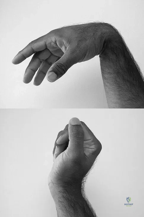


Explanation

The clinical photographs indicate that the tenodesis effect of digit flexion with passive wrist extension and digit extension with passive wrist flexion is intact, indicating no discontinuity of the extensor or flexor tendons. The most likely injury is a laceration of the posterior interosseous nerve.

Question 26

A 25-year-old male presents after a high-speed motorcycle collision with a closed right femur fracture, left tibial shaft fracture, and bilateral pulmonary contusions. The surgical team is debating between early total care (ETC) and damage control orthopedics (DCO). Which of the following serum markers provides the most reliable indicator of adequate resuscitation to safely proceed with definitive intramedullary nailing of both long bones?


Explanation

Serum lactate and base deficit are the most reliable clinical markers for assessing tissue perfusion and the adequacy of resuscitation in polytrauma patients. Normalization of serum lactate (typically less than 2.5 mmol/L) indicates adequate tissue perfusion, allowing the surgical team to safely proceed with early total care (ETC) rather than damage control orthopedics (DCO).

Question 27

A 30-year-old man sustains a closed, isolated, displaced femoral neck fracture. Radiographs demonstrate a vertically oriented fracture line with an angle greater than 50 degrees from the horizontal (Pauwels Type III). What biomechanical construct provides the most stable fixation against the dominant deforming forces for this specific fracture pattern?


Explanation

Pauwels Type III femoral neck fractures have a highly vertical fracture line (>50 degrees), subjecting the fracture to extreme shear forces rather than compressive forces. Biomechanical studies consistently demonstrate that a sliding hip screw (a fixed-angle device) supplemented with a derotational cancellous screw provides superior biomechanical stability against vertical shear forces compared to three parallel cancellous screws.

Question 28

A 42-year-old farmer sustains a severe open tibia fracture after his leg is caught in a piece of agricultural machinery. The wound is 14 cm long with extensive soft tissue stripping, bone loss, and gross contamination with soil and manure. According to current guidelines, which of the following is the most appropriate initial intravenous antibiotic regimen?


Explanation

This is a Gustilo-Anderson Type IIIB open fracture with farm/soil contamination. Standard prophylaxis for Type III open fractures includes a first-generation cephalosporin (for Gram-positive coverage) and an aminoglycoside (for Gram-negative coverage). For farm injuries or gross soil contamination, high-dose penicillin should be added specifically to cover anaerobes, most notably Clostridium perfringens, to prevent gas gangrene.

Question 29

A 28-year-old motorcyclist presents to the trauma bay with an avulsed, flail, and pulseless upper extremity. Radiographs show a massively laterally displaced scapula, an intact clavicle, and complete acromioclavicular separation. What is the most common vascular injury associated with this diagnosis?


Explanation

Scapulothoracic dissociation is a devastating, high-energy injury characterized by complete disruption of the scapulothoracic articulation, marked by lateral displacement of the scapula. It is highly associated with severe neurovascular injuries, most notably complete avulsion of the brachial plexus and disruption of the subclavian artery or vein. Early vascular imaging and intervention are critical.

Question 30

A 35-year-old man falls from a 15-foot ladder and sustains a displaced fracture of the talar neck. Imaging reveals subluxation of both the subtalar and tibiotalar joints, but the talonavicular joint remains reduced. What is the approximate reported risk of developing avascular necrosis (AVN) of the talar body in this injury pattern?


Explanation

This clinical scenario describes a Hawkins Type III talar neck fracture (fracture of the talar neck with dislocation/subluxation of both the subtalar and tibiotalar joints). The blood supply to the talar body (artery of the tarsal canal, artery of the sinus tarsi, and deltoid branches) is severely compromised. The historical risk of avascular necrosis (AVN) for Hawkins Type III fractures is reported to be between 80% and 100%.

Question 31

During the ilioinguinal approach for open reduction and internal fixation of an anterior column acetabular fracture, vigorous arterial bleeding is encountered on the posterior aspect of the superior pubic ramus, approximately 5 cm from the pubic symphysis. Which of the following anatomic structures is the most likely source of this bleeding?


Explanation

The bleeding source is the corona mortis ('crown of death'), which is a critical vascular anastomosis situated on the posterior aspect of the superior pubic ramus. It typically connects the obturator system (internal iliac system) with the external iliac system (via the inferior epigastric vessels). Care must be taken to identify and ligate this structure during the ilioinguinal or modified Stoppa approaches.

Question 32

A 45-year-old roofer undergoes open reduction and internal fixation of a displaced intra-articular calcaneus fracture via an extensile lateral approach. Postoperatively, he complains of significant numbness and burning pain along the lateral aspect of his foot. Which of the following nerves was most likely injured during the surgical approach?


Explanation

The sural nerve is at significant risk of injury during the extensile lateral approach to the calcaneus. It courses posterior to the fibula and supplies sensation to the posterolateral lower leg and the lateral border of the foot. To minimize risk, the horizontal limb of the incision should be placed precisely at the junction of the glabrous and non-glabrous skin, and a full-thickness flap must be elevated without direct retraction on the soft tissues.

Question 33

A 38-year-old unrestrained driver presents with a severely swollen knee after a motor vehicle collision. CT scan reveals a supracondylar distal femur fracture with an associated intra-articular coronal plane fracture of the lateral femoral condyle. Which of the following accurately describes the biomechanically optimal fixation for this coronal plane component?


Explanation

A coronal plane fracture of the femoral condyle is known as a Hoffa fracture, most commonly involving the lateral condyle. Biomechanical studies have demonstrated that anterior-to-posterior directed partially threaded cancellous lag screws provide the strongest fixation. They allow compression perpendicular to the fracture plane and have significantly greater pull-out strength compared to posterior-to-anterior directed screws.

Question 34

A 24-year-old man sustains a closed comminuted tibial shaft fracture. Two hours after presentation, he complains of severe pain out of proportion to the injury that is not relieved by intravenous narcotics. His leg is swollen and tense. Compartment pressures are measured. What pressure differential (Delta P) is considered the absolute threshold for performing an emergency four-compartment fasciotomy?


Explanation

The diagnosis of acute compartment syndrome requires high clinical suspicion. When clinical signs are equivocal or in an unexaminable patient, compartment pressure monitoring is indicated. The widely accepted threshold for performing a fasciotomy is a Delta P (Diastolic Blood Pressure minus Compartment Pressure) of less than or equal to 30 mm Hg. Using absolute pressure values is less reliable and can lead to unnecessary fasciotomies, especially in hypotensive patients.

Question 35

A 28-year-old man sustains a closed spiral fracture of the middle third of the humerus after an arm-wrestling match. On examination in the emergency department, he is unable to extend his wrist or fingers, and he has decreased sensation over the dorsal first web space. What is the most appropriate initial management?


Explanation

A radial nerve palsy associated with a closed humeral shaft fracture (often a Holstein-Lewis fracture in the distal third, but also common in midshaft spiral fractures) has a spontaneous recovery rate of over 85-90%. Initial management is non-operative, utilizing closed reduction and splinting or functional bracing. Operative exploration is reserved for open fractures, penetrating injuries, palsy developing after closed reduction, or failure of clinical or EMG recovery by 3 to 4 months.

Question 36

A 45-year-old polytrauma patient is brought to the trauma bay after a high-speed motor vehicle collision. He presents with a severe head injury (Glasgow Coma Scale score of 7) and a closed midshaft femur fracture. According to current guidelines for Damage Control Orthopedics (DCO), what is the most appropriate initial management of the femur fracture?


Explanation

In a polytrauma patient with a severe head injury and physiological instability, Damage Control Orthopedics (DCO) is indicated to avoid the 'second hit' phenomenon. External fixation allows for rapid stabilization of the fracture without the systemic inflammatory burden, potential for increased intracranial pressure, and prolonged surgical time associated with intramedullary nailing or plating.

Question 37

A 28-year-old man sustains a Pauwels type III (vertical) femoral neck fracture after falling from a ladder. Which of the following fixation constructs provides the most biomechanically stable fixation against shear forces for this specific fracture pattern?


Explanation

Pauwels type III femoral neck fractures have a highly vertical fracture line (angle >50 degrees), which subjects the fracture to high shear forces and increases the risk of varus collapse and nonunion. Biomechanical studies have consistently demonstrated that a fixed-angle construct, such as a sliding hip screw combined with an anti-rotation screw, provides superior stability and resistance to shear forces compared to multiple parallel cancellous screws.

Question 38

A 35-year-old woman sustains a highly comminuted distal femur fracture following a high-energy trauma. Computed tomography (CT) imaging reveals an isolated coronal plane fracture of the lateral femoral condyle. What is the eponym for this specific fracture fragment?


Explanation

A coronal plane fracture of the femoral condyle is known as a Hoffa fracture. It is a severe intra-articular injury that most commonly affects the lateral condyle. It requires anatomic reduction and stable internal fixation, typically with anterior-to-posterior or posterior-to-anterior directed lag screws, to prevent articular incongruity and post-traumatic arthritis.

Question 39

A 30-year-old man sustains an open tibia fracture with massive soft tissue stripping and exposed bone (Gustilo-Anderson IIIB) after a motorcycle collision. After initial thorough surgical debridement and bony stabilization, what is the optimal timeframe for definitive soft tissue coverage to minimize the risk of deep infection and flap failure?


Explanation

Based on Godina's classic principles and subsequent supporting literature, early soft tissue coverage (optimally within 72 hours) of severe open tibia fractures with significant soft tissue loss (Gustilo IIIB) significantly reduces the rate of deep infection, flap failure, and nonunion compared to delayed coverage.

Question 40

A 65-year-old woman presents with an isolated closed, highly displaced distal radius fracture. She undergoes closed reduction and splinting in the emergency department. Which of the following nerves is at greatest risk of developing an acute compression neuropathy following this specific injury and manipulation?


Explanation

Distal radius fractures are a well-known cause of acute carpal tunnel syndrome. The median nerve can be compressed within the carpal tunnel due to initial fracture displacement, fracture hematoma, or post-reduction swelling. Severe pain, paresthesias, or subjective numbness in the median nerve distribution necessitates careful evaluation, release of constricting splints, and potentially urgent carpal tunnel release if symptoms are unremitting.

Question 41

A 25-year-old man falls from a height of 20 feet, sustaining a displaced, intra-articular calcaneus fracture. The Sanders classification system is utilized to guide surgical management based on CT imaging. What is the primary anatomical landmark assessed on coronal CT to determine the Sanders classification?


Explanation

The Sanders classification system is the most widely used scheme for intra-articular calcaneus fractures. It is based on the number and location of primary fracture lines extending through the posterior facet of the calcaneus, as visualized on the widest coronal CT image.

Question 42

A 40-year-old man sustains a Hawkins type III fracture of the talar neck after a severe motor vehicle collision. What is the approximate reported risk of developing avascular necrosis (AVN) of the talar body with this specific injury pattern?


Explanation

Hawkins type III fractures involve a fracture of the talar neck with dislocation of both the subtalar and tibiotalar joints. This injury severely disrupts the delicate, retrograde blood supply to the talar body (artery of the tarsal canal, artery of the sinus tarsi, and deltoid branches). Consequently, the risk of avascular necrosis is exceptionally high, typically cited between 70% and 100%.

Question 43

Which of the following statements is most accurate regarding the initial management of a traumatic, hemodynamically unstable pelvic ring disruption?


Explanation

Preperitoneal pelvic packing is a rapid and highly effective surgical method for controlling the life-threatening venous plexus and cancellous bone bleeding typically seen in unstable pelvic fractures. Pelvic binders must be centered over the greater trochanters, not the iliac crests. Angiography targets arterial (not venous) bleeding. Binders generally reduce pelvic volume as effectively or more effectively than external fixation in the acute setting.

Question 44

A 55-year-old man presents with a closed, highly comminuted tibial shaft fracture. His diastolic blood pressure is 60 mm Hg. What absolute intra-compartmental pressure would meet the widely accepted delta pressure threshold for performing an immediate four-compartment fasciotomy?


Explanation

The delta pressure (ΔP) is calculated as the diastolic blood pressure minus the intra-compartmental pressure. A ΔP of less than 30 mm Hg is a widely accepted threshold indicating inadequate tissue perfusion and mandating an immediate fasciotomy. With a diastolic BP of 60 mm Hg, an absolute compartment pressure of 35 mm Hg results in a ΔP of 25 mm Hg (which is <30 mm Hg).

Question 45

A 22-year-old male sustains a severe pelvic trauma. Radiographs and CT imaging reveal a transverse fracture of the acetabulum combined with a posterior wall fracture. According to the Letournel and Judet classification of acetabular fractures, which of the following categories best describes this injury?


Explanation

The Letournel and Judet classification system divides acetabular fractures into 5 elementary types (anterior wall, anterior column, posterior wall, posterior column, transverse) and 5 associated types (posterior column/posterior wall, transverse/posterior wall, T-shaped, anterior column/posterior hemitransverse, both columns). A transverse plus posterior wall fracture falls under the associated fracture category.

Question 46

A 35-year-old man sustains a severe open tibia fracture (Gustilo-Anderson IIIB) in a motorcycle crash. He is hemodynamically stable. Which of the following factors has been shown in the literature to be the most critical independent predictor for reducing the risk of deep infection in this patient?


Explanation

The most critical and consistently proven factor in reducing infection rates in open fractures is the early administration of systemic antibiotics (ideally within 1 hour of injury). The traditional '6-hour rule' for surgical debridement has been largely disproven as an independent predictor of infection risk, provided adequate debridement occurs within the first 24 hours.

Question 47

A 28-year-old male is admitted with a closed midshaft tibia fracture. Overnight, he develops increasing pain out of proportion to his injury that is poorly responsive to IV opioids. Pain is elicited with passive stretch of his hallux. You measure his compartment pressures. Which of the following parameters represents an absolute indication for emergency fasciotomy?


Explanation

Delta P (diastolic blood pressure minus compartment pressure) < 30 mmHg is the most reliable threshold for diagnosing acute compartment syndrome and is an absolute indication for fasciotomy. This measurement accounts for the tissue perfusion gradient, making it far more accurate than absolute pressure alone, especially in hypotensive patients.

Question 48

A 25-year-old female presents with a high-energy Pauwels type III (vertical shear) femoral neck fracture. Which of the following internal fixation constructs provides the most biomechanical stability for this specific fracture pattern?


Explanation

Pauwels type III fractures are highly unstable due to the vertical orientation of the fracture line, creating immense shear forces. Biomechanical studies have demonstrated that a fixed-angle device, such as a dynamic hip screw (DHS) combined with a derotational screw, provides superior stability and higher failure loads compared to multiple cancellous screws in high-shear, vertical femoral neck fractures.

Question 49

A 40-year-old male motorcyclist experiences a severe lateral traction injury to his right shoulder. He presents with massive shoulder swelling, an absent right radial pulse, and a completely flail right upper extremity. Radiographs reveal a widened scapulothoracic articulation and lateral displacement of the scapula. What is the most likely associated neurologic injury?


Explanation

The clinical scenario describes scapulothoracic dissociation, a highly lethal, high-energy traction injury. It is characterized by complete disruption of the scapulothoracic articulation and is highly associated with catastrophic neurovascular injuries, most notably complete brachial plexus avulsions and subclavian/axillary artery disruptions.

Question 50

In a polytrauma patient with bilateral femur fractures and a pulmonary contusion, 'Damage Control Orthopedics' (DCO) with temporary external fixation is being considered over Early Total Care (ETC) with intramedullary nailing. Which of the following physiological parameters classifies the patient as 'borderline' and supports the use of DCO?


Explanation

According to Pape et al., 'borderline' polytrauma patients are at high risk of ARDS and multiple organ failure if subjected to early total care (e.g., prolonged reaming and intramedullary nailing). Indicators supporting DCO include a base deficit > 8 mmol/L, initial lactate > 2.5 mmol/L, pH < 7.25, hypothermia (< 35°C), and significant bilateral pulmonary contusions.

Question 51

A 22-year-old male football player sustains a spontaneous reduction of a knee dislocation prior to arrival at the emergency department. He has a grossly unstable knee but normal, palpable dorsalis pedis and posterior tibial pulses. What is the most appropriate next step in his management to evaluate for vascular injury?


Explanation

For knee dislocations presenting with normal, palpable pulses, an Ankle-Brachial Index (ABI) should be measured first. An ABI > 0.9 reliably excludes flow-limiting vascular injury and allows for observation. If the ABI is < 0.9 or pulses are asymmetric/absent, advanced imaging such as CT angiography is warranted.

Question 52

A 45-year-old roofer falls 15 feet, sustaining a displaced, intra-articular calcaneus fracture. You review his imaging to determine the Sanders classification. Which specific imaging modality and view dictates the Sanders classification for calcaneus fractures?


Explanation

The Sanders classification is strictly based on coronal CT images through the widest portion of the posterior facet of the calcaneus. It categorizes the fracture based on the number and location of the primary fracture lines dividing the posterior facet, which correlates with surgical prognosis.

Question 53

A 35-year-old male is undergoing open reduction and internal fixation of a transverse posterior wall acetabular fracture via a Kocher-Langenbeck approach. To minimize iatrogenic traction injury to the sciatic nerve during retraction, what is the optimal position of the operative lower extremity?


Explanation

During a Kocher-Langenbeck approach for acetabular fractures, the sciatic nerve is at significant risk of iatrogenic stretch injury from retractors. Keeping the hip extended and the knee flexed introduces maximum slack into the sciatic nerve, thereby minimizing tension and reducing the risk of a post-operative nerve palsy (e.g., foot drop).

Question 54

Six months after open reduction and internal fixation of a volar Barton's distal radius fracture using a volar locked plate, a 65-year-old female presents with sudden inability to actively flex the interphalangeal (IP) joint of her thumb. What is the most likely etiology of this complication?


Explanation

Flexor pollicis longus (FPL) tendon rupture is a well-documented complication of volar locked plating of the distal radius. It typically results from placing the plate too far distal (beyond the watershed line), causing the prominent hardware to chronically irritate and eventually attrite the FPL tendon.

Question 55

A 32-year-old male is brought to the trauma bay after a motorcycle crash. He has an 'open book' pelvic ring injury (APC III) and is hemodynamically unstable. In severe pelvic fractures with massive volume expansion, what is the primary anatomical source of the life-threatening hemorrhage?


Explanation

In severe pelvic ring injuries with massive retroperitoneal volume expansion, approximately 80-90% of the hemorrhage is venous in origin (primarily from the pre-sacral venous plexus) combined with bleeding from the expansive, raw cancellous bone surfaces. While arterial bleeding (such as from the superior gluteal or pudendal arteries) can be catastrophic, it accounts for a much smaller percentage of overall pelvic hemorrhage cases.

Question 56

An 82-year-old man presents with a hip injury after a mechanical fall. Radiographs and CT scan show an anterior column and posterior hemitransverse acetabular fracture with severe superomedial dome impaction (the "gull sign"). Which of the following factors most strongly predicts early failure if open reduction and internal fixation alone is chosen?


Explanation

Superomedial dome impaction (the 'gull sign') in osteopenic patients represents a poor prognostic factor for open reduction and internal fixation (ORIF) alone. It involves severe crushing of the osteoporotic subchondral bone of the acetabular roof. Attempted ORIF in the presence of this sign carries a very high rate of early mechanical failure and progression to post-traumatic arthritis. Thus, acute total hip arthroplasty (often combined with column stabilization) is frequently indicated in this population.

Question 57

A 30-year-old man with a closed midshaft tibia fracture complains of severe leg pain out of proportion to his injury, worsening with passive toe stretch. His blood pressure is 110/60 mm Hg. Intracompartmental pressure monitoring reveals an anterior compartment pressure of 40 mm Hg. What is the most appropriate next step in management?


Explanation

This patient has acute compartment syndrome. The delta pressure (diastolic blood pressure minus intracompartmental pressure) is 60 - 40 = 20 mm Hg. A delta pressure of less than 30 mm Hg, combined with clinical signs of severe pain with passive stretch, is an absolute indication for emergent four-compartment fasciotomies of the leg.

Question 58

A 25-year-old man sustains a displaced, vertically oriented (Pauwels type III) femoral neck fracture. He undergoes open reduction and internal fixation. What biomechanical advantage does a sliding hip screw with a derotation screw provide over three parallel cancellous screws for this specific fracture pattern?


Explanation

Pauwels type III femoral neck fractures are highly vertically oriented, leading to significant vertical shear forces and a high risk of varus collapse and nonunion. A sliding hip screw with a derotation screw provides superior biomechanical resistance to these vertical shear forces compared to three parallel cancellous screws, making it the preferred construct for young patients with this fracture pattern.

Question 59

What is the most critical factor in reducing the risk of infection in a patient who sustains a severe, open tibia fracture in a high-speed motor vehicle collision?


Explanation

While surgical debridement is essential, literature and current guidelines emphasize that the single most important factor in reducing the rate of infection in open fractures is the early administration of systemic antibiotics, ideally within 1 hour of injury.

Question 60

A 22-year-old man sustains a low-velocity gunshot wound to the right distal thigh. Radiographs demonstrate a comminuted fracture of the distal femoral diaphysis with the bullet lodged in the adjacent soft tissues. Distal pulses are palpable, and the ABI is 1.0. Which of the following is the most appropriate initial soft tissue management?


Explanation

Low-velocity gunshot wounds causing fractures, without clinical evidence of vascular compromise or massive soft tissue contamination/destruction, are typically treated similarly to closed fractures regarding the soft tissues. Local wound care, tetanus prophylaxis, and short-course systemic antibiotics are indicated, followed by definitive fracture fixation. Routine exploration and debridement of the bullet track or bullet removal are not necessary.

Question 61

A 35-year-old woman sustains a 'floating shoulder' injury, defined by ipsilateral displaced fractures of the clavicle and the scapular neck. Which of the following represents the primary biomechanical rationale for open reduction and internal fixation of the clavicle in this injury pattern?


Explanation

A 'floating shoulder' results from double disruption of the superior shoulder suspensory complex (SSSC). Fixation of the clavicle restores the strut function of the SSSC, stabilizing the complex and indirectly aligning the scapular neck fracture. This reduces the risk of malunion, drooping shoulder, and subsequent functional deficits.

Question 62

A 68-year-old woman treated with alendronate for 8 years presents with a 3-month history of an unprovoked dull ache in her right thigh. A radiograph of the femur reveals localized lateral cortical thickening and a transverse radiolucent line in the subtrochanteric region. What is the most appropriate next step in orthopaedic management?


Explanation

This patient has symptoms and classic radiographic signs (lateral cortical thickening, transverse radiolucent line, 'beaking') of an impending bisphosphonate-related atypical femur fracture. Because the lesion is symptomatic, she is at high risk for completion of the fracture. Prophylactic intramedullary nailing is indicated to prevent complete displacement and associated morbidity.

Question 63

A 42-year-old man sustains an anteroposterior compression type II (APC II) pelvic ring injury. He is hemodynamically stable. Imaging shows a 3.5 cm symphyseal diastasis and bilateral anterior sacroiliac joint widening. The posterior sacroiliac ligaments are intact. What is the optimal surgical treatment to restore pelvic ring stability?


Explanation

An APC II injury involves disruption of the symphysis pubis (or anterior ring) and the anterior sacroiliac, sacrotuberous, and sacrospinous ligaments, while the robust posterior sacroiliac ligaments remain intact. Because the posterior tension band is intact, restoring the anterior ring with open reduction and internal fixation of the pubic symphysis alone is sufficient to stabilize the entire pelvic ring.

Question 64

A 55-year-old woman undergoes volar locked plating for a displaced intra-articular distal radius fracture. Six weeks postoperatively, she suddenly loses the ability to actively extend her thumb interphalangeal joint. What is the most likely etiology of this complication?


Explanation

Inability to extend the thumb IP joint following volar plating of the distal radius is most commonly due to attritional rupture of the extensor pollicis longus (EPL) tendon. This occurs when screws are too long and protrude through the dorsal cortex into the third extensor compartment. While FPL rupture can also occur from a plate placed distal to the watershed line, it results in a loss of thumb flexion, not extension.

Question 65

A 45-year-old man sustains a closed, highly comminuted tibial pilon fracture with severe soft tissue swelling and multiple clear and blood-filled fracture blisters over the ankle. What is the preferred initial management strategy?


Explanation

High-energy tibial pilon fractures with severe soft tissue compromise (e.g., massive swelling, fracture blisters) are associated with unacceptably high rates of wound complications and deep infection if treated with immediate open reduction and internal fixation. The standard of care is a staged approach: immediate application of a joint-spanning external fixator to restore length and alignment while allowing the soft tissues to heal, followed by definitive internal fixation 10 to 21 days later.

Question 66

A 25-year-old man sustains a low-velocity gunshot wound to the right thigh, resulting in a comminuted midshaft femur fracture. The bullet is retained in the soft tissues adjacent to the fracture. Distal pulses are palpable and symmetrical to the contralateral limb, and there are no expanding hematomas. What is the most appropriate initial management of the wound and fracture?


Explanation

Low-velocity gunshot wounds to the femur without hard signs of vascular injury or severe gross contamination can be safely treated with superficial wound debridement, systemic antibiotics, and antegrade reamed intramedullary nailing. Extensive exploration of the bullet track or formal extraction of the bullet is unnecessary unless the bullet is within the joint space, causing neurovascular compression, or associated with a high-velocity weapon with massive soft tissue destruction.

Question 67

A 32-year-old man is brought to the trauma bay after a fall from a height of 15 feet. Radiographs reveal a displaced basicervical femoral neck fracture. Which of the following internal fixation constructs is considered biomechanically superior for this specific fracture pattern?


Explanation

Basicervical femoral neck fractures are mechanically distinct from subcapital fractures. They behave more like intertrochanteric fractures and are inherently unstable due to the lack of interlocking bony fragments. Multiple cancellous screws and single-screw cephalomedullary nails have a high rate of biomechanical failure in this pattern. A sliding hip screw (SHS), often supplemented with a derotational screw, provides fixed-angle stability and is the preferred construct.

Question 68

A 30-year-old farmer with no known allergies sustains a severe open tibia fracture (Gustilo-Anderson Type IIIA) heavily contaminated with manure and soil. In addition to a first-generation cephalosporin and an aminoglycoside, which of the following antibiotics is most critical to include in his initial prophylactic regimen?


Explanation

Farm injuries, particularly those contaminated with soil or manure, carry a significant risk for anaerobic infections, most notably Clostridium perfringens, which can lead to gas gangrene (myonecrosis). The addition of high-dose intravenous penicillin is strongly recommended for anaerobic coverage in these heavily contaminated farm injuries, on top of the standard gram-positive (cefazolin) and gram-negative (gentamicin) coverage.

Question 69

A 28-year-old construction worker sustains a Hawkins type III talar neck fracture following a fall. At his 8-week postoperative follow-up, an AP radiograph of the ankle demonstrates 'Hawkins sign'. What does this radiographic finding represent?


Explanation

Hawkins sign is characterized by a subchondral radiolucent band seen on an AP radiograph of the ankle, typically appearing 6 to 8 weeks after a talar neck fracture. This radiolucency is secondary to subchondral bone atrophy (disuse osteopenia). For bone resorption to occur, the blood supply to the talar dome must be intact. Therefore, the presence of Hawkins sign is a highly reliable indicator that avascular necrosis (AVN) will not occur.

Question 70

A 40-year-old woman with a highly comminuted Schatzker VI tibial plateau fracture undergoes application of a spanning external fixator. Twelve hours postoperatively, she reports excruciating, unremitting leg pain that is out of proportion to her injury. Passive stretch of her toes elicits severe pain. Intracompartmental pressure testing reveals an anterior compartment pressure of 45 mm Hg. Her current blood pressure is 100/60 mm Hg. What is the most appropriate next step in management?


Explanation

The patient is presenting with classic signs of acute compartment syndrome. The diagnosis is confirmed by measuring the Delta P (Diastolic Blood Pressure - Compartment Pressure). In this case, 60 mm Hg - 45 mm Hg = 15 mm Hg. A Delta P of less than 30 mm Hg indicates critically impaired tissue perfusion and is an absolute indication for urgent four-compartment fasciotomy. Elevation above the heart is contraindicated as it further decreases arterial perfusion to the compartment.

Question 71

A 76-year-old female with advanced osteoporosis and severe preexisting osteoarthritis of the elbow sustains a comminuted, intra-articular distal humerus fracture (AO/OTA 13-C3) after a mechanical fall. Which of the following is the most appropriate definitive surgical intervention?


Explanation

In an elderly patient with poor bone quality (osteoporosis), severe comminution of the articular surface, and preexisting symptomatic osteoarthritis or rheumatoid arthritis, total elbow arthroplasty (TEA) is the treatment of choice for a distal humerus fracture. TEA allows for immediate stability, early range of motion, and predictable pain relief, circumventing the high risk of fixation failure and stiffness associated with ORIF in this demographic.

Question 72

A 22-year-old motorcyclist is involved in a high-speed collision and presents with massive soft tissue swelling over the left shoulder and a flail, insensate left upper extremity. Radiographs reveal lateral displacement of the scapula with widening of the acromioclavicular joint. Which of the following nerve injuries is most strongly associated with this specific injury pattern?


Explanation

The clinical presentation and radiographic findings describe scapulothoracic dissociation, a highly traumatic, limb-threatening injury. It is defined by the disruption of the scapulothoracic articulation with lateral displacement of the scapula. This injury is notorious for massive neurovascular compromise. Complete brachial plexus avulsion occurs in over 80% of these cases, frequently resulting in a permanent flail limb and commonly requiring above-elbow amputation. Subclavian/axillary vascular injuries are also frequently present.

Question 73

A 35-year-old man falls from a height of 30 feet, sustaining a spinopelvic dissociation characterized by a U-shaped sacral fracture on imaging. Upon physical examination, he has decreased perianal sensation, loss of voluntary anal sphincter tone, and urinary retention. This clinical presentation is primarily due to injury at which of the following neurologic levels?


Explanation

A U-shaped sacral fracture involves bilateral longitudinal sacral fractures connected by a transverse fracture line, most commonly occurring at the S1 or S2 level. Because the transverse component crosses the central sacral spinal canal, it highly compromises the sacral nerve roots of the cauda equina. This results in cauda equina syndrome, characterized by saddle anesthesia, loss of sphincter tone, and bowel/bladder dysfunction.

Question 74

A 29-year-old active male falls directly onto his left shoulder, sustaining a completely displaced, shortened midshaft clavicle fracture. Which of the following radiographic or demographic characteristics is recognized as the most significant predictor for nonunion if this injury is treated nonoperatively?


Explanation

The most significant risk factors for nonunion in midshaft clavicle fractures treated nonoperatively include complete fracture displacement (greater than 100% of the width of the clavicle), shortening greater than 2 cm, severe comminution, and advancing patient age. A completely displaced clavicle fracture has a significantly higher rate of nonunion compared to a nondisplaced or minimally displaced fracture, making it a strong relative indication for operative fixation in active patients.

Question 75

A 21-year-old collegiate football player suffers a high-energy knee dislocation during a tackle. Upon arrival at the emergency department, his knee has spontaneously reduced. Pedal pulses are palpable, but his Ankle-Brachial Index (ABI) is measured at 0.8. What is the most appropriate next step in his management?


Explanation

A knee dislocation carries a high risk of popliteal artery injury. Current guidelines recommend measuring the Ankle-Brachial Index (ABI) after reduction of a knee dislocation. An ABI of less than 0.9 is abnormal and highly suspicious for a vascular injury, even if pulses are palpable. It mandates further objective vascular imaging, typically a CT angiogram or conventional angiogram. Immediate operative exploration is reserved for 'hard signs' of vascular injury, such as absent pulses, expanding hematoma, or pulsatile bleeding.

Question 76

In the context of damage control orthopaedics for a polytrauma patient, when is it most appropriate to convert an external fixator of a femoral shaft fracture to a reamed intramedullary nail?


Explanation

In damage control orthopaedics, conversion from external fixation to intramedullary nailing of a femur fracture is typically done when the patient's systemic physiology has improved and stabilized. Reliable indicators of adequate resuscitation include normalization of lactate and base deficit, as well as stable hemodynamics, coagulation profiles, and pulmonary function. Proceeding too early in a physiologically unresuscitated patient risks triggering a lethal secondary hit.

Question 77

A 35-year-old man sustains an isolated closed tibial shaft fracture treated with a reamed intramedullary nail. Postoperatively, he develops severe pain out of proportion to the injury, which is markedly exacerbated by passive stretch of the hallux. His leg is tense and swollen. What is the most appropriate next step in management?


Explanation

The clinical presentation of severe pain out of proportion, a tense extremity, and pain with passive muscle stretch in an awake and alert patient with a recently nailed tibia fracture is the classic diagnostic triad for acute compartment syndrome. When the clinical diagnosis is clear, immediate four-compartment fasciotomies are required. Measuring pressures is unnecessary and delays definitive treatment when the diagnosis is clinically unequivocal.

Question 78

A 45-year-old man presents to the emergency department with a midshaft clavicle fracture after falling off his bicycle. Which of the following is considered an absolute indication for operative fixation of this injury?


Explanation

Absolute indications for operative treatment of a clavicle fracture include open fracture, displaced fracture with compromised or threatened skin (skin tenting with impending necrosis), vascular injury requiring repair, and progressive neurologic deficit. Shortening >2 cm, complete displacement, and high athletic demands are considered relative indications for surgery.

Question 79

A 25-year-old male sustains a low-velocity gunshot wound to the thigh, resulting in a minimally displaced midshaft femur fracture. The bullet completely traversed the thigh. Examination reveals normal distal pulses and no signs of compartment syndrome. What is the most appropriate management?


Explanation

Low-velocity gunshot wounds resulting in femur fractures without vascular injury, massive tissue destruction, severe contamination, or compartment syndrome can be treated similarly to closed fractures. Local wound care, tetanus prophylaxis, a short course of antibiotics (usually a first-generation cephalosporin), and reamed intramedullary nailing represents the standard of care. Formal I&D of the bullet track is not routinely indicated for low-velocity injuries.

Question 80

Which of the following injury patterns represents the classic 'terrible triad' of the elbow?


Explanation

The terrible triad of the elbow consists of an elbow dislocation, a radial head or neck fracture, and a coronoid fracture. It is invariably associated with a tear of the lateral collateral ligament (LCL) complex, which is avulsed from its origin on the lateral epicondyle during the dislocation. The mechanism typically involves a fall on an outstretched hand with the elbow in extension, applying valgus, axial, and posterolateral rotatory forces.

Question 81

In the initial management of a severe Type II open tibia fracture, what is the single most important factor in decreasing the risk of deep infection?


Explanation

The single most critical factor in preventing infection in open fractures is the early and adequate administration of systemic intravenous antibiotics. While thorough surgical debridement is absolutely essential, the classic '6-hour rule' for timing of debridement has been challenged by modern literature, which places primary emphasis on antibiotic timing (ideally within 1 hour of presentation) and the thoroughness of the debridement, rather than an arbitrary 6-hour surgical window.

Question 82

A 30-year-old male sustains a talar neck fracture with subluxation of the subtalar joint (Hawkins Type II) after a fall from a height. Which of the following vascular structures provides the primary blood supply to the talar body and is most at risk of injury in this specific fracture pattern?


Explanation

The artery of the tarsal canal, a branch of the posterior tibial artery, provides the majority of the blood supply to the body of the talus. Talar neck fractures put this retrograde blood supply at significant risk, leading to avascular necrosis (AVN). Hawkins Type II fractures (talar neck fracture with subtalar dislocation) have an AVN rate of approximately 20-50% due to the disruption of this vascular anastamosis.

Question 83

A 65-year-old female sustains a completely displaced, off-ended subtrochanteric femur fracture. She is scheduled for cephalomedullary nailing. To prevent the most common malreduction during this procedure, what intraoperative consideration is most critical?


Explanation

Subtrochanteric fractures are subject to strong deforming forces: the proximal fragment is flexed (iliopsoas), abducted (gluteus medius/minimus), and externally rotated (short external rotators), while the distal fragment is pulled proximally and into varus. Nailing in a varus malreduction leads to high rates of hardware failure and nonunion. Therefore, these fractures must be anatomically reduced (often utilizing percutaneous clamps, blocking screws, or an open reduction) prior to reaming and nail passage.

Question 84

A 40-year-old man presents with a pelvic ring injury after a high-speed motorcycle crash. An AP pelvis radiograph demonstrates widening of the pubic symphysis of 3.5 cm and widening of the left sacroiliac joint. He remains hemodynamically unstable (BP 70/40) despite receiving 2 liters of crystalloid and 2 units of PRBCs. What is the most appropriate next step in orthopedic management?


Explanation

In a hemodynamically unstable patient with an anteroposterior compression (APC) pelvic ring injury, immediate mechanical stabilization must be achieved to reduce pelvic volume and promote venous tamponade. This is best accomplished emergently with a pelvic binder or sheet placed correctly and centered over the greater trochanters. Placement over the iliac crests is incorrect and can act as a fulcrum, paradoxically exacerbating the pubic symphysis diastasis.

Question 85

A 50-year-old woman is evaluated for a nonunion of a midshaft humerus fracture 6 months after injury. She was originally treated with a Sarmiento cast brace. Radiographs show atrophic bone ends with no callus formation. Laboratory markers for infection are negative. What is the most reliable surgical treatment?


Explanation

Atrophic nonunions occur due to a lack of biology and require both mechanical stability and biological stimulation to heal. For an atrophic humeral shaft nonunion, the gold standard treatment is open reduction and internal fixation (typically using heavy compression plating) combined with autologous bone grafting (e.g., from the iliac crest) to provide the necessary osteoinductive and osteoconductive properties.

Question 86

Which of the following inflammatory markers is considered the most reliable early predictor for the development of acute respiratory distress syndrome (ARDS) and multiple organ dysfunction syndrome (MODS) in a polytrauma patient, guiding the decision between early total care and damage control orthopedics?


Explanation

Interleukin-6 (IL-6) is a pro-inflammatory cytokine that has been shown to be the best early marker for the magnitude of the systemic inflammatory response following major trauma. Peak IL-6 levels correlate closely with the extent of tissue injury, the systemic inflammatory response syndrome (SIRS), and the subsequent development of ARDS, MODS, and mortality. While IL-1 and TNF-alpha are also part of the inflammatory cascade, they have shorter half-lives and are more difficult to measure consistently. CRP and ESR are non-specific and peak much later.

Question 87

A 32-year-old man sustains a subtrochanteric femur fracture. During attempts at closed reduction, the proximal fracture fragment is noted to be severely flexed, abducted, and externally rotated. The abduction deformity of the proximal fragment is primarily caused by the pull of which of the following muscles?


Explanation

In a subtrochanteric femur fracture, the proximal fragment is subjected to distinct deforming forces from the muscles attaching to the proximal femur. The iliopsoas (attaching to the lesser trochanter) pulls the fragment into flexion. The short external rotators pull it into external rotation. The gluteus medius and minimus (attaching to the greater trochanter) pull the fragment into abduction. The distal fragment is typically shortened and adducted by the pull of the adductor musculature.

Question 88

A 45-year-old farmer is brought to the emergency department after his leg was caught in a tractor mechanism. He has sustained a Gustilo-Anderson Type III open tibia fracture that is heavily contaminated with soil and organic material. In addition to prompt surgical debridement, what is the most appropriate initial intravenous antibiotic regimen according to classic trauma guidelines?


Explanation

For Gustilo-Anderson Type III open fractures, standard prophylaxis includes a first-generation cephalosporin (e.g., cefazolin) for Gram-positive coverage and an aminoglycoside (e.g., gentamicin) for expanded Gram-negative coverage. When the injury involves a farm environment, gross soil contamination, or standing water, there is a significantly increased risk of Clostridium infection (gas gangrene). In such high-risk scenarios, high-dose penicillin must be added to the regimen to provide coverage against anaerobic organisms, specifically Clostridium species.

Question 89

A 40-year-old man sustains an anteroposterior compression type III (APC III) pelvic ring injury in a high-speed motorcycle collision. In the trauma bay, a pelvic binder is applied, and he receives massive transfusion protocol. His systolic blood pressure remains 70 mm Hg. The Focused Assessment with Sonography for Trauma (FAST) examination is negative, and a chest radiograph shows no abnormalities. What is the most appropriate next step in the management of this patient?


Explanation

The patient remains hemodynamically unstable despite a pelvic binder and fluid resuscitation, with a negative FAST exam indicating the absence of massive intra-abdominal hemorrhage. The bleeding is most likely retroperitoneal from the highly unstable APC III pelvic injury. Pelvic angiography is the most appropriate next step to identify and embolize actively bleeding arterial vessels (most commonly branches of the internal iliac artery, such as the superior gluteal or pudendal arteries). CT scanning is contraindicated in hemodynamically unstable patients.

Question 90

A 30-year-old woman undergoes closed reduction and percutaneous pinning of a Hawkins type II talar neck fracture. At her 8-week follow-up appointment, an anteroposterior radiograph of the ankle reveals a subchondral radiolucent band extending across the dome of the talus. What is the clinical significance of this radiographic finding?


Explanation

The radiographic finding described is known as the 'Hawkins sign'. It consists of a subchondral radiolucent band in the talar dome typically seen 6 to 8 weeks after a talar neck fracture. This radiolucency represents subchondral osteopenia, which occurs as a result of disuse atrophy. For this bone resorption to happen, the talar body must have an intact blood supply. Therefore, the presence of a positive Hawkins sign is a highly reliable indicator that the talar body remains vascularized and has not undergone avascular necrosis (AVN).

Question 91

A 28-year-old man is brought to the trauma center following a severe motor vehicle accident. On physical examination, his left upper extremity is flail, pale, and completely pulseless. Radiographs reveal massive lateral displacement of the left scapula, an intact clavicle, and complete disruption of the acromioclavicular joint. Which of the following associated injuries carries the highest risk of acute mortality in this patient?


Explanation

This clinical presentation is classic for scapulothoracic dissociation, a severe injury resulting from massive traction force to the shoulder girdle. It is characterized by lateral displacement of the scapula with an intact clavicle or clavicular fracture, and disruption of the AC or SC joint. While complete brachial plexus avulsions are extremely common and cause severe long-term morbidity, the highest risk of acute mortality is due to concomitant disruption of the subclavian or axillary vessels, which can lead to rapid, life-threatening exsanguination.

Question 92

A 6-year-old boy sustains a completely displaced, extension-type Gartland type III supracondylar humerus fracture. Prior to operative intervention, a thorough neurologic examination reveals that he is unable to actively flex the interphalangeal joint of his thumb and the distal interphalangeal joint of his index finger. Which of the following nerves has most likely been injured?


Explanation

The patient demonstrates an inability to flex the interphalangeal joint of the thumb (innervated by the flexor pollicis longus) and the distal interphalangeal joint of the index finger (innervated by the flexor digitorum profundus). This motor deficit, often tested by asking the patient to make an 'A-OK' sign, is indicative of an anterior interosseous nerve (AIN) palsy. The AIN is a branch of the median nerve and is the most commonly injured nerve in pediatric extension-type supracondylar humerus fractures.

Question 93

A 68-year-old woman with a history of osteoporosis treated with alendronate for the past 10 years presents with 3 months of progressive, severe right thigh pain worsened by weight-bearing. Plain radiographs show cortical thickening of the lateral cortex of the right subtrochanteric femur with a visible transverse radiolucent line. What is the most appropriate management of this patient's condition?


Explanation

The patient's history of long-term bisphosphonate use, prodromal thigh pain, and radiographic findings of lateral cortical thickening with a transverse radiolucent line are characteristic of an incomplete atypical femur fracture. Because the patient is highly symptomatic and has a visible radiolucent line indicating a stress fracture, the lesion is at very high risk of progressing to a complete, displaced fracture. The standard of care for a symptomatic incomplete atypical femur fracture is prophylactic intramedullary nailing.

Question 94

A 35-year-old man undergoes reamed intramedullary nailing for a closed diaphyseal tibia fracture. Twelve hours postoperatively, he complains of severe leg pain that is completely out of proportion to his injury and is unresponsive to escalating doses of intravenous opioids. On examination, he experiences severe pain upon passive flexion of his great toe. This finding specifically suggests compartment syndrome involving which of the following compartments of the leg?


Explanation

Pain out of proportion to the injury and increased pain with passive stretch of the muscles within a compartment are the hallmark clinical signs of acute compartment syndrome. Passive flexion of the great toe stretches the extensor hallucis longus (EHL) muscle. The EHL, along with the tibialis anterior, extensor digitorum longus, and peroneus tertius, resides in the anterior compartment of the lower leg. Thus, pain on passive great toe flexion indicates anterior compartment ischemia.

Question 95

A 42-year-old construction worker falls from a scaffolding, sustaining a severely displaced, closed, intra-articular calcaneus fracture. While evaluating the patient to determine whether operative or nonoperative management is indicated, which of the following patient-specific factors is considered a strong relative or absolute contraindication to open reduction and internal fixation through an extensile lateral approach?


Explanation

Surgical treatment of intra-articular calcaneus fractures via an extensile lateral approach carries a high risk of wound complications and infection. Poor tissue healing and immune response make certain patient populations highly unsuitable for this surgery. Insulin-dependent diabetes mellitus, especially when complicated by peripheral neuropathy, is a strong relative (and in many centers, absolute) contraindication to operative management due to the exceptionally high rates of wound breakdown, deep infection, amputation, and subsequent Charcot arthropathy.

Question 96

A 28-year-old polytrauma patient presents to the emergency department after a high-speed motorcycle collision. He has bilateral closed femoral shaft fractures, severe pulmonary contusions, and a traumatic brain injury. His initial vital signs include a blood pressure of 85/50 mm Hg and a heart rate of 125 bpm. Laboratory studies reveal a lactate of 5.5 mmol/L, pH 7.20, and base excess of -8 mmol/L. According to the principles of damage control orthopedics (DCO), what is the most appropriate initial management of his bilateral femur fractures?


Explanation

Damage control orthopedics (DCO) is indicated in polytrauma patients who are physiologically unstable or 'in extremis'. Objective parameters indicating physiological exhaustion include pH < 7.25, base excess < -5.5 mmol/L, serum lactate > 2.5 mmol/L, and hypothermia (< 35°C). Performing early total care (e.g., bilateral reamed intramedullary nailing) in this patient exposes him to a massive 'second hit' of systemic inflammation, significantly increasing the risk of acute respiratory distress syndrome (ARDS), multiple organ dysfunction syndrome (MODS), and death. Therefore, rapid, temporary stabilization with external fixation is the standard of care to control hemorrhage and restore bone length without overwhelming the patient's physiologic reserves.

Question 97

A 24-year-old man sustains a closed fracture of the middle third of the humeral shaft during an arm-wrestling match. On physical examination, he is unable to actively extend his wrist or fingers, and has decreased sensation over the dorsal first web space of the ipsilateral hand. Radiographs confirm a transverse midshaft humerus fracture. What is the most appropriate initial management?


Explanation

Primary radial nerve palsy occurs in up to 18% of closed humeral shaft fractures. The vast majority (70-90%) of these injuries represent neurapraxia and will recover spontaneously within 3 to 4 months. Therefore, the standard initial management for a closed humerus fracture with a primary radial nerve palsy is nonoperative treatment using a coaptation splint or functional brace. Immediate surgical exploration is generally reserved for open fractures, severe soft tissue compromise, vascular injury, or secondary palsy (loss of radial nerve function that occurs after a closed reduction attempt). A hanging arm cast is relatively contraindicated for transverse fractures due to the risk of fracture distraction leading to nonunion. EMG is usually not indicated until 3 to 4 weeks post-injury if no clinical recovery is observed.

Question 98

A 30-year-old man sustains a low-velocity gunshot wound to the right leg. Radiographs reveal a comminuted midshaft tibia fracture. There is a 1-cm entrance wound on the anteromedial aspect of the leg and no exit wound. The bullet is lodged in the deep posterior compartment soft tissues. Distal pulses are strong, and compartment compartments are soft and compressible. What is the most appropriate initial management of the wound and fracture?


Explanation

Low-velocity gunshot wounds (GSWs) causing long bone fractures are generally managed similarly to Gustilo-Anderson Type I or II open fractures. The bullet itself does not necessarily drag vast amounts of contaminated debris into the wound, and extensive formal operating room debridement is not routinely indicated unless the wound is grossly contaminated, there is substantial devitalized tissue, or the bullet is intra-articular. Standard treatment includes superficial local wound care in the emergency department, 24 hours of a first-generation cephalosporin (e.g., cefazolin), and standard fracture stabilization. For a length-unstable, comminuted midshaft tibia fracture, reamed intramedullary nailing is the treatment of choice. Routine bullet extraction from soft tissues is unnecessary and may cause further iatrogenic injury.

Question 99

A 45-year-old male is brought to the trauma bay after being crushed by heavy machinery. His pelvis is unstable to manual compression, and radiographs confirm an anteroposterior compression (APC-III) pelvic ring injury. During the secondary survey, he complains of a severe inability to void, and the examining physician notes blood at the urethral meatus. What is the next most appropriate step in the evaluation of his urologic injury?


Explanation

In a patient with a high-energy pelvic ring injury, the presence of blood at the urethral meatus, a high-riding or non-palpable prostate on digital rectal examination, or a scrotal/perineal hematoma is highly suspicious for a urethral injury. Attempting to insert a standard Foley catheter in this setting is absolutely contraindicated as it may convert a partial urethral tear into a complete transection. The mandatory next diagnostic step is a retrograde urethrogram (RUG). Once the urethra is confirmed to be intact via RUG, a bladder catheter can be safely placed. If a urethral tear is identified, urology consultation for a suprapubic catheter is typically required.

Question 100

A 52-year-old farmer sustains an open tibial shaft fracture after his leg becomes entangled in an agricultural tiller. In the emergency department, the wound measures 12 cm in length with significant muscle crush injury, periosteal stripping, and visible heavy soil and manure contamination. According to classic evidence-based guidelines for open fractures, what is the most appropriate initial intravenous antibiotic regimen for this patient?


Explanation

The classic management of open fractures relies on the Gustilo-Anderson classification to guide antibiotic therapy. Type I and II fractures are typically treated with a first-generation cephalosporin (e.g., cefazolin). Type III fractures require broader coverage, classically achieved by adding an aminoglycoside (e.g., gentamicin) to cover Gram-negative organisms. In situations involving heavy soil contamination, farm-related injuries, or potential bowel contamination, high-dose penicillin is added to provide explicit coverage against Clostridium species, which are responsible for gas gangrene. While modern institutional protocols sometimes substitute these with ceftriaxone and metronidazole, the classic board-tested regimen for a heavily contaminated farm injury remains a first-generation cephalosporin, an aminoglycoside, and penicillin.

None

Clinic OS
Medically Verified Content by
Prof. Clinic OS
Consultant Orthopedic & Spine Surgeon
Chapter Index