العربية
Part of the Master Guide

Orthopedic Surgery Board Prep: Set 403 (100 MCQs on Fractures & Casting)

Orthopedic Trauma 2026 MCQs: Board Review Questions & Answers (Part 4)

23 Apr 2026 57 min read 79 Views
Figure for Trauma 2000 MCQs - Part 4 - Question 76

Key Takeaway

For anyone wondering about Orthopedic Trauma 2026 MCQs: Board Review Questions & Answers (Part 4), Top-rated Orthopedic Trauma 2026 MCQs bank. Practice with clinical case questions, orthopedic surgery board review, and evidence-based answers updated for 2026.

Orthopedic Trauma 2026 MCQs: Board Review Questions & Answers (Part 4)

Comprehensive 100-Question Exam


00:00

Start Quiz

Question 1

A 47-year-old man ruptured his left patellar tendon and twisted his right ankle in a fall. Initial radiographs of the ankle are unremarkable. One week following repair of the left patellar tendon, he reports increased pain with weight bearing in his right ankle. A follow-up radiograph is shown in Figure 38. Management of the ankle injury should consist of


Explanation

The radiograph reveals disruption of the syndesmosis with lateral displacement of the talus and widening of the medial ankle clear space. No fibular fracture is noted, although radiographs of the entire tibia and fibula are necessary to rule out a more proximal fibula fracture. There is clear instability of the syndesmosis, and surgical stabilization is needed, either by direct repair of the ligaments or more commonly with surgical stabilization of the fibula to the tibia with screws. Functional rehabilitation and early range of motion are indicated with anterior-lateral ankle sprains but not with true instability of the syndesmosis. In anterior syndesmotic injuries in which there are no signs of instability on plain radiographs or with stressing, cast immobilization and protected weight bearing until tenderness subsides is warranted. Long leg cast immobilization is unlikely to be adequate in maintaining reduction of the syndesmosis. Repair of the talofibular ligaments or fibular osteotomy does not address the pathology at the syndesmosis. Chronic syndesmotic disruption is likely to lead to chronic ankle pain and early arthrosis. Wuest TK: Injuries to the distal lower extremity syndesmosis. J Am Acad Orthop Surg 1997;5:172-181.

Question 2

A 45-year-old man reports severe discomfort following a twisting injury to his right ankle and foot. Plain radiographs are negative; however, the CT scans shown in Figures 39a and 39b reveal a fracture. Management should consist of


Explanation

39b The CT scans show a fracture of the anterior process of the calcaneus that involves less than 25% of the joint surface with minimal to no displacement. The preferred treatment is external immobilization in either a walking cast or, more typically, a removable cast boot. For larger fractures that involve more than 25% of the articular surface with joint incongruity, open reduction and internal fixation may be indicated. Primary calcaneocuboid joint arthrodesis is not warranted because symptoms are rare in most patients. Delayed excision of the fragment is a late reconstructive option if painful nonunion develops. Percutaneous pin fixation is not indicated beceause there tends to be inherent stability in this fracture. Heckman JD: Fractures and dislocations in the foot, in Rockwood CA Jr, Green DP, Bucholz RW, Heckman JD (eds): Rockwood and Green's Fractures in Adults, ed 4. Philadelphia, PA, Lippincott-Raven, 1996, pp 2267-2405.

Question 3

Which of the following complications occurs more commonly after antegrade femoral nail insertion when compared with retrograde insertion?


Explanation

There is no difference between the rates of union, malunion, range of motion of the hip or knee, muscle weakness, or infection for the two types of femoral nail insertion. The only difference is the location of the morbidity, which is around the insertion point of the rod. The antegrade technique has more morbidity about the hip, and the retrograde insertion technique has more morbidity about the knee. Morgan E, Ostrum RF, DiCicco J, McElroy J, Poka A: Effects of retrograde femoral intramedullary nailing on the patellofemoral articulation. J Orthop Trauma 1999;13:13-16. Ricci WM, Bellabarba C, Evanoff B, Herscovici D, DiPasquale T, Sanders R: Retrograde versus antegrade nailing of femoral shaft fractures. J Orthop Trauma 2001;15:161-169. Ostrum RF, Agarwal A, Lakatos R, Poka A: Prospective comparison of retrograde and antegrade femoral intramedullary nailing. J Orthop Trauma 2000;14:496-501.

Question 4

A 24-year-old man has right forearm pain after sliding head first into home plate. Examination reveals that the arm is swollen, but there are no neurovascular deficits or skin lacerations. Radiographs reveal a both-bone forearm fracture. The ulna has an oblique fracture with a 30% butterfly fragment, and the radius is comminuted over 75% of its circumference. In addition to reduction and plate fixation of both bones, management should consist of


Explanation

The patient has a both-bone fracture with a comminuted radial shaft. Open reduction and internal fixation of both bones is the treatment of choice. In the past, Chapman and associates recommended bone grafting radial shaft fractures with more than 30% comminution of the circumference. This has remained the recommendation in most textbooks. More recent studies, where modern biologic plating techniques were used, found that the addition of bone graft to comminuted fractures was not necessary because the union rate did not differ from that of nongrafted comminuted fractures. Anderson LD, Sisk TD, Tooms RE, Park WI III: Compression-plate fixation in acute diaphyseal fractures of the radius and ulna. J Bone Joint Surg Am 1975;57:287-297. Chapman MW, Gordon JE, Zissimos AG: Compression-plate fixation of acute fractures of the diaphyses of the radius and ulna. J Bone Joint Surg Am 1989;71:159-169. Wright RR, Schmeling GJ, Schwab JP: The necessity of acute bone grafting in diaphyseal forearm fractures: A retrospective review. J Orthop Trauma 1997;11:288-294.

Question 5

A 32-year-old woman has an isolated left posterior wall acetabular fracture in which about 25% of the wall surface is involved. Which of the following criteria would indicate the need for surgical reduction and fixation?


Explanation

Fractures with a posterior wall fragment that makes up less than one third of the surface generally are stable. Conversely, fractures with a fragment making up more than 50% of the surface are unstable. Patients with an intermediate fracture fragment should undergo a fluoroscopic examination under sedation or anesthesia to determine if the fragment is truly stable. If so, the patient can be treated nonoperatively and safely mobilized. Tornetta P III: Non-operative management of acetabular fractures: The use of dynamic stress views. J Bone Joint Surg Br 1999;81:67-70.

Question 6

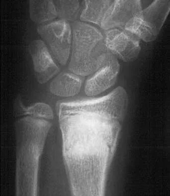
A 25-year-old man reports wrist pain following a motorcycle accident. Examination reveals minimal swelling, slightly limited active range of motion, and point tenderness in the snuff box region. AP and oblique radiographs are shown in Figures 40a and 40b. Management should consist of


Explanation

40b The radiographs reveal a scaphoid fracture with displacement and comminution and an unstable fracture pattern. Treatment should consist of open reduction and internal fixation. In displaced scaphoid fractures and fractures with unstable fracture patterns, closed reduction is ineffective and is likely to lead to nonunion. Limited intercarpal fusion and proximal row carpectomy are used to correct a variety of traumatic and posttraumatic problems of the wrist. Amadio PC, Taleisnik J: Fractures of the carpal bone, in Green DP, Hotchkiss RN, Pederson WC (eds): Green's Operative Hand Surgery, ed 4. Philadelphia, PA, 1999, pp 809-823. Rettig ME, Kozin SH, Cooney WP: Open reduction and internal fixation of acute displaced scaphoid waist fractures. J Hand Surg Am 2001;26:271-276. Cooney WP, Dobyns JH, Linscheid RL: Fractures of the scaphoid: A rational approach to management. Clin Orthop 1980;149:90-97.

Question 7

A 42-year-old woman reports that she has low back pain and had a transient loss of consciousness after falling off a horse. She denies having neck pain but notes that she was involved in a motor vehicle accident 2 years ago and had neck pain at that time. Examination reveals full range of motion of the neck and no localized tenderness. The neurologic examination is normal. A lateral radiograph of the cervical spine is obtained. Figures 41a and 41b show CT and MRI scans. What is the most likely diagnosis?


Explanation

41b The examination findings do not correlate with an acute injury (full range of cervical motion and the absence of pain). Radiographically, the fracture appears old based on the smooth contour of the fracture fragments and the absence of soft-tissue swelling. Flexion-extension radiographs can be obtained to determine potential instability; if present, stabilization and fusion should be considered. Schatzker J, Rorabeck CH, Waddell JP: Non-union of the odontoid process: An experimental investigation. Clin Orthop 1975;108:127-137.

Question 8

What neurologic structure is most at risk when performing intramedullary screw fixation of a fifth metatarsal base fracture?


Explanation

The sural nerve and its terminal branches course through the lateral hindfoot and midfoot area and are directly at risk in surgeries involving the peroneal tendon complex and the fifth metatarsal. The first branch of the lateral plantar nerve originates in the tarsal tunnel region and courses across the plantar heel area to innervate the abductor digiti minimi; it is not at direct risk with fifth metatarsal surgery. The saphenous, superficial peroneal, and deep peroneal nerves are not at risk anatomically with a lateral midfoot incision. Donley BG, McCollum MJ, Murphy GA, Richardson EG: Risk of sural nerve injury with intramedullary screw fixation of fifth metatarsal fractures: A cadaver study. Foot Ankle Int 1999;20:182-184.

Question 9

A 25-year-old man sustained an L1 compression fracture in a fall from his roof. He is neurologically intact and has no other injuries. Radiographs reveal a 25% loss of height anteriorly and 5 degrees of kyphosis at the fracture site. A CT scan reveals no compromise of the posterior column. Management should consist of


Explanation

The patient has a stable fracture that can be initially treated with bed rest, followed by bracing and quick mobilization. The outcome is good and surgery is not required. These fractures can be treated nonsurgically if there is less than 50% compression, 15 degrees of angulation, and intact posterior structures. Cantor JB, Lebwohl NH, Garvey T, Eismont FJ: Nonoperative management of stable thoracolumbar burst fractures with early ambulation and bracing. Spine 1993;18:971-976.

Question 10

A 35-year-old man sustained a 10% compression fracture of the C5 vertebra in a diving accident. Radiographs show good alignment, and examination reveals no neurologic compromise. An MRI scan reveals no significant soft-tissue disruption posteriorly. Management should consist of


Explanation

The patient has a stable flexion-compression injury of the cervical spine. The fracture occurs as a result of compression failure of the vertebral body. If the force continues, a tension failure of the posterior structures occurs, leading to potential dislocation. Immobilization in a rigid cervical orthosis will allow this fracture to heal. Clark CR (ed): The Cervical Spine, ed 3. Philadelphia, PA, Lippincott-Raven, 1998, pp 457-464.

Question 11

Figures 42a and 42b shows the radiographs of a 20-year-old man who sustained a hyperextension injury to his little finger. Multiple attempts at closed reduction have been unsuccessful. Management should now consist of


Explanation

42b The radiographs show a complex dislocation of the little finger metacarpophalangeal joint. This is characterized by obvious dislocation on the AP and lateral views and a type of bayonet apposition best visualized on the lateral view. Irreducibility of this injury is caused by displacement of the volar plate that has been traumatically avulsed from its origin on the metacarpal, with subsequent displacement into the metacarpophalangeal joint. This abnormal position of the volar plate causes irreducibility that can be corrected only by open reduction. This can be effected either by dorsal or palmar approaches. Stern PJ: Fractures of the metacarpals and phalanges, in Green DP, Hotchkiss RN, Pederson WC (eds): Green's Operative Hand Surgery, ed 4. Philadelphia, PA, 1999, pp 711-771. Becton JL, Christian JD Jr, Goodwin HN, Jackson JG III: A simplified technique for treating the complex dislocation of the index metacarpophalangeal joint. J Bone Joint Surg Am 1975;57:698-700.

Question 12

A 34-year-old man sustains an extra-articular fracture of the proximal phalanx of his right index finger in a fall. Examination reveals that the fracture is closed and oblique in orientation. Closed reduction and splinting fail to maintain the reduction. Management should now consist of


Explanation

The patient has an unstable oblique fracture of the proximal phalanx that is easily reducible but unstable; therefore, the treatment of choice is closed reduction and percutaneous pin fixation, followed by casting. Closed reduction and percutaneous pin fixation offers a better functional result than open reduction and plate fixation. Repeat closed reduction and buddy taping is inadequate because of the inherently unstable fracture pattern. Buddy taping will allow the dislocation to recur. The other options represent more aggressive surgical techniques than are necessary to treat this fracture. Stern PJ: Fractures of the metacarpals and phalanges, in Green DP, Hotchkiss RN, Pederson WC (eds): Green's Operative Hand Surgery, ed 4. Philadelphia, PA, 1999, pp 711-771.

Question 13

Figures 43a and 43b show the AP and lateral radiographs of the radius and ulna of a 9-year-old patient. The fracture is manipulated and placed in a long arm cast with the elbow flexed to 90 degrees and the forearm to neutral rotation. Figures 43c and 43d show the alignment of the fracture after the manipulation. What is the next most appropriate step in management?


Explanation

43b 43c 43d By placing the forearm at neutral rotation, as shown in Figures 43c and 43d, the distal fragment has become malrotated by 90 degrees. This is evident by the fact that the bicipital tuberosity is rotated 90 degrees to the radial styloid. Normally, it should be directly opposite (180 degrees) to the radial styloid. The correct alignment was present in the original radiographs shown in Figures 43a and 43b. Another clue to the malrotation in the postreduction radiographs is the difference in the diameters of the opposing radial shafts. To correct this rotational malalignment, the distal fragment needs to be remanipulated into supination so that it is correctly aligned with the supinated proximal radius. Evans EM: Fractures of the radius and ulna. J Bone Joint Surg Br 1951;33:548-561.

Question 14

Which of the following findings is an indication for adjunctive use of high-dose steroids?


Explanation

According to NASCIS III, the high-dose steroid protocol involves infusion of 30 mg/kg methylprednisolone followed by 5.4 mg/kg/h for 24 hours if the patient has sustained a spinal cord injury within the last 3 hours. The drip is continued for 48 hours if administration is started between 3 and 8 hours of the onset of neurologic deficit. No benefit has been conclusively demonstrated with steroids administered beginning 8 hours or longer after injury. Steroid use is not indicated for nerve root deficits, brachial plexus deficits, or gunshot wounds. Kellam JF, Fischer TJ, Tornetta P III, Bosse MJ, Harris MB (eds): Orthopaedic Knowledge Update: Trauma 2. Rosemont, IL, American Academy of Orthopaedic Surgeons, 2000, pp 319-328.

Question 15

A 22-year-old man sustained a stable pelvic fracture, bilateral femur fractures, and a left closed humeral shaft fracture in a motor vehicle accident. Examination 24 hours after injury reveals that the patient is confused and has shortness of breath. A clinical photograph of his conjunctiva is shown in Figure 44. He has a temperature of 101 degrees F (38.3 degrees C) and a pulse rate of 120/min. Laboratory studies show a hemoglobin level of 8 g/dL, a platelet count of 50,000/mm3, and a PaO2 of 57 mm Hg on 2L of oxygen. What is the most likely diagnosis?


Explanation

The major criteria for the diagnosis of fat embolism syndrome include hypoxemia (PaO2 of less than 60 mm Hg), central nervous system depression, and a petechial rash that is most often located in the axillae, conjunctivae, and palate. The rash is often transient. Tachycardia, pyrexia, anemia, thrombocytopenia, and the presence of fat in the urine are all considered minor criteria. To establish the diagnosis of fat embolism syndrome, one major and four minor signs should be present. Pulmonary embolism, which is the major differential diagnosis, usually is not associated with conjunctival petechia or thrombocytopenia.

Question 16

Figure 45 shows the current radiograph of an 11-year-old girl who sustained a simple nondisplaced fracture of the distal radius 4 weeks ago. Management at the time of injury consisted of application of a short arm cast but no manipulation. What is the major concern at this time?


Explanation

The fracture pattern represents a Peterson type I physeal injury, which is a comminuted metaphyseal fracture in which the fracture lines extend up to the physis. Because there is no displacement of the physis and the fracture lines do not cross the physis, there may be a tendency to dismiss this injury as a simple metaphyseal fracture with no significant sequelae. A small percentage of patients (3% in Peterson's series) experience growth arrest. In this patient, a disabling ulnar plus deformity, defined as increased ulnar length in relationship to the distal radius, developed. Peterson HA: Physeal fractures: Part 2. Two previously unclassified types. J Pediatr Orthop 1994;14:431-438.

Question 17

Which of the following is considered the best measure of the adequacy of resuscitation in the first 6 hours after injury?


Explanation

The end point of resuscitation is adequate tissue perfusion and oxygenation. Blood lactate is the end point of anaerobic metabolism. The level of blood lactate reflects global hypoperfusion and is directly proportional to oxygen debt. Two separate prospective studies have verified a significant difference in mortality when blood lactate was used as a measure of resuscitation when compared to traditional parameters (mean arterial pressure, urine output, central venous pressure, and heart rate). Base deficit is a direct measure of metabolic acidosis and an indirect measure of blood lactate levels. It correlates well with organ dysfunction, mortality, and adequacy of resuscitation. It is easy to measure, can be obtained rapidly, and is an excellent assessment of the adequacy of resuscitation. Porter JM, Ivatury RR: In search of the optimal end points of resuscitation in trauma patients: A review. J Trauma 1998;44:908-914.

Question 18

A 26-year-old man sustains a displaced bimalleolar fracture by sliding into second base while playing baseball. Following initial closed reduction and splinting of the fracture, moderate swelling is noted. What is the safest time to perform surgery?


Explanation

Following any closed fracture, the most important determinant for the timing of surgery is the condition of the soft tissues and especially the skin. The best determinant of appropriate soft-tissue condition is the presence of wrinkling of the skin (wrinkle sign) at the site of the incision. A wrinkle sign is present when all the interstitial edema has left the skin; this may take up to 14 to 21 days of elevation. Any abrasion must be epithelialized so that there are no bacteria left at the site. To date, no other method of soft-tissue viability measurement has been shown to be of any clinical benefit. Stover MD, Kellam JF: Articular fractures: Principles, in Ruedi TP, Murphy WM (eds): AO Principles of Fracture Management. Stuttgart, Thieme, 2000, pp 105-119. Hahn DM, Colton CL, Malleolar fractures, in Ruedi TP, Murphy WM (eds): AO Principles of Fracture Management. Stuttgart, Thieme, 2000, pp 559-581.

Question 19

A 28-year-old woman sustained an injury to her dominant right arm after falling off her porch. Examination reveals a deformity at the elbow. She is neurovascularly intact. Figures 46a and 46b show the radiographs obtained before closed reduction, and postreduction radiographs are shown in Figure 46c and 46d. What is the most likely early complication?


Explanation

00 46a 46b 46c 46d The patient has a complex fracture-dislocation of the elbow. The radial head is fractured, and there is a displaced coronoid fracture. These associated fractures indicate that the elbow is at high risk for recurrent instability after initial treatment. To prevent this complication, surgical treatment will most likely be required and will consist of some or all of the following: radial head open reduction and internal fixation or replacement, coronoid open reduction and internal fixation, medial and lateral ligament repairs, and even articulated external fixation. This patient was treated with open reduction and internal fixation of the radial head, and the elbow redislocated postoperatively. Ring D, Jupiter JB: Reconstruction of posttraumatic elbow instability. Clin Orthop 2000;370:44-56. O'Driscoll SW: Classification and evaluation of recurrent instability of the elbow. Clin Orthop 2000;370:34-43.

Question 20

What is the most likely long-term sequela of the injury shown in Figures 47a and 47b?


Explanation

47b The imaging studies show a comminuted lateral talar process fracture. This injury is often missed on plain radiographs; therefore, CT provides the best method of diagnostic evaluation. The most likely long-term sequela of this injury is subtalar joint arthrosis. Although this injury involves the fibular gutter region, progression to true ankle arthritis is unlikely. There does not appear to be any association with this injury and chronic mechanical instability of the ankle or disruption of the superior peroneal retinaculum and subsequent peroneal tendon instability. Entrapment of the flexor hallucis longus tendon may occur with fractures of the sustentaculum tali but not with injuries of the lateral talar process. Surgical management includes open reduction and internal fixation versus excision; the goal is preservation of the large articular surface fragments. In this patient, there is significant comminution and early fragment excision may be the best option for acute treatment. Tucker DJ, Feder JM, Boylan JP: Fractures of the lateral process of the talus: Two case reports and a comprehensive literature review. Foot Ankle Int 1998;19:641-646.

Question 21

A 16-year-old high school football player has diffuse pain with attempted digital flexion after injuring the ring finger of the dominant hand 1 week ago. Examination reveals that he is unable to flex the distal interphalangeal joint. Management should consist of


Explanation

The patient has an avulsion of the flexor digitorum profundus. Treatment should include surgical exploration and tendon reinsertion. This is not an avulsion of the flexor digitorum superficialis because the patient's deficiency is the inability to flex the distal interphalangeal joint, not the proximal interphalangeal joint. Surgical release of the anterior interosseous nerve is not indicated because the flexor digitorum profundus of the ring finger is innervated by the ulnar nerve. A median nerve contusion causes wrist pain and/or numbness and tingling in the median nerve distribution. Strickland JW: Flexor tendons: Acute injuries, in Green DP, Hotchkiss RN, Pederson WC (eds): Green's Operative Hand Surgery, ed 4. Philadelphia, PA, 1999, pp 1851-1897.

Question 22

A 25-year-old construction worker lands on his outstretched hand in a fall. The position of his wrist at the time of impact causes a force that leads to hyperextension, ulnar deviation, and intercarpal supination. Radiographs are shown in Figures 48a and 48b. What type of injury pattern is shown?


Explanation

48b The patient has a transscaphoid dorsal perilunate dislocation. The radiographs clearly define a dorsal dislocation of the capitolunate joint, and the scaphoid fracture component is easily visible on the AP view. A scaphoid fracture alone is an unlikely diagnosis because of the midcarpal dislocation component. The radiocarpal joint is not dislocated because the lunate is sitting in the lunate fossa of the radius. Isolated radiocarpal dislocations are not associated with a midcarpal disruption. While a midcarpal dislocation is a component of a dorsal perilunate dislocation, this diagnosis does not address the scaphoid fracture. A volar lunate dislocation is not seen because the lunate is reduced in the lunate fossa of the distal radius. Volar lunate dislocations are in the spectrum of injury of perilunate dislocations and fracture-dislocations; however, the radiographs show a transscaphoid dorsal perilunate dislocation. Mayfield JK, Johnson RP, Kilcoyne RK: Carpal dislocations: Pathomechanics and progressive perilunar instability. J Hand Surg Am 1980;5:226-241.

Question 23

A 25-year-old construction worker lands on his outstretched hand in a fall. The position of his wrist at the time of impact causes a force that leads to hyperextension, ulnar deviation, and intercarpal supination. Radiographs are shown in Figures 48a and 48b. Management should consist of


Explanation

48b Open reduction and internal fixation is the treatment of choice for accurate reduction of the disrupted intercarpal ligaments. In addition, the displaced scaphoid fracture will require open reduction and internal fixation and possible bone grafting. Closed reduction and long arm casting will not allow accurate reduction of the dislocated intracarpal intervals, and it is unlikely to allow accurate reduction of the scaphoid. The maneuver required to effect closed reduction of a displaced scaphoid fracture will most likely cause the scaphoid lunate interval to displace. Closed reduction with percutaneous pin fixation or with an external fixator is unable to effect anatomic reduction of the injury. Proximal row carpectomy is used as a salvage procedure for a variety of degenerative and posttraumatic problems of the wrist. Kozin SH: Perilunate injuries: Diagnosis and treatment. J Am Acad Orthop Surg 1998;6:114-120. Herzberg G, Comtet JJ, Linscheid RL, Amadio PC, Cooney WP, Stalder J: Perilunate dislocations and fracture-dislocations: A multicenter study. J Hand Surg Am 1993;18:768-779.

Question 24

A 17-year-old boy who fell on a pitchfork in a barn 1 day ago now has a painful, swollen forearm. Examination reveals erythema, exquisite tenderness, and crepitus to palpation of the forearm. He has a pulse rate of 110/min and a blood pressure of 80/60 mm Hg. Radiographs show subcutaneous air and no fractures. Gram stain of wound drainage reveals a gram-positive bacillus. The next most appropriate step in management should consist of


Explanation

The successful treatment of necrotizing soft-tissue infections such as clostridial myonecrosis depends on prompt recognition and aggressive surgical debridement of all involved muscle, fascia, and soft tissue, resecting to a clearly normal healthy, viable margin. The effective antibiotic regimen for clostridial infection is high-dose penicillin; however, necrotizing infections are frequently polymicrobial so initially broad-spectrum antibiotics are indicated. Hyperbaric oxygen therapy may be used as an adjunct to surgical treatment but is insufficient as a primary therapy. Prolonged application of tourniquets and wound closure should be avoided. Pellegrini VD, Evarts CM: Complications, in Rockwood CA Jr, Green DP (eds): Fractures in Adults, ed 3. Philadelphia, PA, JB Lippincott, 1991, pp 365-370. Gerding DN, Peterson LR: Infections caused by anaerobic bacteria, in Shulman ST, Phair JP, Peterson LR, Warren JR (eds): Infectious Diseases, ed 5. Philadelphia, PA, WB Saunders, 1997, pp 416-417.

Question 25

In the management of an open tibia fracture, what factor is considered most important in preventing deep infection?


Explanation

The most important aspect of management of any open fracture, and in particular the tibia, is the degree and the completeness of the debridement of the soft tissue and most importantly, the muscle. The ultimate function is determined by the amount of muscle left, as well as the ability to heal. The amount of necrotic muscle left in the wound also determines the predisposition to infection. The method of fixation, the size of the wound, and the amount of contamination are controlled by the surgeon or the injury and have little to do with the long-term outcome. Initial wound cultures have little predictive value. Clifford P: Open fractures, in Ruedi TP, Murphy WM (eds): AO Principles of Fracture Management. Stuttgart, Thieme, 2000, pp 617-638.

Question 26

A 35-year-old man presents with a hemodynamically unstable pelvic ring injury following a motorcycle collision. A pelvic binder is applied. To optimally reduce the pelvic volume in an anteroposterior compression (APC) injury, over which anatomic structure should the pelvic binder be centered?


Explanation

Pelvic binders should be centered over the greater trochanters to effectively close the pelvic ring and reduce pelvic volume in APC-type injuries. Placement over the iliac crests is less effective and may cause paradoxical opening of the pelvic floor.

Question 27

Following a high-energy closed tibial shaft fracture, a 28-year-old patient develops severe pain out of proportion to the injury. On examination, he has pain with passive extension of the great toe. Which muscle compartment of the leg is most likely affected based on this specific clinical finding?


Explanation

Pain with passive extension of the great toe stretches the flexor hallucis longus, which is located in the deep posterior compartment. The anterior compartment would present with pain on passive plantarflexion of the toes.

Question 28

A 25-year-old man sustains a completely displaced, vertically oriented (Pauwels type III) femoral neck fracture. Which of the following fixation constructs provides the most biomechanically stable construct to resist shear forces in this specific fracture pattern?


Explanation

For high-shear vertically oriented (Pauwels type III) femoral neck fractures in young adults, a fixed-angle device such as a dynamic hip screw (with a derotation screw) provides superior biomechanical stability against vertical shear forces compared to multiple cancellous screws.

Question 29

A 45-year-old woman sustains a complex bicondylar tibial plateau fracture with a large, displaced posteromedial fragment. Which surgical approach provides the most direct access for buttress plating of the posteromedial fragment?


Explanation

A posteromedial approach (often via a medial gastrocnemius interval) provides direct access to the posteromedial tibial plateau, allowing for optimal placement of an anti-glide or buttress plate. This is essential to prevent varus collapse.

Question 30

A 50-year-old man sustains a high-energy closed tibial pilon fracture with severe soft tissue swelling and fracture blisters. What is the optimal initial management strategy?


Explanation

The standard of care for high-energy pilon fractures with severe soft tissue compromise is damage control orthopedics using a spanning external fixator. Definitive internal fixation is delayed until soft tissue swelling resolves, typically 10 to 21 days later.

Question 31

A 32-year-old man sustains a Gustilo-Anderson IIIB open tibial shaft fracture. After adequate debridement and skeletal stabilization, what is the optimal timeframe for soft tissue coverage to minimize infection rates?


Explanation

Current literature supports early soft tissue coverage (flap reconstruction) for Gustilo IIIB open tibia fractures, ideally within 3 to 7 days, to significantly decrease the risk of deep infection and flap failure.

Question 32

During open reduction and internal fixation of a transverse posterior wall acetabular fracture utilizing the Kocher-Langenbeck approach, the knee is maintained in flexion and the hip in extension. This positioning is primarily utilized to protect which of the following structures?


Explanation

Maintaining the hip in extension and the knee in flexion during the Kocher-Langenbeck approach relaxes the sciatic nerve. This minimizes tension and reduces the risk of iatrogenic sciatic nerve palsy during retraction.

Question 33

A 29-year-old motorcyclist sustains a severely comminuted distal femur fracture. Preoperative CT imaging reveals an independent coronal plane fracture of the medial condyle. Which of the following justifies the use of a supplemental medial surgical approach?


Explanation

A medial Hoffa fracture (coronal shear fracture of the medial condyle) often requires a separate medial arthrotomy or approach to obtain anatomic reduction and place lag screws directly, as it is exceedingly difficult to reduce and fix from a standard lateral approach.

Question 34

Intraoperatively, following fixation of a Weber C ankle fracture, the cotton test demonstrates widening of the syndesmosis. Which of the following radiographic parameters best assesses the adequacy of syndesmotic reduction on a standard AP or mortise radiograph?


Explanation

Tibiofibular overlap is a key radiographic parameter to assess syndesmotic integrity. On a proper AP radiograph, it should be greater than 10 mm, and on a mortise view, greater than 1 mm.

Question 35

Six weeks following open reduction and internal fixation of a displaced talar neck fracture, an AP radiograph of the ankle demonstrates a subchondral radiolucent line in the talar dome. What does this radiographic finding indicate?


Explanation

A subchondral radiolucent line in the talar dome (Hawkins sign) visible at 6 to 8 weeks post-injury indicates subchondral osteopenia due to an intact vascular supply. It is a reliable predictor that avascular necrosis will not occur.

Question 36

The Sanders classification for intra-articular calcaneus fractures is based on the number and location of fracture lines in which specific anatomic area as seen on coronal CT imaging?


Explanation

The Sanders classification is based on coronal plane CT scans showing the number and location of fracture lines through the posterior articular facet of the calcaneus, which guides operative treatment and prognosis.

Question 37

In a purely ligamentous Lisfranc injury with instability demonstrated on weight-bearing radiographs, what is the most appropriate surgical treatment to maximize long-term functional outcomes?


Explanation

For purely ligamentous Lisfranc injuries, primary arthrodesis of the medial column (1st, 2nd, and 3rd tarsometatarsal joints) has been shown to have better functional outcomes and lower reoperation rates compared to open reduction and internal fixation.

Question 38

Which of the following is considered an absolute indication for operative fixation of a midshaft clavicle fracture?


Explanation

Absolute indications for operative fixation of a clavicle fracture include open fractures, associated neurovascular compromise, and severe skin tenting threatening skin integrity. Displacement alone is a relative indication.

Question 39

A 72-year-old woman sustains a displaced 4-part proximal humerus fracture. Examination reveals loss of sensation over the lateral deltoid. Which nerve is most likely injured?


Explanation

The axillary nerve provides sensation to the skin overlying the lateral deltoid (Sergeant's patch) and motor innervation to the deltoid and teres minor. It is the most commonly injured nerve in proximal humerus fractures and shoulder dislocations.

Question 40

A 30-year-old male sustains a high-energy motor vehicle collision resulting in a Hawkins type III fracture of the talar neck with an extruded talar body. What is the approximate risk of developing avascular necrosis (AVN) of the talar body?


Explanation

A Hawkins type III fracture involves the talar neck with subluxation or dislocation of both the subtalar and tibiotalar joints. The disruption of the major blood supplies results in an AVN rate historically reported between 80% and 100%.

Question 41

A 28-year-old polytrauma patient with a closed tibia fracture is intubated in the intensive care unit. His diastolic blood pressure is 65 mmHg. Intracompartmental pressure monitoring is placed due to swelling. What is the minimum compartment pressure that would mandate a 4-compartment fasciotomy based on the delta P concept?


Explanation

The delta P is defined as the diastolic blood pressure minus the intracompartmental pressure. A delta P of less than 30 mmHg is the accepted threshold for diagnosing acute compartment syndrome and indicates the need for fasciotomy. With a diastolic BP of 65 mmHg, an absolute pressure of 36 mmHg results in a delta P of 29 mmHg.

Question 42

A 32-year-old man sustains a closed tibial shaft fracture. Two hours later, he complains of severe pain out of proportion to the injury. Which of the following is the most reliable criterion for diagnosing acute compartment syndrome in a patient who is awake and alert?


Explanation

The delta P (diastolic blood pressure minus compartment pressure) < 30 mm Hg is the most objective and reliable criterion for diagnosing compartment syndrome. Pain with passive stretch is an early clinical sign but remains highly subjective.

Question 43

A 45-year-old woman is brought to the emergency department after a motor vehicle collision. Her blood pressure is 80/50 mm Hg. Pelvic radiographs show a widely displaced anteroposterior compression (APC) type III pelvic ring injury. After initial fluid resuscitation, a pelvic binder is applied. What is the optimal anatomic landmark for the proper placement of the pelvic binder?


Explanation

A pelvic binder should be placed at the level of the greater trochanters to effectively reduce pelvic volume and stabilize the fracture. Placement over the iliac crests is incorrect and can exacerbate the deformity or fail to reduce the volume.

Question 44

A 28-year-old man sustains a subtrochanteric femur fracture. Preoperative radiographs demonstrate the classic flexion, abduction, and external rotation deformity of the proximal fragment. Which of the following muscles is primarily responsible for the external rotation deformity of the proximal fragment?


Explanation

In a subtrochanteric fracture, the proximal fragment is flexed by the iliopsoas, abducted by the gluteus medius and minimus, and externally rotated by the short external rotators.

Question 45

A 34-year-old man sustains a Hawkins type II talar neck fracture and undergoes open reduction and internal fixation. At his 8-week follow-up, an anteroposterior radiograph of the ankle reveals subchondral radiolucency in the talar dome. What does this radiographic finding indicate?


Explanation

The Hawkins sign is subchondral radiolucency in the talar dome observed 6 to 8 weeks after injury, indicating subchondral bone resorption secondary to an intact vascular supply, thereby ruling out avascular necrosis.

Question 46

During an anterior intrapelvic (modified Stoppa) approach for an acetabular fracture, significant hemorrhage is encountered over the superior pubic ramus. Which of the following vascular structures is most likely injured?


Explanation

The corona mortis is a vascular anastomosis between the obturator and external iliac (or inferior epigastric) systems, located over the superior pubic ramus. It is at high risk of injury during anterior approaches to the pelvis and acetabulum.

Question 47

A 38-year-old woman is involved in a high-speed motor vehicle collision and sustains a distal femur fracture. CT scan reveals a coronal plane fracture of the lateral femoral condyle. What is the recommended surgical treatment for this specific fragment?


Explanation

A coronal plane fracture of the femoral condyle is known as a Hoffa fracture. It requires anatomic reduction and internal fixation, typically using anterior-to-posterior or posterior-to-anterior directed lag screws to compress the fracture.

Question 48

A 24-year-old man sustains a Denis Zone 3 sacral fracture following a fall. Which of the following neurologic deficits is most commonly associated with this specific injury zone?


Explanation

Denis Zone 3 sacral fractures involve the central sacral canal and are highly associated with cauda equina injury, leading to bowel, bladder, and sexual dysfunction.

Question 49

A 21-year-old motorcyclist is thrown from his bike and presents with a massively swollen shoulder and a pulseless upper extremity. Radiographs show lateral displacement of the scapula and a widely displaced clavicle fracture. Which of the following is the most likely neurologic injury associated with this condition?


Explanation

Scapulothoracic dissociation is characterized by lateral displacement of the scapula, clavicle fracture, and severe neurovascular injury. It is frequently associated with subclavian artery disruption and complete brachial plexus avulsion.

Question 50

A 40-year-old roofer falls and sustains a displaced intra-articular calcaneus fracture. He undergoes open reduction and internal fixation via an extensile lateral approach. Which of the following is the most common complication associated with this specific surgical approach?


Explanation

The extensile lateral approach to the calcaneus has a high rate of wound complications, particularly wound edge necrosis, due to the tenuous vascular supply of the L-shaped flap. Sural nerve injury is also possible but less frequent than wound healing issues.

Question 51

A 55-year-old man sustains a tibial plateau fracture. Radiographs and CT demonstrate a bicondylar tibial plateau fracture with complete dissociation of the articular surface from the tibial diaphysis. According to the Schatzker classification, what is the correct grade?


Explanation

A Schatzker Type VI fracture is defined by the complete dissociation of the tibial plateau from the underlying tibial diaphysis. Type V involves both condyles but retains continuity between the metaphysis and diaphysis.

Question 52

A 30-year-old man sustains a Gustilo-Anderson Type IIIB open tibia fracture. Following serial debridement, a 6 cm soft tissue defect over the middle third of the tibia exposes bare bone. What is the most appropriate option for soft tissue coverage?


Explanation

For soft tissue defects over the middle third of the tibia exposing bare bone, a soleus rotational flap is the workhorse local flap. The medial gastrocnemius is used for the proximal third, and free flaps are typically required for the distal third.

Question 53

A 22-year-old football player sustains a hyperplantarflexion injury to his foot. On an anteroposterior radiograph of the foot, a small bony avulsion is seen in the first intermetatarsal space. This 'fleck sign' represents an avulsion of the Lisfranc ligament from which of the following structures?


Explanation

The Lisfranc ligament connects the medial cuneiform to the base of the second metatarsal. The 'fleck sign' represents a bony avulsion of this ligament, most commonly from the base of the second metatarsal.

Question 54

A 65-year-old woman is managed conservatively in a cast for a non-displaced distal radius fracture. Eight weeks later, she reports the sudden inability to actively extend her thumb interphalangeal joint. Rupture of which of the following tendons is the most likely cause?


Explanation

Extensor pollicis longus (EPL) tendon rupture is a known complication of non-displaced distal radius fractures due to ischemia or mechanical attrition at the Lister tubercle. It presents with an inability to actively extend the thumb interphalangeal joint.

Question 55

In the pre-hospital and emergency department management of a hemodynamically unstable patient with a suspected pelvic ring injury, what is the proper anatomical placement of a circumferential pelvic binder?


Explanation

A pelvic binder should be centered directly over the greater trochanters. Placement over the iliac crests is a common error that can paradoxically widen the true pelvis and fail to adequately reduce pelvic volume.

Question 56

A 32-year-old man sustains a closed tibial shaft fracture. Two hours later, he complains of severe leg pain out of proportion to the injury. His diastolic blood pressure is 70 mmHg, and his anterior compartment pressure measures 45 mmHg. What is the most appropriate next step in management?


Explanation

The patient has a delta pressure (diastolic BP minus compartment pressure) of 25 mmHg. A delta pressure of less than 30 mmHg is an absolute indication for emergent four-compartment fasciotomy.

Question 57

A 25-year-old man sustains a highly vertical (Pauwels Type III) femoral neck fracture. To maximize biomechanical stability and resist vertical shear forces, which fixation construct is most appropriate?


Explanation

For vertically oriented (Pauwels III) fractures in young adults, a sliding hip screw with a derotational screw provides superior biomechanical stability against shear forces compared to multiple cannulated screws.

Question 58

A 28-year-old presents after a high-energy knee dislocation, which is immediately reduced in the emergency department. The patient has palpable pedal pulses. The Ankle-Brachial Index (ABI) is measured at 0.8. What is the most appropriate next step in management?


Explanation

An Ankle-Brachial Index (ABI) of less than 0.9 after a knee dislocation is highly suspicious for a popliteal artery injury and warrants immediate further imaging, typically with a CT angiogram.

Question 59

A 40-year-old sustains a Gustilo-Anderson Type IIIB open tibia fracture. Following thorough debridement and skeletal stabilization, what is the optimal timing for soft tissue coverage to minimize the risk of deep infection?


Explanation

Soft tissue coverage (flap) of Gustilo Type IIIB fractures within 72 hours is strongly associated with significantly lower infection rates and improved functional outcomes compared to delayed coverage.

Question 60

A 22-year-old motorcycle crash victim sustains a scapulothoracic dissociation. Which of the following associated injuries is the strongest clinical predictor for the necessity of an early forequarter amputation?


Explanation

Complete brachial plexus avulsion results in a flail, insensate upper extremity. This catastrophic neurologic injury is the strongest predictor for early forequarter amputation in patients with scapulothoracic dissociation.

Question 61

A trauma patient sustains a U-type sacral fracture (spinopelvic dissociation). Which neurological complication is most specifically associated with this classic fracture pattern?


Explanation

U-type sacral fractures typically involve the central sacral canal, frequently compressing the sacral nerve roots. This results in cauda equina syndrome, characterized by bowel, bladder, and sexual dysfunction.

Question 62

A 35-year-old male sustains a purely ligamentous Lisfranc injury with dynamic instability. Based on recent literature, which surgical intervention yields the best long-term functional outcomes and lowest revision rates?


Explanation

Current evidence demonstrates that purely ligamentous Lisfranc injuries treated with primary arthrodesis of the first three TMT joints have superior functional outcomes and lower revision rates compared to ORIF.

Question 63

Three months following volar plate fixation of a distal radius fracture, a patient experiences a spontaneous rupture of the flexor pollicis longus (FPL) tendon. What is the most likely technical error leading to this complication?


Explanation

Volar plates placed too distally, beyond the watershed line of the distal radius, can impinge on the flexor tendons. This friction leads to tenosynovitis and subsequent rupture, most commonly affecting the FPL.

Question 64

A 29-year-old sustains a Hawkins Type III talar neck fracture. Disruption of which of the following arteries is the primary cause of the high rate of avascular necrosis seen in this injury?


Explanation

The artery of the tarsal canal, a branch of the posterior tibial artery, provides the predominant blood supply to the talar body. Its disruption in displaced talar neck fractures is the primary driver of avascular necrosis.

Question 65

A 45-year-old patient presents with a Bado Type I Monteggia fracture. Intraoperatively, after achieving rigid plate fixation of the ulna, the radial head remains anteriorly dislocated. What is the most appropriate next step?


Explanation

Persistent radial head dislocation after ulnar fixation in a Monteggia fracture is almost always due to malreduction (residual shortening or angulation) of the ulna. The ulnar fixation must be revised to restore appropriate length and bow.

Question 66

A 75-year-old woman sustains a periprosthetic femur fracture around a cemented total hip arthroplasty. Radiographs reveal the fracture is around the stem, the stem is loose, but the proximal femoral bone stock is adequate. What is the Vancouver classification and optimal treatment?


Explanation

A fracture around a loose stem with adequate proximal bone stock is a Vancouver B2 injury. The standard of care is revision arthroplasty using a long cementless stem that bypasses the fracture.

Question 67

A 30-year-old unrestrained driver sustains a traumatic spondylolisthesis of C2 with severe angulation and minimal translation (Levine-Edwards Type IIa). What is the most appropriate initial management?


Explanation

Type IIa Hangman's fractures involve a flexion-distraction mechanism. Axial traction is strictly contraindicated as it worsens the deformity; treatment requires gentle extension and compression, typically in a halo vest.

Question 68

A 42-year-old sustains a posterior wall acetabular fracture. During a dynamic fluoroscopic examination under anesthesia (EUA), the hip subluxates posteriorly. Involvement of what minimum percentage of the posterior wall articular surface typically correlates with this instability?


Explanation

Posterior wall fractures involving greater than 20-25% of the articular surface generally compromise the structural integrity of the joint, leading to hip instability and serving as an indication for surgical fixation.

Question 69

A polytrauma patient is diagnosed with a "floating knee" (ipsilateral femoral and tibial shaft fractures). Due to the nature of this specific combination of injuries, the patient is at significantly increased risk for which of the following early systemic complications?


Explanation

Floating knee injuries expose the marrow of two major long bones simultaneously, drastically increasing the load of marrow fat into the venous system and making Fat Embolism Syndrome highly probable.

Question 70

A 24-year-old active male has a completely displaced, shortened midshaft clavicle fracture. If he chooses to proceed with nonoperative management, he should be counseled that his risk of nonunion is approximately:


Explanation

While historically thought to heal reliably, modern prospective studies show that completely displaced and shortened midshaft clavicle fractures have a nonunion rate of approximately 15% when treated nonoperatively.

Question 71

A 22-year-old sustains a low-velocity civilian gunshot wound to the midshaft femur, resulting in a comminuted fracture. There is no neurovascular injury, and the entry/exit wounds are small and clean. What is the optimal surgical management?


Explanation

Low-velocity civilian gunshot fractures of the femur without severe soft-tissue contamination can be safely treated with superficial wound care, IV antibiotics, and early intramedullary nailing without formal open debridement of the fracture site.

Question 72

A 45-year-old smoker presents with a symptomatic aseptic nonunion of a tibial shaft fracture 9 months after initial intramedullary nailing. The implant is intact. What is the most appropriate next step in surgical management?


Explanation

Exchange nailing with a larger reamed nail provides improved mechanical stability and biological stimulation (autograft generated from reamings), making it the gold standard for aseptic tibial nonunions.

Question 73

A 50-year-old patient presents with a massive, fluctuant Morel-Lavallée lesion over the greater trochanter that has been present for 4 weeks following a blunt trauma. What is the most definitive management?


Explanation

Chronic Morel-Lavallée lesions form an epithelialized pseudocapsule that prevents resorption. Simple aspiration has a very high recurrence rate; therefore, definitive treatment requires open excision or aggressive debridement with sclerodesis.

Question 74

A 38-year-old falls from a significant height, sustaining a high-energy, severely displaced tibial pilon fracture with massive soft tissue swelling and fracture blisters. What is the most appropriate initial treatment approach?


Explanation

High-energy pilon fractures with massive soft tissue compromise require staged management. Initial spanning external fixation allows the vulnerable soft tissues to recover prior to delayed definitive internal fixation.

Question 75

During the ilioinguinal approach for a transverse acetabular fracture, massive hemorrhage is encountered while dissecting near the superior pubic ramus. This bleeding is most likely due to injury of an anastomosis between which two vascular structures?


Explanation

The corona mortis is an anatomical vascular anastomosis between the obturator and external iliac (or inferior epigastric) systems located over the superior pubic ramus. It is highly susceptible to injury during the ilioinguinal approach to the pelvis.

Question 76

A 28-year-old woman is evaluated 8 weeks following closed reduction and percutaneous pinning of a Hawkins type II talar neck fracture. An AP radiograph of the ankle demonstrates a subchondral radiolucent band in the talar dome. What does this radiographic finding indicate?


Explanation

Hawkins sign is a subchondral radiolucent band seen in the talar dome on an AP or mortise radiograph 6 to 8 weeks post-injury. It indicates subchondral atrophy from disuse, which confirms that the vascular supply to the talar body remains intact.

Question 77

In the evaluation of a displaced 4-part proximal humerus fracture, which of the following anatomic variables is the strongest predictor of subsequent avascular necrosis of the humeral head?


Explanation

A metaphyseal head extension (calcar length) of less than 8 mm, disruption of the medial hinge, and a basicervical fracture pattern are the most reliable predictors of ischemia and subsequent avascular necrosis in proximal humerus fractures.

Question 78

A 30-year-old man sustains a closed distal third spiral humerus fracture. Examination reveals a complete radial nerve palsy present immediately after the injury. After closed reduction and application of a coaptation splint, the fracture is acceptably aligned but the palsy persists. What is the most appropriate initial management of the nerve injury?


Explanation

Primary radial nerve palsies associated with closed humeral shaft fractures should be observed, as 70-90% will spontaneously resolve. Surgical exploration is reserved for open fractures, penetrating injuries, or failure to recover clinically or electromyographically by 3 to 4 months.

Question 79

A 25-year-old man sustains a subtrochanteric femur fracture. During closed intramedullary nailing, the proximal fragment is typically difficult to reduce due to the deforming forces of local musculature. The proximal fragment is classically pulled into which of the following positions?


Explanation

The proximal fragment in a subtrochanteric fracture is deformed into flexion by the iliopsoas, abduction by the gluteus medius and minimus, and external rotation by the short external rotators. Proper reduction requires neutralizing these forces before nailing.

Question 80

A 22-year-old man sustains a vertical, shear-type (Pauwels type III) femoral neck fracture. Which of the following fixation constructs provides the most biomechanically stable fixation for this specific fracture pattern?


Explanation

Pauwels type III fractures are high-angle, vertical fractures with significant shear forces. A sliding hip screw (fixed-angle device) combined with an anti-rotation cancellous screw offers superior biomechanical stability compared to multiple cancellous screws for this pattern.

Question 81

A 28-year-old man sustains a closed femoral shaft fracture and bilateral pulmonary contusions in a motor vehicle collision. On arrival, his lactate is 4.5 mmol/L, base deficit is 8 mEq/L, and pH is 7.21. After initial fluid resuscitation, his lactate improves to 3.0 mmol/L and base deficit to 6 mEq/L. What is the most appropriate management of his femoral shaft fracture?


Explanation

This polytrauma patient remains 'borderline' or 'unstable' based on elevated lactate (>2.5 mmol/L) and base deficit (>5 mEq/L) despite resuscitation. Damage control orthopedics (external fixation) is favored to minimize the 'second hit' inflammatory response associated with reamed intramedullary nailing in the setting of severe chest trauma.

Question 82

A 65-year-old woman undergoes evaluation for a 4-part proximal humerus fracture. According to Hertel's criteria, which of the following radiographic findings is the most reliable predictor for the development of humeral head osteonecrosis?


Explanation

Hertel described specific criteria for predicting ischemia and subsequent avascular necrosis in proximal humerus fractures. The most reliable predictors include a metaphyseal head extension (calcar segment) of less than 8 mm and disruption of the medial hinge.

Question 83

A 34-year-old man presents with a purely ligamentous Lisfranc injury of the midfoot following a fall from a horse. The injury involves the 1st, 2nd, and 3rd tarsometatarsal joints. Comparing primary arthrodesis to open reduction and internal fixation (ORIF), primary arthrodesis in this specific injury pattern is associated with:


Explanation

Studies comparing ORIF to primary arthrodesis for purely ligamentous Lisfranc injuries demonstrate that primary arthrodesis yields similar or slightly superior functional outcomes. Arthrodesis significantly lowers the rates of subsequent surgeries, specifically hardware removal and secondary salvage fusions.

Question 84

An extensile lateral approach is planned for open reduction and internal fixation of a displaced intra-articular calcaneus fracture. To prevent wound slough and edge necrosis, the surgeon must be aware that the full-thickness fasciocutaneous flap's viability is primarily dependent on which of the following arteries?


Explanation

The extensile lateral approach creates a 'no-touch' full-thickness fasciocutaneous flap containing the sural nerve and peroneal tendons. Its vascular supply is primarily based on the lateral calcaneal artery, a terminal branch of the peroneal artery.

Question 85

A 42-year-old man sustains a high-energy Schatzker VI tibial plateau fracture. During the initial evaluation, he has a tense, swollen calf and decreased sensation in the first dorsal webspace. Passive plantarflexion of the hallux elicits severe pain. Which compartment of the lower leg is most likely experiencing critically elevated pressures?


Explanation

Decreased sensation in the first dorsal webspace (deep peroneal nerve distribution) and severe pain with passive toe plantarflexion (stretching the extensor hallucis longus) are classic signs of anterior compartment syndrome. The deep peroneal nerve and anterior tibial artery reside within this compartment.

Question 86

A 25-year-old man sustains a displaced midshaft clavicle fracture after being thrown over the handlebars of his bicycle. Which of the following is considered an absolute indication for operative fixation over nonoperative management?


Explanation

Absolute indications for operative fixation of a clavicle fracture include open fractures, associated vascular injuries requiring repair, and progressive neurologic deficits. Severe shortening (>2 cm) and marked displacement are generally considered relative indications depending on patient activity level.

None

Clinic OS
Medically Verified Content by
Prof. Clinic OS
Consultant Orthopedic & Spine Surgeon
Chapter Index