العربية
Part of the Master Guide

Orthopedic Surgery Board Prep: Set 403 (100 MCQs on Fractures & Casting)

Orthopedic Trauma 2026 MCQs: Board Review Questions & Answers (Part 3)

23 Apr 2026 86 min read 85 Views
Figure for Trauma 2006 MCQs - Part 3 - Question 51

Key Takeaway

Learn more about Orthopedic Trauma 2026 MCQs: Board Review Questions & Answers (Part 3) and how to manage it. Top-rated Orthopedic Trauma 2026 MCQs bank. Practice with clinical case questions, orthopedic surgery board review, and evidence-based answers updated for 2026.

Orthopedic Trauma 2026 MCQs: Board Review Questions & Answers (Part 3)

Comprehensive 100-Question Exam


00:00

Start Quiz

Question 1

A 54-year-old man sustained a small superficial abrasion over the left acromioclavicular joint after falling from his bicycle. Examination reveals no other physical findings. Radiographs show a displaced fracture of the lateral end of the clavicle distal to a line drawn vertically to the coracoid process. Management should consist of


Explanation

Displaced clavicular fractures lateral to the coracoid process (Neer type II and III) are best managed nonsurgically with sling immobilization and physical therapy, starting with pendulum exercises and progressing to active-assisted exercises when comfortable. Supervised therapy should be performed for 3 months or until full painless motion is achieved. In one study by Robinson and Cairns, this form of treatment provided patients with a 86% chance of avoiding a secondary reconstructive procedure. Robinson CM, Cairns DA: Primary nonoperative treatment of displaced lateral fractures of the clavicle. J Bone Joint Surg Am 2004;86:778-782.

Question 2

A 47-year-old man sustained a degloving injury over the pretibial surface and anterior ankle region in a motor vehicle accident. After debridement and irrigation, there is inadequate tissue for closure of the exposed anterior tibial tendon and tibia. Prior to definitive soft-tissue coverage, management should consist of


Explanation

With soft-tissue loss, local or free flap coverage may be necessary to treat exposed tendon and bone. However, a vacuum-assisted closure device is a good temporizing dressing. It prevents external contamination, reduces edema around the wound, increases oxygen tension in the wound, and promotes the formation of granulation tissue. The use of this negative pressure device has been described in both acute traumatic and in chronic wound scenarios. If sufficient granulation tissue forms, closure may be by split graft, avoiding a more complex coverage procedure. Immediate skin grafting over the exposed anterior tibial tendon and tibia would have a low likelihood of success. Dressing changes with sulfasalazine may be beneficial in a burn wound to assist with removal of skin slough; however, in a granulating wound, the material may be toxic to early epithelialization. Xenograft is a foreign body and should not be applied to an acute contaminated open wound. Historically, a cross-leg flap was a treatment alternative for lower extremity soft-tissue loss; however, its current applications are extremely limited. Webb LX: New techniques in wound management: Vacuum assisted wound closure. J Am Acad Orthop Surg 2002;10:303-311.

Question 3

The humeral nonunion shown in Figure 27 is most likely to unite when using what method of treatment?


Explanation

The radiograph shows an atrophic nonunion of the humeral shaft. The management of humeral nonunions has been studied with compression plates and bone graft, as well as intramedullary nailing and bone graft. Compression plating with bone graft results in the highest rate of union. Compression plating by itself is not adequate, given the bone loss and lack of callous in this nonunion. Pulsed electromagnetic fields is a viable option for hypertrophic nonunions where there is inherent stability. Intramedullary nailing does not provide as much compression and stability as that achieved with compression plating. Pugh DM, McKee MD: Advances in the management of humeral nonunion. J Am Acad Orthop Surg 2003;11:48-59.

Question 4

An adult with a distal humeral fracture underwent open reduction and internal fixation. What is the most common postoperative complication?


Explanation

Most patients lose elbow range of motion after open reduction and internal fixation of a distal humeral fracture. Ulnar nerve dysfunction, nonunion, and infection all occur less commonly. Webb LX: Distal humerus fractures in adults. J Am Acad Orthop Surg 1996;4:336-344.

Question 5

The radiographs and CT scan seen in Figures 28a through 28d reveal what type of acetabular fracture pattern?


Explanation

28b 28c 28d The AP, obturator oblique, and iliac oblique views of the pelvis reveal a fracture that disrupts the iliopectineal and ilioischial lines, indicating a fracture that involves both anterior and posterior columns. However, it does not have the other features of anterior or posterior column fracture patterns. A displaced posterior wall fracture is also present, best seen on the obturator oblique view. The anterior to posterior directed fracture line on the CT scan indicates a transverse fracture; therefore, the patient has a transverse with posterior wall fracture pattern. A T-type fracture would be similar but would have a break into the obturator ring. Tile M: Describing the injury: Classification of acetabular fractures, in Tile M, Helfet DL, Kellam JF (eds): Fractures of the Pelvis and Acetabulum, ed 3. Philadelphia, PA, Lippincott Williams & Wilkins, 2003, pp 427-475.

Question 6

A 26-year-old man sustained an isolated injury to his left hip joint in a motor vehicle accident. Closed reduction was performed, and the postreduction radiograph is shown in Figure 29. Management should now consist of


Explanation

The patient has a posterior fracture-dislocation of the hip and following reduction, an incarcerated fragment of bone resulted in an incongruent reduction. Whereas expedient removal of the fragment is required to limit articular cartilage damage, this situation is not an emergency and the procedure may be performed when the appropriate surgical team is available and the patient is stabilized. Skeletal traction through either the femur or tibia may relieve some pressure on the joint and prevent articular damage. Nonsurgical care for incarcerated fragments is contraindicated. Tile M, Olson SA: Decision making: Non operative and operative indications for acetabular fractures, in Tile M, Helfet DL, Kellam JF (eds): Fractures of the Pelvis and Acetabulum. Philadelphia, PA, Lippincott Williams and Wilkins, 2003, pp 496-532.

Question 7

A 35-year-old man is brought to the emergency department following a motorcycle accident. He is breathing spontaneously and has a systolic blood pressure of 80 mm Hg, a pulse rate of 120/min, and a temperature of 98.6 degrees F (37 degrees C). Examination suggests an unstable pelvic fracture; AP radiographs confirm an open book injury with vertical displacement on the left side. Ultrasound evaluation of the abdomen is negative. Despite administration of 4 L of normal saline solution, he still has a systolic pressure of 90 mm Hg and a pulse rate of 110. Urine output has been about 20 mL since arrival 35 minutes ago. What is the next best course of action?


Explanation

The patient is at risk for a pelvic vascular injury and major hemorrhage. This type of complication of pelvic trauma is highest in motorcyclists. Once it is recognized that the pelvic ring has opened, it is important to close that ring to tamponade any venous bleeding with a pelvic binder and to add a skeletal traction pin to the limb on the involved side. This will correct any translational displacement. The noninvasive pelvic binders or sheets are easy to apply and are very effective. They do not compromise future care and allow the surgeons access to the abdomen. External fixation or pelvic resuscitation clamps require a certain amount of skill to apply and are not always available. If the pelvic stabilization does not improve the hemodynamic parameters in 10 to 15 minutes, angiography is necessary.

Question 8

A healthy 25-year-old man sustains a grade IIIB open tibial fracture. Following appropriate debridement, irrigation, and stabilization with an external fixator, the soft-tissue injury is shown in Figure 30. What is the most appropriate definitive soft-tissue coverage procedure?


Explanation

This is a very large near circumferential defect with posterior as well as anterior skin and muscle injury. Bone is exposed. The posterior muscles cannot be rotated since they are part of the zone of injury. The bone and other poorly vascularized areas of this wound would not accept a skin graft. The best chance for limb salvage will be to obtain soft-tissue coverage with a free tissue transfer using the latissimus dorsi. Mathes SJ, Nahai F: Vascular anatomy of muscle: Classification and applications, in Mathes SJ, Nahai F (eds): Clinical Application for Muscle and Musculocutaneous Flaps. St Louis, MO, CV Mosby, 1982, p 20.

Question 9

A 25-year-old woman undergoes surgical treatment of a displaced proximal humeral fracture via a deltopectoral approach. At the first postoperative visit, she reports a tingling numbness along the anterolateral aspect of the forearm. What structure is most likely injured?


Explanation

Sensation along the anterolateral aspect of the forearm is supplied by the lateral antebrachial cutaneous nerve, the terminal branch of the musculocutaneous nerve. The musculocutaneous nerve can be injured by proximal humeral fractures or dislocations, and is also at risk during surgical exposure if excessive retraction is placed on the conjoint tendon. The musculocutaneous nerve enters the conjoint tendon 1 cm to 5 cm distal to the coracoid process. McIlveen SJ, Duralde XA, D'Alessandro DF, et al: Isolated nerve injuries about the shoulder. Clin Orthop 1994;306:54-63.

Question 10

A 7-year-old girl has pain and swelling of the right elbow after falling off her bicycle. Radiographs are shown in Figure 31. What is the most appropriate initial step in management?


Explanation

Lateral condylar fractures are challenging to treat because of late displacement and development of a nonunion that may lead to valgus instability, pain, or tardy ulnar nerve palsy. Fractures such as this one with more than 2 mm of displacement on any radiographic view are prone to nonunion and should be stabilized. Fractures with less than 2 mm of displacement usually are stable and may be treated nonsurgically. In these patients, careful follow-up is recommended within several days of casting to check for fracture displacement. Arthrography or MRI may be helpful in these minimally displaced fractures. Fractures with an intact articular cartilage surface, such as noted on these studies, are unlikely to displace further. Finnbogason T, Karlsson G, Lindberg L, et al: Nondisplaced and minimally displaced fractures of the lateral humeral condyle in children: A prospective radiographic investigation of fracture stability. J Pediatr Orthop 1995;15:422-425. Attarian DE: Lateral condyle fractures: Missed diagnoses in pediatric elbow injuries. Mil Med 1990;155:433-434. Flynn JC: Nonunion of slightly displaced fractures of the lateral humeral condyle in children: An update. J Pediatr Orthop 1989;9:691-696.

Question 11

A 32-year-old man sustained a fracture of his upper arm in a motor vehicle accident. Radiographs are shown in Figure 32. Because of other associated injuries, surgical stabilization is chosen. What technique will result in the least complications and the best outcome?


Explanation

Most humeral fractures will heal with nonsurgical functional brace management. When the initial pain has subsided in a coaptation splint, the patient is converted to a functional brace and allowed to use the arm for activities. The fracture should heal within 6 weeks to 12 weeks with acceptable results. Surgery is indicated if there is vascular injury, open injury, floating elbow, chest injury, bilateral humeral fractures, or if a reduction cannot be obtained or maintained. The surgical treatment of choice is either antegrade reamed locked intramedullary nailing or plate osteosynthesis. Plate osteosynthesis appears to offer better results with respect to union, function, and risk of complications. Schemitsch EH, Bhandari M: Fractures of the humeral shaft, in Browner BD: Skeletal Trauma, ed 3. Philadelphia, PA, WB Saunders, 2003, pp 1481-1511.

Question 12

A 56-year-old man sustained a nondisplaced extra-articular fracture of the proximal aspect of the third metatarsal after dropping a heavy object on his left foot. Management should consist of


Explanation

This injury pattern is one of a direct trauma to the mid aspect of the foot. Without additional forces involved, capsular ligamentous injury is not anticipated; therefore, the injury should be a stable pattern. Treatment should consist of protected weight bearing as tolerated in a walking boot or walking cast. Surgical intervention with open reduction and internal fixation, percutaneous pinning, or open reduction and internal fixation with primary tarsometatarsal joint fusion is not indicated with this pattern of injury. The use of external bone stimulation in this acute fracture setting is not indicated. With injuries to the midfoot area where the exact mechanism of injury is uncertain, there should be a high index of suspicion for an associated injury to the tarsometatarsal joint, and standing radiographs or stress radiographs should be obtained. Myerson MS: Foot and Ankle Disorders. Philadelphia, PA, WB Saunders, 2000, pp 1265-1296.

Question 13

During a posterior approach to the glenoid with retraction as shown in Figure 33, care should be taken during superior retraction to avoid injury to which of the following structures?


Explanation

During a posterior approach to the shoulder for either a scapular fracture, glenoid fracture, or posterior shoulder pathology, the interval between the teres minor and infraspinatus is split. Excessive superior retraction on the infraspinatus, or excessive dissection superomedially under the infraspinatus muscle and tendon can cause injury to the suprascapular nerve and/or artery. During dissection in this interval, the axillary artery and axillary nerve are well protected. A branch of the circumflex scapular artery ascends between the teres minor and infraspinatus muscle, but it is at risk during dissection on the scapula in the mid portion of the interval and not during superior retraction. The profunda brachii artery is not present in this interval. Jerosch JJ, Greig M, Peuker ET, et al: The posterior subdeltoid approach: A modified access to the posterior glenohumeral joint. J Shoulder Elbow Surg 2001;10:265-268. Judet R: Surgical treatment of scapular fractures. Acta Orthop Belg 1964;30:673-678.

Question 14

A 42-year-old woman sustained a closed, displaced talar neck fracture in a motor vehicle accident. Which of the following is an avoidable complication of surgical treatment?


Explanation

Malunion of the talus is a devastating complication that leads to malpositioning of the foot and subsequent arthrosis of the subtalar joint complex. This is considered an avoidable complication in that accurate surgical reduction will minimize its development. Posttraumatic arthritis of the subtalar joint, osteonecrosis of the talus, posttraumatic arthritis of the ankle joint, and complex regional pain syndrome all may develop as a result of the initial traumatic event and may not be avoidable despite anatomic reduction. Rockwood and Green's Fractures in Adults, ed 5. Philadelphia, PA, Lippincott, Williams and Wilkins, 2001, pp 2091-2132.

Question 15

Figures 34a through 34c show the radiographs of a 51-year-old woman who injured her elbow in a fall from standing height. Examination reveals that elbow range of motion is limited by pain only. Management should consist of


Explanation

34b 34c The radiographs show a small minimally displaced radial head fracture that is amenable to nonsurgical management. Early range-of-motion exercises will best restore function and minimize stiffness. A long arm cast for any length of time will result in severe elbow stiffness. Morrey BF: Radial head fracture, in Morrey BF (ed): The Elbow and Its Disorders, ed 3. Philadelphia, PA, WB Saunders, 2000, pp 341-364.

Question 16

Figure 35 shows the radiograph of a 12-year-old boy who fell off a snowmobile and landed on his left shoulder. He has a closed injury. Management should consist of


Explanation

Proximal humeral fractures in children are classified as metaphyseal or Salter-Harris type I or II fractures, and most of these fractures are treated with closed methods. Eighty percent of the growth of the humerus comes from the proximal physis; therefore, tremendous remodeling potential is present. Indications for open reduction include open fractures or severely displaced fractures in adolescents with minimal growth remaining. Acceptable limits of reduction in adolescent proximal humeral fractures include bayonet apposition and angulation of less than 35 degrees. Common blocks to reduction in adolescents include the biceps tendon and periosteum. For this fracture, use of a shoulder sling without reduction will lead to healing and an excellent result as the proximal humerus remodels. Kohler R, Trillaud JM: Fracture and fracture separation of the proximal humerus in children: Report of 136 cases. J Pediatr Orthop 1983;3:326-332. Beaty JH: Fractures of the proximal humerus and shaft in children. Instr Course Lect 1992;41:369-372. Dobbs MB, Luhmann SL, Gordon JE, et al: Severely displaced proximal humeral epiphyseal fractures. J Pediatr Orthop 2003;23:208-215. Beringer DC, Weiner DS, Noble JS, et al: Severely displaced proximal humeral epiphyseal fractures: A follow-up study. J Pediatr Orthop 1998;18:31-37.

Question 17

What is the most common complication requiring reoperation after dorsal plating for a distal radius fracture?


Explanation

The most common complication of dorsal plating is extensor tenosynovitis, which often causes pain and is a frequent reason for hardware removal. Other less frequent complications include loss of reduction and extensor tendon ruptures, with flexor tendon ruptures occuring to an even lesser degree. Rozental TD, Beredjiklian PK, Bozentka DJ: Functional outcome and complications following two types of dorsal plating for unstable fractures of the distal part of the radius. J Bone Joint Surg Am 2003;85:1956-1960.

Question 18

Figures 36a and 36b show the radiographs of a 48-year-old woman who smokes cigarettes and sustained a segmental femoral shaft fracture in a motor vehicle accident 9 months ago. Initial management consisted of stabilization with a reamed statically locked intramedullary nail. She now reports lower leg pain that increases with activity. In addition to advising the patient to quit smoking, management should include


Explanation

36b The patient has an oligotrophic nonunion of the distal femoral fracture. Although the proximal fracture appears incompletely united, it was stable at exchange nailing. The treatment of choice is exchange reamed nailing to at least 2 mm above the nail in place. Bone grafting is debatable. Recent studies have shown a 70% to 75% success rate with exchange nailing only, so in nonhypertrophic nonunions, bone grafting can be considered. Nonsurgical management consisting of observation or external stimulation runs the risk of implant failure. Plate fixation is acceptable but is considered a second choice because of the need to consider stabilization of the proximal fracture until union is achieved. Also, plate fixation definitely requires bone grafting. Webb LX, Winquist RA, Hansen ST: Intramedullary nailing and reaming for delayed union or nonunion of the femoral shaft: A report of 105 consecutive cases. Clin Orthop 1986;212:133-141. Weresh MJ, Hakanson R, Stover MD, et al: Failure of exchange reamed intramedullary nailing for ununited femoral shaft fractures. J Orthop Trauma 2000;14:335-338.

Question 19

A 5-year-old boy has a deformity of his right arm after falling from a jungle gym. A radiograph is shown in Figure 37. Management should consist of


Explanation

Monteggia fractures in children must be recognized. Early appropriate treatment is much easier than delayed reconstruction for a missed radial head dislocation. In younger children, attempts should be made to reduce the ulna fracture and radial head dislocation with traction and manual manipulation. Anterior Monteggia fractures are the most common, and in this variety the radius is much better stabilized in elbow flexion. Posterior Monteggia fractures are less common and may be managed in elbow extension. Closed reduction is much more successful in younger children; ulnar fixation with a rod or plate may be needed in older patients with unstable fractures. Annular ligament repair is rarely needed in the acute fracture. Wilkins KE: Changes in the management of Monteggia fractures. J Pediatr Orthop 2002;22:548-554. Kay RM, Skaggs DL: The pediatric Monteggia fracture. Am J Orthop 1998;27:606-609.

Question 20

What is the most important factor in determining recovery after surgical repair of a complete laceration of a nerve at the wrist?


Explanation

All other factors being equal, a patient's age is the most important factor in determining outcome after peripheral nerve injury. Repair of a nerve laceration within the first 2 weeks is generally considered appropriate. Fascicular repair may be of benefit in larger proximal nerves to reapproximate appropriate nerve bundles; distally perineural or epineural repair is sufficient. Use of a fibrin tissue sealant for nerve repair does not result in improved outcomes over suture repair. Nerve conduits have shown promise in digital nerves but do not have proven benefit in larger caliber nerves. Sunderland S: Nerve Injuries and Their Repair: A Critical Appraisal. New York, NY, Churchill Livingstone, 1991. Wilgis ES, Brushart TM: Nerve repair and grafting, in Green DP, Hotchkiss RN (eds): Operative Hand Surgery, ed 3. New York, NY, Churchill Livingstone, 1993, p 1325. Narakas A: The use of fibrin glue in repair of peripheral nerves. Orthop Clin North Am 1988;19:187-199.

Question 21

A 39-year-old woman fell onto her flexed elbow and sustained a comminuted displaced radial head and neck fracture. Radiographs confirm concentric reduction of the ulnohumeral joint. Examination reveals pain with compression of the radius and ulna at the wrist. What is the best treatment for the radial head fracture?


Explanation

Patients with comminuted radial neck and head fractures and associated wrist pain have a significant injury to the elbow and forearm. Nonsurgical management is an option, but initial casting will result in stiffness and early range of motion is likely to be unsuccessful secondary to pain. Surgical treatment with open reduction and internal fixation, although possible, is technically demanding and results are unpredictable with comminuted fractures. Excision alone in the face of wrist pain may lead to radial shortening. The treatment of choice is excision and metallic radial head arthroplasty. Silastic implants have been associated with synovitis and wear debris. Furry KL, Clinkscales CM: Comminuted fractures of the radial head: Arthroplasty versus internal fixation. Clin Orthop 1998;353:40-52.

Question 22

A 25-year-old laborer sustains a transverse fracture of the proximal 25% of the scaphoid. CT reconstructions reveal a 1-mm fracture gap. What is the most appropriate treatment?


Explanation

A higher risk of nonunion and the need for prolonged immobilization is seen after nonsurgical management of proximal pole fractures of the scaphoid. Because of the relatively poor blood supply of the proximal pole, surgical treatment with a compression screw is advocated for fractures of the proximal third of the scaphoid. Clay NR, Dias JJ, Costigan PS, et al: Need the thumb be immobilized in scaphoid fractures? A randomised prospective trial. J Bone Joint Surg Br 1991;73:828-832.

Question 23

A 34-year-old man sustained a tibial fracture in a motorcycle accident. What perioperative variable is associated with the greatest relative risk for reoperation to achieve bone union?


Explanation

In a recent analysis of 200 patients with tibial fractures, Bhandari and associates attempted to identify variables that were predictive of reoperation. The variables in the study were type of injury (fracture pattern), degree of open injury, mechanism of injury, cortical bone contact, postoperative complications, polytrauma, anti-inflammatory drug use, nail insertion technique (reamed versus nonreamed), smoking history, alcohol use, diabetes mellitus, peripheral vascular disease, age, disability status pre-injury, gender, surgeon, time to surgery, steroid use, phenytoin use, antibiotic use, anticoagulant use, and type of fixation used. Three variables were statistically significant predictors of reoperation to achieve bone union in the first postinjury year: transverse fracture pattern, open fracture, and cortical contact of 50% or less. Using these three variables, four reoperation risk groups were identified based on the number of these three variables present: 0, 1, 2, or 3. The risk for reoperation was 0%, 18%, 47%, and 94%, respectively. The authors concluded that these statistics can provide prognostic information to patients and help identify those high-risk patients where early intervention to achieve union is indicated. In addition, the data highlights the significance of achieving cortical contact at the time of initial fixation.

Question 24

Figure 38a shows the radiograph of a 12-year-old boy who underwent a reamed intramedullary nailing for a closed femoral shaft fracture. One year after rod removal, he reports groin pain. A current radiograph is shown in Figure 38b. The findings are most likely the result of


Explanation

38b Osteonecrosis of the femoral head is a known complication from the use of rigid intramedullary nails for femoral fractures in adolescents. When the nails are placed through the piriformis fossa, the lateral ascending vessels of the femoral neck may be injured, resulting in osteonecrosis of the femoral head in 1% to 2% of patients. Rigid reamed nails placed into the piriformis fossa are contraindicated in children with open growth plates because the physis is a barrier to blood supply and the ligamentum teres does not provide sufficient vascularity. Alternative fixation methods for femoral fractures in adolescents include external fixation and open reduction and internal fixation. Nailing through the tip of the trochanter may decrease the incidence of this serious complication. Letts M, Jarvis J, Lawton L, et al: Complications of rigid intramedullary rodding of femoral shaft fractures in children. J Trauma 2002;52:504-516. Stans AA, Morrissy RT, Renwick SE: Femoral shaft fracture treatment in patients age 6 to 16 years. J Pediatr Orthop 1999;19:222-228. Buckley SL: Current trends in the treatment of femoral shaft fractures in children and adolescents. Clin Orthop 1997;338:60-73.

Question 25

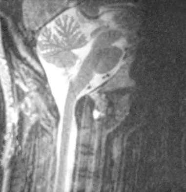
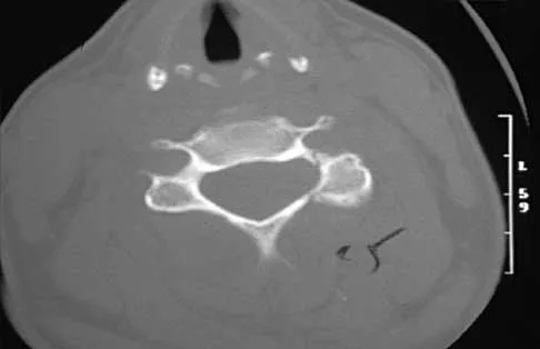
A 36-year-old woman has neck pain in the upper cervical region and occipital discomfort after being involved in a motor vehicle accident. Examination reveals no forehead or scalp lacerations. The neurologic examination is normal. A CT scan shows no evidence of bony injury. Figures 39a and 39b show a lateral radiograph and an MRI scan. Management should consist of


Explanation

39b The lateral radiograph shows 8 mm of atlantoaxial translation. In the absence of a bony injury, this represents rupture of the transverse atlantal ligament. The MRI scan reveals soft-tissue swelling posterior to the odontoid and a high intensity zone in the atlanto-dens interval consistent with acute injury. These injuries require arthrodesis because nonsurgical measures will not provide stability. Techniques for C1-2 fusion include Gallie, Brooks, or triple wiring. Transarticular screw fixation across the C1-2 articulation provides the most rigid means of fixation and the highest arthrodesis rates but is technically demanding. Anterior C2-3 arthrodesis will not address the level of instability. The normal atlanto-dens interval is 3 mm in an adult and 4 mm in a child. Kurz LT: Transverse atlantal ligament insufficiency, in Clark CR (ed): The Cervical Spine. Philadelphia, PA, Lippincott-Raven, 1998, pp 401-407.

Question 26

A 35-year-old male is brought to the ED after a motorcycle collision. He is hypotensive with a systolic BP of 70 mmHg. Primary survey reveals an unstable pelvis. Radiographs show a widened pubic symphysis of 4 cm and disruption of the anterior and posterior sacroiliac ligaments. A pelvic binder is to be applied to temporarily stabilize the pelvis. To achieve optimal reduction of pelvic volume and mechanical stability, where should the pelvic binder be centered?


Explanation

To optimally reduce pelvic volume and control hemorrhage in anterior-posterior compression (APC) pelvic ring injuries, a pelvic binder or sheet must be centered directly over the greater trochanters. Placement over the iliac crests is a common error and can paradoxically increase the pelvic volume by pushing the iliac wings inward at the top and outward at the bottom, worsening bleeding.

Question 27

A 28-year-old man sustains a closed midshaft tibia fracture. Four hours post-injury, he complains of worsening leg pain out of proportion to the injury, not relieved by intravenous opioids. On examination, there is severe pain with passive stretch of the hallux. The most reliable diagnostic parameter for acute compartment syndrome is:


Explanation

The most reliable parameter for diagnosing acute compartment syndrome is a delta pressure (diastolic blood pressure minus absolute compartment pressure) of less than 30 mmHg. Relying on an absolute pressure threshold (e.g., 30 mmHg) can lead to overdiagnosis and unnecessary fasciotomies, especially in hypertensive or hypotensive patients.

Question 28

A 25-year-old man sustains a high-energy Pauwels type III (vertical shear) femoral neck fracture. To maximize biomechanical stability and minimize the risk of varus collapse, which of the following fixation constructs is most appropriate?


Explanation

Pauwels type III femoral neck fractures in young adults are characterized by a vertically oriented fracture line, predisposing the hip to high shear forces and varus collapse. Biomechanical studies have consistently shown that a fixed-angle construct, such as a sliding hip screw (often supplemented with an anti-rotation screw), provides superior construct stiffness and resistance to vertical shear forces compared to multiple parallel cancellous screws.

Question 29

A 42-year-old construction worker sustains a Gustilo-Anderson IIIB open tibia fracture with a 10 cm soft tissue defect over the middle third of the tibia, exposing the bone devoid of periosteum. Following serial debridement, skeletal stabilization, and negative pressure wound therapy, definitive soft-tissue coverage is planned. The most appropriate local flap choice for this specific defect is:


Explanation

For soft tissue coverage of the lower extremity, the leg is classically divided into thirds. The proximal third is typically covered by a gastrocnemius rotational flap. The middle third is best covered by a soleus rotational flap. The distal third generally requires a free tissue transfer or a reverse sural artery flap, as local muscle bellies do not provide adequate distal reach.

Question 30

A 30-year-old female presents with midfoot pain after falling from a horse. Her foot was plantarflexed at the time of impact. Radiographs demonstrate a widening of the space between the first and second metatarsal bases with a distinct "fleck sign." Based on current literature, which of the following treatments results in better functional outcomes and lower revision rates for purely ligamentous Lisfranc injuries?


Explanation

For purely ligamentous Lisfranc injuries, multiple prospective randomized studies have demonstrated that primary arthrodesis of the medial column (first, second, and third tarsometatarsal joints) yields superior functional outcomes, less pain, and lower revision hardware removal rates compared to standard ORIF. ORIF is typically preferred for bony Lisfranc fracture-dislocations.

Question 31

A 45-year-old male sustains a distal femur fracture following a motor vehicle collision. CT imaging reveals an isolated coronal plane fracture of the lateral femoral condyle. Which surgical approach provides the best visualization to anatomically reduce this specific articular fracture fragment?


Explanation

A coronal plane fracture of the femoral condyle is known as a Hoffa fracture, with the lateral condyle being the most commonly affected. To adequately visualize the intra-articular surface and ensure anatomic reduction of the lateral Hoffa fragment, the "Swashbuckler" approach (a modified lateral approach involving reflection of the vastus lateralis from the lateral intermuscular septum) is utilized. A standard direct lateral approach is insufficient for viewing the complex intra-articular reduction.

Question 32

A 68-year-old woman with a 10-year history of alendronate therapy presents with progressively worsening right thigh pain for 3 months. Radiographs demonstrate focal cortical thickening of the lateral cortex of the subtrochanteric right femur with a transverse radiolucent "dreaded black line." The patient denies any trauma. What is the most appropriate next step in management?


Explanation

This patient presents with a symptomatic impending atypical femur fracture (AFF) associated with long-term bisphosphonate use. The "dreaded black line" indicates a stress fracture. Due to the high risk of completion, symptomatic impending AFFs should be treated with prophylactic full-length intramedullary nailing. Alendronate must be discontinued, and because these lesions are frequently bilateral, imaging of the contralateral femur is absolutely mandatory.

Question 33

A 50-year-old male is involved in a high-speed MVC. Pelvic radiographs and CT demonstrate a fracture involving both the anterior and posterior columns of the acetabulum. On the obturator oblique plain radiograph, a prominent "spur sign" is identified. This radiographic sign represents:


Explanation

The "spur sign" on an obturator oblique radiograph is pathognomonic for an associated both-column acetabular fracture. It represents the lowest margin of the intact, unfractured superior ilium (which remains anatomically attached to the axial skeleton via the sacroiliac joint) protruding posteriorly relative to the medially displaced articular segments of the acetabulum.

Question 34

A 22-year-old male motorcyclist is struck by a car and presents with massive swelling of the right shoulder girdle and an entirely flail, pulseless right upper extremity. Chest radiograph shows severe lateral displacement of the scapula. Which of the following neurologic injuries is most commonly associated with this specific pattern of high-energy trauma?


Explanation

Scapulothoracic dissociation is a devastating, high-energy injury characterized by lateral displacement of the scapula and massive soft-tissue trauma to the shoulder girdle. It is highly associated with complete brachial plexus avulsion (occurring in up to 80-90% of cases) and subclavian or axillary artery disruption. This frequently results in an ischemic, flail limb that may necessitate early forequarter amputation.

Question 35

A 34-year-old male falls from a height of 15 feet and sustains a displaced talar neck fracture with subluxation of the subtalar joint, while the tibiotalar and talonavicular joints remain located. According to the Hawkins classification, this is a Type II injury. The primary blood supply to the talar body, which is at highest risk in this injury, is derived from the:


Explanation

The talus has a tenuous, retrograde blood supply, predisposing it to avascular necrosis following neck fractures. The primary blood supply to the vast majority of the talar body is the artery of the tarsal canal, which is a branch of the posterior tibial artery. It forms an anastomotic sling with the artery of the tarsal sinus. While deltoid branches supply the medial aspect, the artery of the tarsal canal provides the most significant vascular contribution.

Question 36

A 42-year-old man sustains a severe closed pelvic fracture following a motorcycle collision. Clinical examination reveals a large, fluctuant area over the greater trochanter with intact, but hypoesthetic overlying skin. Aspiration yields serosanguineous fluid. Which of the following best describes the pathophysiology of this specific soft-tissue injury?


Explanation

The scenario describes a Morel-Lavallée lesion, a closed degloving injury. It is caused by traumatic shearing forces that separate the subcutaneous tissue from the underlying deep fascia. This separation shears the trans-fascial perforating vessels, leading to an accumulation of hemolymphatic fluid, blood, and necrotic fat in the newly created potential space. Hypoesthesia is common due to shearing of the cutaneous nerves.

Question 37

A 28-year-old man sustains a closed midshaft tibia fracture. Four hours post-injury, he complains of severe leg pain that is unresponsive to opioids. His blood pressure is 110/65 mmHg. Intracompartmental pressures are measured: anterior 42 mmHg, lateral 38 mmHg, deep posterior 45 mmHg, and superficial posterior 30 mmHg. What is the most appropriate management based on the objective data?


Explanation

Delta pressure is calculated as the Diastolic Blood Pressure minus the highest intracompartmental pressure. In this case, 65 mmHg - 45 mmHg = 20 mmHg. A delta pressure of 30 mmHg or less is widely accepted as an absolute indication for emergency four-compartment fasciotomy to prevent irreversible muscle and nerve ischemia associated with acute compartment syndrome.

Question 38

A 30-year-old woman is involved in a high-speed motor vehicle collision and sustains a Hawkins Type III talar neck fracture. Which of the following blood vessels provides the dominant blood supply to the talar body, which is critically at risk in this injury?


Explanation

The artery of the tarsal canal, a branch of the posterior tibial artery, provides the dominant blood supply to the talar body. In a Hawkins Type III fracture (talar neck fracture with subtalar and tibiotalar dislocation), the three main sources of blood supply (artery of the tarsal canal, artery of the sinus tarsi, and dorsal neck vessels) are typically disrupted, leading to an exceptionally high rate of avascular necrosis (AVN), often exceeding 80%.

Question 39

A 22-year-old man sustains a low-velocity gunshot wound to the distal thigh. Radiographs demonstrate a comminuted distal femur fracture and a retained bullet lodged within the knee joint space. The entry wound is 1 cm, clean, without massive soft-tissue destruction. What is the most appropriate initial management regarding the retained bullet?


Explanation

Retained bullets within a synovial joint space must be surgically removed. Synovial fluid dissolves the lead over time, which can lead to systemic lead toxicity (plumbism) as well as severe intra-articular third-body wear and mechanical cartilage damage. Extensile debridement of low-velocity entry tracts is generally not required unless heavily contaminated.

Question 40

A 19-year-old motorcyclist is thrown from his bike. He presents with a completely flail, pulseless left upper extremity. Radiographs reveal lateral displacement of the left scapula by 3 cm compared to the right, a displaced clavicle fracture, and acromioclavicular joint disruption. Which of the following injuries is most strongly associated with this clinical picture?


Explanation

The patient has sustained a scapulothoracic dissociation, characterized by lateral displacement of the scapula, clavicle fracture or AC/SC joint disruption, and severe soft tissue injury. It is considered a closed, traumatic forequarter amputation and is frequently associated with complete brachial plexus avulsions (up to 80-90%) and severe subclavian/axillary vascular injuries. The limb is often neurologically flail and may require early amputation.

Question 41

A 45-year-old construction worker sustains an open tibial shaft fracture with a 6 cm laceration, significant periosteal stripping, and exposed bone, but adequate soft tissue for coverage without requiring a flap (Gustilo-Anderson IIIA). Which of the following interventions is the most critical factor in preventing deep infection in this patient?


Explanation

Early administration of systemic antibiotics (ideally within 1 hour of injury) is the single most critical and strongly evidence-based factor in reducing the risk of infection in open fractures. High-pressure lavage is no longer recommended due to the risk of driving debris and bacteria deeper into tissues or causing further soft-tissue damage (as demonstrated in the FLOW trial).

Question 42

A 25-year-old male falls from a height of 15 feet, sustaining a vertically oriented, displaced intracapsular femoral neck fracture (Pauwels type III). What biomechanical force is most responsible for the high rate of internal fixation failure in this specific fracture pattern?


Explanation

Pauwels type III femoral neck fractures have a vertical fracture line (angle > 50 degrees to the horizontal). This vertical orientation subjects the fracture to profoundly high shear forces and varus instability during weight-bearing. This leads to a significantly higher rate of fixation failure, nonunion, and osteonecrosis compared to more horizontally oriented fractures (Pauwels I), which experience primarily stable compressive forces.

Question 43

A 76-year-old woman with a well-functioning cruciate-retaining (CR) total knee arthroplasty (TKA) falls and sustains a displaced supracondylar femur fracture (Lewis-Rorabeck Type II). Radiographs demonstrate a closed fracture with the distal component ending 1 cm superior to the intact, well-fixed femoral component. Which of the following is the most appropriate surgical treatment?


Explanation

For a periprosthetic distal femur fracture above a well-fixed femoral component (Lewis-Rorabeck Type II), lateral locked plating is the standard of care. A cruciate-retaining (CR) TKA femoral component typically lacks an intercondylar box or sufficient intercondylar notch space to permit the insertion of a standard retrograde intramedullary nail. Distal femoral replacement is reserved for loose components, severe comminution in the elderly, or exceedingly poor bone stock.

Question 44

A 22-year-old collegiate football player sustains a hyperplantarflexion injury to his midfoot. Weight-bearing radiographs demonstrate 3 mm of widening between the base of the first and second metatarsals, with a subtle "fleck sign" adjacent to the medial cuneiform. What is the anatomical path of the primarily injured ligament?


Explanation

The Lisfranc ligament is an interosseous ligament that connects the lateral aspect of the medial cuneiform to the medial aspect of the base of the second metatarsal. It is the critical stabilizer of the tarsometatarsal joint complex. A "fleck sign" represents an avulsion fracture of this ligament. There is no direct transverse ligament connecting the bases of the first and second metatarsals.

Question 45

In the initial ATLS resuscitation of a hemodynamically unstable patient with an anteroposterior compression (APC) III pelvic ring injury, what is the correct anatomical landmark for the optimal placement of a circumferential pelvic binder?


Explanation

A pelvic binder or sheet must be centered directly over the greater trochanters to effectively reduce the pelvic volume and stabilize the fracture via indirect compression of the pelvic ring. Placing the binder too high (e.g., over the iliac crests or abdomen) is a common error that fails to close the pelvic ring adequately and can paradoxically increase bleeding or limit diaphragmatic excursion.

Question 46

A 35-year-old male is brought to the emergency department after a high-speed motor vehicle collision. He is hypotensive with a blood pressure of 75/40 mmHg. A pelvic binder is immediately applied. The FAST exam is negative. An anteroposterior pelvic radiograph shows an anteroposterior compression (APC) type III injury with a widened pubic symphysis (>3 cm) and disrupted sacroiliac joints. Despite ongoing fluid and blood resuscitation, the patient remains hemodynamically unstable. What is the most appropriate next step in management?


Explanation

In a hemodynamically unstable patient with an unstable pelvic ring injury and a negative FAST exam, the source of bleeding is predominantly retroperitoneal, typically from the venous plexus or arterial branches of the internal iliac system. Once a pelvic binder has been applied to reduce pelvic volume, if the patient remains hemodynamically unstable, ATLS and AAOS guidelines recommend either preperitoneal pelvic packing or pelvic angiography with embolization. A CT scan is contraindicated in a hemodynamically unstable patient.

Question 47

A 28-year-old female sustains a displaced, highly vertical (Pauwels type III) femoral neck fracture after falling from a horse. Which of the following internal fixation constructs provides the greatest biomechanical stability for this specific fracture pattern?


Explanation

Pauwels type III femoral neck fractures are characterized by a highly vertical fracture line (angle >50 degrees), which subjects the fracture to significant shear forces rather than compressive forces. Biomechanical studies have consistently shown that fixed-angle constructs, specifically a sliding hip screw combined with an anti-rotation (derotation) screw, provide superior stability, higher load-to-failure, and better resistance to shear compared to multiple parallel cannulated screws in young adults with vertical femoral neck fractures.

Question 48

A 45-year-old man sustains an open fracture of the proximal third of the tibia, resulting in a 6 x 6 cm anterior soft-tissue defect with exposed bone lacking periosteum. After serial debridements and adequate skeletal stabilization, there is no evidence of infection, but the bone remains exposed. Which of the following soft-tissue coverage options is the most appropriate definitive management?


Explanation

The lower extremity is traditionally divided into thirds for the purpose of soft-tissue coverage planning over exposed bone. The proximal third of the tibia is most reliably covered by a medial (or lateral) gastrocnemius rotational muscle flap. The middle third is typically covered by a soleus muscle flap. The distal third generally lacks adequate local muscle bulk and requires a free tissue transfer (such as an anterolateral thigh or latissimus dorsi free flap) or local fasciocutaneous flaps (like the reverse sural flap) for smaller defects. A split-thickness skin graft will not survive on bare cortical bone devoid of periosteum.

Question 49

An 82-year-old woman presents with severe knee pain and inability to bear weight after a mechanical fall. She underwent a total knee arthroplasty 10 years ago. Radiographs demonstrate a displaced supracondylar distal femur fracture. Careful radiographic evaluation shows no evidence of osteolysis, and the femoral component remains rigidly fixed to the bone.

What is the most appropriate surgical management?


Explanation

This patient has a Lewis-Rorabeck type II periprosthetic fracture, defined as a displaced fracture around a well-fixed prosthesis. The standard of care for a displaced periprosthetic distal femur fracture above a well-fixed total knee arthroplasty component is internal fixation. This is typically achieved with a lateral locking plate or a retrograde intramedullary nail (if the intercondylar box of the prosthesis is open and accommodates a nail). Revision arthroplasty to a distal femoral replacement is reserved for loose components (Lewis-Rorabeck type III) or profoundly deficient bone stock not amenable to any fixation.

Question 50

A 24-year-old professional football player sustains an acute, purely ligamentous Lisfranc injury involving the first, second, and third tarsometatarsal joints. Which of the following surgical treatments has been shown to yield the best long-term functional outcomes for this specific injury pattern?


Explanation

Purely ligamentous Lisfranc injuries exhibit a higher rate of hardware failure, loss of reduction, and subsequent post-traumatic midfoot arthritis when treated with open reduction and internal fixation (ORIF) compared to primarily bony avulsion fractures. High-quality prospective studies (e.g., Ly and Coetzee) have demonstrated that primary arthrodesis of the medial column (first, second, and third tarsometatarsal joints) for purely ligamentous injuries results in superior functional outcome scores, a more reliable return to pre-injury activity levels, and significantly lower revision rates than ORIF.

Question 51

A 32-year-old man underwent unreamed intramedullary nailing of a closed diaphyseal tibia fracture. He was discharged on postoperative day 3. Six months later, he complains of pain in the calf and the development of severe clawing of his lesser toes (flexion at the PIP and DIP joints). He notes the deformity worsens when his ankle is passively dorsiflexed. Ischemic contracture of which of the following compartments is the most likely cause of this deformity?


Explanation

The patient is presenting with late sequelae of an unrecognized deep posterior compartment syndrome. The deep posterior compartment of the leg contains the flexor hallucis longus (FHL), flexor digitorum longus (FDL), and tibialis posterior muscles, as well as the tibial nerve. Ischemic contracture of the FHL and FDL tendons results in fixed flexion of the interphalangeal joints of the toes, causing a claw toe deformity. This deformity typically becomes more pronounced with ankle dorsiflexion due to the tenodesis effect on the shortened tendons.

Question 52

A 48-year-old construction worker falls from scaffolding, sustaining a high-energy, highly comminuted closed distal tibia pilon fracture. On presentation to the emergency department, the ankle is grossly deformed, and the overlying skin is tight, shiny, and demonstrates multiple fracture blisters over the medial malleolus. What is the most appropriate initial step in the operative management?


Explanation

High-energy pilon fractures are notorious for severe surrounding soft-tissue injury. Performing immediate open reduction and internal fixation (ORIF) through swollen, compromised skin is associated with unacceptably high rates of wound dehiscence, deep infection, and hardware exposure. The current gold standard for initial management is the "span, scan, and plan" approach. This entails urgent application of a joint-spanning external fixator to restore length, alignment, and allow the soft tissues to rest. Definitive fixation is delayed (usually 10 to 21 days) until the soft-tissue envelope has sufficiently recovered, indicated by the resolution of edema and the reappearance of skin wrinkles (the "wrinkle sign").

Question 53

During surgical management of a 'terrible triad' injury of the elbow (elbow dislocation, radial head fracture, and coronoid fracture), you perform fixation of the coronoid fragment, replace the non-reconstructible radial head with a metallic arthroplasty, and repair the lateral ulnar collateral ligament (LUCL) to its anatomic origin on the lateral epicondyle. Upon fluoroscopic examination through a full arc of motion, the elbow subluxates posteriorly when extended past 30 degrees. What is the most appropriate next step to restore stability?


Explanation

The standard treatment algorithm for a terrible triad injury involves restoring the anterior column (coronoid), lateral column (radial head), and lateral soft-tissue stabilizers (LUCL). If concentric stability is not achieved after addressing these structures, it usually indicates persistent medial-sided instability or an inadequate restoration of the coronoid buttress. The recommended next step to address persistent instability (especially in extension) is to either repair the medial collateral ligament (MCL) or to apply a hinged elbow external fixator. Casting for 6 weeks is contraindicated due to the high risk of severe, permanent joint stiffness.

Question 54

A 29-year-old man undergoes open reduction and internal fixation for a displaced Hawkins type II fracture of the talar neck. At his 8-week postoperative visit, an anteroposterior radiograph of the ankle demonstrates a subchondral radiolucent band beneath the dome of the talus. What does this specific radiographic finding indicate regarding the patient's prognosis?


Explanation

The finding described is the Hawkins sign. It appears as a subchondral radiolucent band in the talar dome on an AP (or mortise) radiograph, typically becoming visible 6 to 8 weeks following a talar neck fracture. This radiolucency represents subchondral osteopenia, which is a consequence of disuse coupled with hyperemic resorption. The presence of the Hawkins sign signifies that the talar body has an intact blood supply (or has successfully revascularized). A positive Hawkins sign is a highly reliable negative predictor for the development of avascular necrosis (AVN).

Question 55

A 25-year-old man complains of persistent radial-sided wrist pain 10 months after falling onto an outstretched hand. Radiographs demonstrate a nonunion of the proximal pole of the scaphoid. A subsequent MRI reveals lack of enhancement in the proximal pole, consistent with avascular necrosis (AVN). There is no radiographic evidence of radiocarpal or midcarpal osteoarthritis.

Which of the following is the most appropriate surgical intervention?


Explanation

The patient has a scaphoid proximal pole nonunion complicated by avascular necrosis (AVN), but without the presence of scaphoid nonunion advanced collapse (SNAC) arthritis. Standard non-vascularized bone grafting has an unacceptably high failure rate in the setting of proximal pole AVN. The treatment of choice to achieve union in this specific scenario is a vascularized bone graft (such as the 1,2-intercompartmental supraretinacular artery [1,2-ICSRA] graft from the distal radius or a free vascularized medial femoral condyle graft) paired with rigid internal fixation. Salvage procedures like a four-corner fusion or proximal row carpectomy are reserved for patients who have already developed established carpal arthritis.

Question 56

A 28-year-old male sustains a vertically oriented, displaced femoral neck fracture (Pauwels type III) after a fall from a height. To maximize biomechanical stability and reduce the risk of varus collapse and nonunion, which of the following internal fixation constructs is most appropriate?


Explanation

A sliding hip screw (SHS) provides superior biomechanical stability for vertically oriented, high-shear (Pauwels type III) femoral neck fractures compared to parallel cancellous screws. The SHS acts as a fixed-angle device that resists varus shear forces, and the addition of a derotational screw helps prevent rotation of the femoral head during lag screw insertion and physiological loading.

Question 57

A 32-year-old male sustains a closed comminuted midshaft tibia fracture. He is complaining of out-of-proportion pain, and physical examination reveals intact pedal pulses. His diastolic blood pressure is 70 mm Hg and his MAP is 85 mm Hg. Direct continuous pressure measurements reveal an anterior compartment pressure of 35 mm Hg, lateral 25 mm Hg, superficial posterior 20 mm Hg, and deep posterior 45 mm Hg. Which of the following is the most appropriate next step in management?


Explanation

The diagnosis of acute compartment syndrome is confirmed when the delta pressure (Diastolic Blood Pressure minus compartment pressure) is less than 30 mm Hg. In this patient, the deep posterior compartment delta pressure is 70 - 45 = 25 mm Hg. This is an absolute indication for emergent fasciotomy. When releasing the leg for compartment syndrome, all four compartments must be released, typically utilizing a two-incision technique.

Question 58

A 45-year-old female presents with a complex intra-articular distal femur fracture (OTA/AO 33-C3). A CT scan reveals a coronal plane fracture of the lateral femoral condyle (Hoffa fracture). What is the most biomechanically appropriate fixation strategy for this specific coronal fragment?


Explanation

A Hoffa fracture is a coronal plane fracture of the femoral condyle. Achieving absolute stability and anatomic reduction of the articular surface is critical. This requires interfragmentary compression using lag screws directed perpendicular to the fracture line (anterior-to-posterior or posterior-to-anterior). A laterally applied locking plate alone will not adequately capture or compress a coronal fragment.

Question 59

A 40-year-old male sustains a displaced intra-articular calcaneus fracture (Sanders type III) with significant soft tissue swelling and fracture blisters over the lateral hindfoot. If an extensile lateral approach for open reduction and internal fixation (ORIF) is planned, which of the following patient factors is the most significant predictor of postoperative wound complications?


Explanation

Smoking is one of the most significant risk factors for wound complications following the extensile lateral approach for calcaneus fractures. Studies have shown a markedly increased incidence of wound edge necrosis, dehiscence, and deep infection in patients who actively smoke.

Question 60

A 25-year-old male is involved in a motor vehicle collision and sustains a Gustilo-Anderson IIIB open midshaft tibia fracture with extensive periosteal stripping and exposed bone that requires a free soft-tissue transfer. According to the Lower Extremity Assessment Project (LEAP) study, which of the following factors is the most significant predictor of poor long-term functional outcome in this patient?


Explanation

The landmark LEAP (Lower Extremity Assessment Project) study demonstrated that poor outcomes in severe lower extremity trauma were most closely correlated with patient characteristics such as poor socioeconomic status, lack of health insurance, lower educational level, and psychological factors (e.g., depression, self-efficacy), rather than the specific reconstructive technique, amputation vs. salvage, or initial lack of plantar sensation.

Question 61

A 38-year-old male requires open reduction and internal fixation of a posterior wall acetabular fracture via a Kocher-Langenbeck approach. During the deep exposure, what is the proper anatomical management of the short external rotators to protect the medial femoral circumflex artery (MFCA)?


Explanation

During the Kocher-Langenbeck approach, releasing the short external rotators (piriformis, obturator internus, and gemelli) close to their greater trochanter insertion while preserving the quadratus femoris muscle protects the main ascending branch of the medial femoral circumflex artery (MFCA), which provides the critical blood supply to the femoral head.

Question 62

A 55-year-old woman undergoes volar locked plating for a displaced distal radius fracture.

Six months postoperatively, she presents with a new-onset inability to actively flex the interphalangeal joint of her thumb. Which of the following technical errors during the initial surgery most likely caused this complication?


Explanation

The patient is presenting with a rupture of the flexor pollicis longus (FPL) tendon, a known complication of volar plating of the distal radius. Placement of the plate distal to the watershed line of the distal radius causes the prominent distal edge of the plate to rub against the FPL tendon during wrist motion, leading to tenosynovitis and subsequent attrition rupture.

Question 63

A 29-year-old male sustained a Hawkins type II talar neck fracture and underwent open reduction and internal fixation. At 8 weeks postoperatively, an AP radiograph of the ankle

demonstrates a subchondral radiolucent band in the talar dome. This radiographic finding most accurately indicates:


Explanation

A subchondral radiolucent band in the talar dome visible on AP mortise radiographs at 6 to 8 weeks post-injury is known as the Hawkins sign. It represents subchondral osteopenia secondary to disuse and hyperemia. The presence of this hyperemia indicates that the vascular supply to the talar body is intact, thus avascular necrosis (AVN) is highly unlikely.

Question 64

A 35-year-old male involved in a high-speed collision presents with bilateral closed femoral shaft fractures, a grade IV liver laceration, and a severe closed head injury (GCS 7). His initial laboratory values show a lactate of 6.5 mmol/L, pH of 7.15, and his core temperature is 34.5°C. According to Damage Control Orthopedics (DCO) principles, what is the most appropriate initial orthopedic management for his femur fractures?


Explanation

This polytrauma patient is physiologically unstable ('in extremis') with a severe head injury, hypothermia, acidosis, and elevated lactate, fulfilling the lethal triad. Damage Control Orthopedics (DCO) dictates that prolonged, physiologically demanding surgeries (like reamed IM nailing) should be avoided to prevent a 'second hit' of systemic inflammation. Rapid stabilization with bilateral external fixators is indicated.

Question 65

A 24-year-old female sustains a closed spiral fracture of the distal third of the humeral shaft (Holstein-Lewis fracture).

On initial evaluation in the emergency department, she is unable to extend her wrist or digits, and has decreased sensation in the first dorsal web space. What is the most appropriate initial management?


Explanation

A primary radial nerve palsy associated with a closed humeral shaft fracture (including the Holstein-Lewis type) is typically a neuropraxia. The standard of care is expectant management with closed reduction and functional fracture bracing (Sarmiento brace). Surgical exploration is generally indicated only if the palsy occurs after closed reduction, in open fractures, or if there is no clinical or electromyographic sign of recovery after 3 to 6 months.

Question 66

A 35-year-old male is brought to the ED after a motorcycle collision. He is hypotensive (BP 75/40 mmHg) and tachycardic (HR 130). AP pelvis radiograph shows a widened pubic symphysis of 4 cm and widened bilateral sacroiliac joints. A pelvic binder has been applied appropriately but he remains hemodynamically unstable after 2 liters of crystalloid and 2 units of packed red blood cells. What is the most appropriate next step in management?


Explanation

In a hemodynamically unstable patient with an unstable pelvic ring injury (such as an APC-III), if the patient remains unstable despite initial resuscitation and pelvic binder application, emergent hemorrhage control is indicated. This is typically achieved via preperitoneal pelvic packing, pelvic angiography with embolization, or a combination of both. Exploratory laparotomy is generally reserved for intraperitoneal bleeding (e.g., positive FAST). External fixation takes time and does not stop venous bleeding as effectively as packing, nor arterial bleeding as well as embolization.

Question 67

A 42-year-old skier presents with a complex bicondylar tibial plateau fracture (Schatzker VI). Radiographs and CT scan reveal a large, displaced posteromedial fragment in addition to lateral plateau depression. When planning surgical fixation, which of the following approaches and patient positioning is most appropriate to optimally address the posteromedial fragment?


Explanation

A displaced posteromedial fragment in a bicondylar tibial plateau fracture cannot be adequately visualized or reduced through a standard anterolateral approach and is biomechanically unstable if not buttressed. A posteromedial approach, often performed with the patient in a prone or lateral position (or 'floppy lateral'), allows direct visualization, reduction, and application of a posterior buttress plate. This counteracts the shear forces and prevents settling of the medial condyle into varus.

Question 68

A 25-year-old man sustains a completely displaced, vertically oriented (Pauwels type III) femoral neck fracture after a fall from a height. Which of the following fixation constructs provides the most biomechanically stable fixation for this specific fracture pattern?


Explanation

Pauwels type III fractures are highly vertical and subjected to significant shear forces rather than compressive forces, leading to higher rates of nonunion and varus collapse. Biomechanical studies have shown that a sliding hip screw (a fixed-angle device) supplemented with a derotation screw provides superior resistance to vertical shear forces and better clinical outcomes for vertical femoral neck fractures in young adults compared to three parallel cancellous screws.

Question 69

A 38-year-old male sustains a comminuted distal femur fracture. CT scan reveals a coronal plane fracture of the lateral femoral condyle (Hoffa fragment). Which of the following principles is most critical when fixing this specific coronal fragment?


Explanation

A Hoffa fracture is a coronal shear fracture of the femoral condyle, most commonly the lateral condyle. The standard and biomechanically most robust method of fixation involves anterior-to-posterior directed lag screws placed perpendicular to the fracture plane. Due to the intra-articular nature of the injury, the screw heads should be countersunk if placed through the articular cartilage, or placed just proximal to the articular surface.

Question 70

A 65-year-old woman undergoes open reduction and internal fixation of a 3-part proximal humerus fracture with a locked plating system. Which of the following technical factors is most strongly associated with failure of fixation and subsequent varus collapse?


Explanation

In locked plating of proximal humerus fractures, the medial hinge is often disrupted. Failure to restore medial cortical contact or, more importantly, failure to place inferiorly directed 'calcar' screws into the inferomedial quadrant of the humeral head, significantly increases the risk of varus collapse, screw cut-out, and overall fixation failure. Medial calcar screws act as a rigid biomechanical buttress.

Question 71

A 22-year-old motorcyclist sustains severe high-energy right shoulder trauma. Radiographs reveal lateral displacement of the scapula, a completely displaced clavicle fracture, and acromioclavicular joint separation. His right upper extremity is pulseless, and he has a complete motor and sensory deficit of the right arm. What is the most likely long-term functional outcome for the injured extremity despite optimal initial management?


Explanation

Scapulothoracic dissociation is a massive injury characterized by disruption of the scapulothoracic articulation, lateral displacement of the scapula, and severe associated neurovascular injuries. The neurological injury is nearly always a complete preganglionic brachial plexus avulsion. Even with successful vascular repair to save the limb from ischemia, complete preganglionic avulsions portend a dismal prognosis for function, often resulting in a flail, insensate limb that frequently requires late amputation.

Question 72

A 28-year-old male sustains a Hawkins Type III fracture of the talar neck. What is the approximate risk of developing avascular necrosis (AVN) of the talar body, and what is the characteristic finding of the Hawkins sign?


Explanation

A Hawkins Type III talar neck fracture involves displacement of the talar body from both the subtalar and tibiotalar joints, disrupting all three major blood supplies. The risk of AVN is exceptionally high, ranging from 70% to 100%. The Hawkins sign is a subchondral radiolucent band seen in the talar dome on an AP radiograph 6 to 8 weeks post-injury. It represents subchondral osteopenia secondary to disuse atrophy. For osteopenia to occur, there must be active bone resorption, which requires an intact blood supply; thus, a positive Hawkins sign indicates intact vascularity.

Question 73

A 45-year-old farmer sustains a severe open tibia fracture after his leg was caught in a tractor. The wound is 12 cm long with extensive muscle damage and gross agricultural contamination. The foot is well-perfused with palpable pulses. According to current evidence-based guidelines, what is the most appropriate initial antibiotic regimen?


Explanation

This is a Gustilo-Anderson Type IIIA open fracture heavily contaminated with agricultural debris. Patients with farm injuries or gross soil contamination are at exceptionally high risk for Clostridium perfringens (gas gangrene) and other virulent anaerobes. Current guidelines recommend broad-spectrum coverage (often a 3rd-generation cephalosporin, or 1st-generation cephalosporin plus an aminoglycoside) and the essential addition of high-dose Penicillin specifically for targeted anaerobic coverage.

Question 74

A 30-year-old male undergoes intramedullary nailing of a closed diaphyseal tibia fracture. In the recovery room, he complains of severe leg pain out of proportion to the injury, unrelieved by IV opioids. On examination, he has pain with passive stretch of the hallux and paresthesias in the first web space. Absolute compartment pressures are measured at 35 mmHg, and his diastolic blood pressure is 60 mmHg. What is the most appropriate next step?


Explanation

The clinical presentation is classic for acute compartment syndrome. The diagnosis is confirmed by a Delta P (diastolic blood pressure minus compartment pressure) of less than 30 mmHg. In this patient, Delta P = 60 - 35 = 25 mmHg, which dictates immediate surgical intervention. The definitive treatment is urgent four-compartment fasciotomy of the leg. Elevating the leg above the heart decreases local arterial perfusion pressure and exacerbates ischemia.

Question 75

A 40-year-old roofer falls 15 feet, sustaining a closed, displaced intra-articular calcaneus fracture (Sanders Type III) with significant blistering over the lateral heel. Surgery via an extensile lateral approach is planned. Which of the following factors is most predictive of wound healing complications following this surgical approach?


Explanation

Wound complications are a major and potentially devastating concern with the extensile lateral approach for calcaneus fractures. While operating through blistered, swollen tissue increases risk, smoking is widely recognized as the single most significant modifiable patient risk factor for postoperative wound edge necrosis and deep infection following this approach. Patients who smoke have complication rates exponentially higher than non-smokers.

Question 76

A 28-year-old polytrauma patient presents with a hemodynamically unstable pelvic ring injury. A pelvic binder is applied in the trauma bay. To be maximally effective in reducing pelvic volume and controlling hemorrhage, the binder should be centered over which of the following anatomic landmarks?


Explanation

Pelvic binders are most effective in reducing pelvic volume when centered over the greater trochanters. Placement over the iliac crests or higher can paradoxically open the true pelvis in certain fracture patterns and is less effective at achieving symphyseal reduction.

Question 77

A 35-year-old male is involved in a high-speed motor vehicle collision and sustains a 'terrible triad' injury of the elbow. Which of the following describes the most appropriate sequence of surgical reconstruction to restore stability?


Explanation

The standard sequence for addressing a terrible triad injury of the elbow (elbow dislocation, radial head fracture, coronoid fracture) typically proceeds from deep to superficial and medial to lateral (when approached laterally). The optimal sequence is coronoid fixation first, followed by radial head repair or arthroplasty, and finally LCL complex repair.

Question 78

A 40-year-old female sustains an isolated closed midshaft tibia fracture treated with a reamed intramedullary nail. Twelve hours postoperatively, she requires escalating doses of IV pain medication and reports severe pain with passive stretch of her hallux. Which compartment of the lower leg is most commonly affected in this scenario?


Explanation

Acute compartment syndrome most commonly affects the anterior compartment of the lower leg following tibial shaft fractures. The hallmark physical exam finding is severe pain with passive stretch of the involved muscles; pain with passive stretch of the great toe (extensor hallucis longus) indicates anterior compartment involvement.

Question 79

A 65-year-old woman with a history of osteoporosis and 7 years of alendronate therapy presents with progressively worsening thigh pain. Radiographs reveal a transverse, non-comminuted fracture of the lateral cortex of the subtrochanteric femur with localized periosteal thickening ('beaking'). What is the most appropriate prophylactic surgical treatment?


Explanation

This patient has an impending atypical femur fracture (AFF) associated with long-term bisphosphonate use. Cephalomedullary (full-length) nailing is the prophylactic and therapeutic treatment of choice for subtrochanteric atypical femur fractures. Plates have a significantly higher rate of failure.

Question 80

In the management of open fractures, current literature demonstrates that the most critical factor in reducing the risk of deep infection is:


Explanation

Evidence consistently demonstrates that the administration of systemic antibiotics as early as possible (ideally within 1 hour of injury) is the single most important factor in reducing infection rates in open fractures. The traditional '6-hour rule' for surgical debridement has been shown to be less critical than the timing of antibiotic administration.

Question 81

A 25-year-old male sustains a Hawkins Type III talar neck fracture following a fall from height. Which of the following best describes the pathoanatomy of a Hawkins Type III fracture and its associated risk of avascular necrosis (AVN)?


Explanation

The Hawkins classification for talar neck fractures is prognostic for AVN. Type I: nondisplaced (0-15% AVN). Type II: subtalar dislocation (20-50% AVN). Type III: subtalar and tibiotalar dislocation, where the AVN risk approaches 80-100%. Type IV includes talonavicular dislocation.

Question 82

A 32-year-old motorcyclist sustains a completely displaced, highly comminuted intra-articular fracture of the calcaneus (Sanders Type IV). If open reduction and internal fixation (ORIF) is attempted via an extensile lateral approach, which nerve is at greatest risk of iatrogenic injury?


Explanation

The sural nerve courses posterior to the lateral malleolus and crosses the lateral aspect of the hindfoot. It is at significant risk of iatrogenic injury during the extensile lateral approach to the calcaneus. A 'no-touch' technique and full-thickness flap retraction are utilized to protect it.

Question 83

A 45-year-old male is undergoing closed reduction and percutaneous pinning for a displaced proximal humerus fracture. To minimize the risk of injury to the axillary nerve, lateral pins should be inserted avoiding a specific zone. The axillary nerve typically courses transversely at what distance distal to the lateral edge of the acromion?


Explanation

The axillary nerve courses circumferentially from posterior to anterior on the deep surface of the deltoid, typically 5 to 7 cm distal to the lateral edge of the acromion. Lateral percutaneous pins or drill bits for plating should strictly avoid this 'danger zone'.

Question 84

A 22-year-old male sustains a vertically oriented, displaced femoral neck fracture (Pauwels Type III) after a fall from a roof. The plan is for urgent open reduction and internal fixation. To maximize biomechanical stability and resist shear forces in this specific fracture pattern, the optimal fixation construct consists of:


Explanation

Pauwels Type III fractures are highly vertically oriented (>50 degrees), leading to significant shear forces across the fracture site. A fixed-angle device, such as a sliding hip screw (DHS) with a derotational screw, provides superior biomechanical stability against shear forces compared to multiple cancellous screws.

Question 85

In a patient with multiple severe injuries including bilateral femur fractures and a severe traumatic brain injury (TBI) with elevated intracranial pressure, the concept of Damage Control Orthopedics (DCO) suggests which of the following initial management strategies for the femur fractures?


Explanation

In polytrauma patients who are physiologically unstable or have a severe TBI with elevated intracranial pressure ('borderline' or 'in extremis'), early total care (reamed IM nailing) can exacerbate systemic inflammation and cause secondary brain injury ('second hit'). Damage control orthopedics with rapid external fixation is indicated to stabilize the fractures rapidly while minimizing physiologic burden.

Question 86

A 35-year-old male presents with an open diaphyseal tibia fracture with a 12 cm soft tissue defect and exposed bone after a motorcycle crash. The limb is vascularly intact. After initial debridement, what is the most appropriate timing and method for soft tissue coverage?


Explanation

Gustilo-Anderson IIIB open fractures require soft tissue coverage with a rotational or free flap. For large or distal third tibia defects, free tissue transfer (e.g., anterolateral thigh or latissimus dorsi) is the standard of care. Early coverage within 3 to 7 days has been shown to significantly reduce infection rates, minimize flap failure, and improve overall functional outcomes compared to delayed coverage.

Question 87

A 42-year-old hemodynamically unstable male presents after a high-speed motor vehicle collision. A pelvic AP radiograph reveals a symphyseal diastasis of 4 cm and disruption of the anterior sacroiliac ligaments bilaterally. He is tachycardic (130 bpm) and hypotensive (80/50 mmHg). FAST exam is negative. What is the most appropriate initial step in management?


Explanation

In a hemodynamically unstable patient with an anteroposterior compression (APC) type pelvic ring injury (often presenting as an 'open book' pelvis), the immediate priority is mechanical stabilization to reduce pelvic volume. A pelvic binder, properly centered over the greater trochanters (not the iliac crests), is the most rapid and effective initial non-invasive step. If instability persists despite adequate volume reduction and fluid resuscitation, angiography with embolization or pre-peritoneal packing is indicated.

Question 88

A 28-year-old female sustains a displaced, vertical (Pauwels type III) femoral neck fracture. She is brought to the operating room for internal fixation. Which of the following biomechanical constructs provides the most stable fixation for this specific fracture pattern?


Explanation

Pauwels type III femoral neck fractures are characterized by a vertical fracture line (angle > 50 degrees from horizontal), resulting in high shear forces and marked instability. Biomechanical studies have consistently demonstrated that a fixed-angle device, such as a sliding hip screw (SHS), provides superior resistance to vertical shear compared to multiple parallel cancellous screws. A derotational screw is often added superiorly to prevent rotational displacement of the head during SHS insertion.

Question 89

A 45-year-old male sustains a closed, displaced intra-articular calcaneus fracture. A CT scan shows a Sanders Type III fracture. The surgeon plans an extensile lateral approach for open reduction and internal fixation. To minimize the risk of wound complications, when should the surgery ideally be performed?


Explanation

The extensile lateral approach to the calcaneus is associated with a high rate of wound healing complications (up to 25%). To mitigate this risk, surgery must be delayed until post-traumatic soft tissue swelling has adequately subsided. The appearance of the 'wrinkle sign' (skin wrinkling on the lateral aspect of the hindfoot) typically occurs between 10 to 14 days post-injury and is a highly reliable clinical indicator that the soft tissue envelope is ready for surgical intervention.

Question 90

A 72-year-old female with osteoporosis presents with a closed, displaced extra-articular distal femur fracture (OTA/AO 33-A). She is scheduled for open reduction and internal fixation with a lateral locking plate. What is the primary mechanical advantage of a locked plating construct over conventional non-locked plating in this specific scenario?


Explanation

Locking plates are fixed-angle constructs where the screw head threads lock into the plate, creating a single beam construct. This design bypasses the need for friction between the plate and bone for stability. This offers a distinct biomechanical advantage in osteoporotic bone by significantly increasing resistance to screw pullout and toggle. Non-locking screws rely on cortical bite and plate-to-bone compression, which frequently fail in weak, osteoporotic bone.

Question 91

A 30-year-old male falls from a height and sustains a Hawkins type III fracture of the talar neck. Which of the following blood vessels provides the primary residual blood supply to the talar body, and what is the approximate rate of avascular necrosis (AVN) for this fracture type?


Explanation

A Hawkins type III talar neck fracture involves displacement of the neck with dislocation of both the subtalar and tibiotalar joints. This severe injury disrupts the arteries of the sinus tarsi and tarsal canal, leaving only the deltoid branches of the posterior tibial artery (supplying the medial aspect of the body) if they remain intact. Due to this catastrophic vascular insult, the rate of avascular necrosis (AVN) of the talar body approaches 80% to 100%.

Question 92

A 22-year-old gymnast sustains a hyperplantarflexion injury to her midfoot. Weight-bearing radiographs reveal widening of the interval between the first and second metatarsal bases. In a purely ligamentous Lisfranc injury, which of the following best describes the anatomical origin and insertion of the primarily injured ligament?


Explanation

The Lisfranc ligament is an essential and robust interosseous stabilizing structure of the midfoot. It originates on the lateral aspect of the medial cuneiform and courses obliquely to insert on the medial aspect of the base of the second metatarsal. There is no transverse ligament directly connecting the first and second metatarsal bases; therefore, the Lisfranc ligament provides the critical link between the medial and middle columns of the foot.

Question 93

A 29-year-old male is admitted with a closed midshaft tibia fracture. Twelve hours post-injury, he complains of severe leg pain out of proportion to the injury, exacerbated by passive toe stretch. His pedal pulses are palpable. Intracompartmental pressure testing reveals a pressure of 45 mmHg with a diastolic blood pressure of 65 mmHg. Which of the following is the most appropriate definitive management?


Explanation

Acute compartment syndrome is a surgical emergency. The diagnosis is confirmed clinically and supported by intracompartmental pressure measurements. A Delta P (diastolic blood pressure minus intracompartmental pressure) of less than 30 mmHg is an absolute indication for emergent fasciotomy. In this patient, the Delta P is 20 mmHg (65 - 45). Elevation above the heart is contraindicated because it further decreases local arterial perfusion pressure, worsening ischemia.

Question 94

A 38-year-old female presents with a spiral fracture of the distal third of the humeral shaft. On examination, she is unable to extend her wrist or fingers. Radiographs confirm a Holstein-Lewis fracture. What is the most appropriate initial management of her nerve palsy?


Explanation

The Holstein-Lewis fracture is a spiral fracture of the distal one-third of the humeral shaft, which places the radial nerve at high risk for injury as it passes through the lateral intermuscular septum. However, a primary radial nerve palsy in the setting of a closed humeral shaft fracture is typically a neuropraxia. The standard management is observation and functional bracing. Surgical exploration is reserved for open fractures, secondary palsies (occurring after closed reduction), or failure to clinically or electromyographically improve after 3 to 4 months.

Question 95

A 50-year-old male presents with a posterior hip dislocation and an associated posterior wall acetabular fracture after a dashboard injury. The hip is reduced in the emergency department. Post-reduction CT scan reveals a single, large posterior wall fragment comprising 45% of the posterior wall, with 3 mm of displacement and evidence of marginal impaction. What is the most appropriate definitive management?


Explanation

Posterior wall acetabular fractures require open reduction and internal fixation (ORIF) if the fragment constitutes greater than 40% of the posterior wall (invariably causing instability), if there is clinical or radiographic hip instability, or if there is associated marginal impaction. In this scenario, the presence of a large fragment (45%) combined with marginal impaction mandates surgical intervention to elevate and graft the impacted articular segment and rigidly fix the wall, thereby minimizing the risk of early post-traumatic osteoarthritis and instability.

Question 96

A 25-year-old intubated polytrauma patient is admitted to the intensive care unit with a comminuted closed tibial shaft fracture. The nursing staff notes the leg is exquisitely swollen and tense. Intracompartmental pressure testing of the anterior compartment reveals a pressure of 35 mm Hg. The patient's current blood pressure is 90/50 mm Hg (mean arterial pressure 63 mm Hg). What is the most appropriate next step in management?


Explanation

The diagnosis of acute compartment syndrome in an obtunded or intubated patient relies heavily on objective pressure measurements. The classic threshold for surgical intervention is a delta pressure (ΔP) of less than 30 mm Hg. Delta pressure is calculated as the Diastolic Blood Pressure minus the Intracompartmental Pressure. In this patient, the ΔP is 50 mm Hg - 35 mm Hg = 15 mm Hg. A ΔP < 30 mm Hg is a definitive indication for an immediate four-compartment fasciotomy to prevent irreversible muscle and nerve necrosis.

Question 97

A 45-year-old male sustains a high-energy Schatzker VI tibial plateau fracture. He is initially managed with a spanning external fixator. At 2 weeks, the soft-tissue envelope improves, and he undergoes definitive open reduction and internal fixation utilizing an anterolateral locking plate and a direct medial plate. Three months postoperatively, radiographs demonstrate a varus collapse of the medial plateau. What technical error is most likely responsible for this failure mechanism?


Explanation

Medial tibial plateau fractures frequently involve a posteromedial shear fragment with a vertical fracture line that exits the posteromedial cortex. A direct medial or anteromedial plate will often fail to adequately capture or buttress this posterior fragment. Biomechanical and clinical studies have shown that failure to specifically support this fragment with a posteromedial anti-glide or buttress plate leads to proximal migration of the fragment and subsequent varus collapse of the joint.

Question 98

A 65-year-old woman is treated nonoperatively in a short arm cast for a nondisplaced extra-articular fracture of the distal radius. Six weeks later, the cast is removed, and radiographs show a healed fracture. However, she suddenly notes an inability to actively extend her thumb interphalangeal joint. The tenodesis effect is absent for the thumb. What is the most appropriate surgical treatment for this complication?


Explanation

The patient has sustained an extensor pollicis longus (EPL) tendon rupture, a known complication following distal radius fractures, even those that are nondisplaced. The rupture typically occurs at Lister's tubercle due to vascular ischemia in the tendon's watershed zone or mechanical attrition from fracture callus. Because the tendon ends are typically frayed, retracted, and degenerated, primary repair is usually impossible. The gold standard treatment is a tendon transfer utilizing the extensor indicis proprius (EIP) to the EPL.

Question 99

A 28-year-old agricultural worker sustains an open midshaft tibia fracture heavily contaminated with soil and manure after being pinned by a tractor. The soft-tissue wound is 12 cm long with extensive muscle devitalization, corresponding to a Gustilo-Anderson Type IIIB injury. According to established orthopedic trauma guidelines, what is the most appropriate initial empiric antibiotic regimen?


Explanation

In the setting of a severe open fracture heavily contaminated with soil or organic farm material (a classic high-risk environment for Clostridium species), empiric antibiotic coverage must be broadened. Standard coverage for Type III open fractures includes a first-generation cephalosporin (for Gram-positive organisms) and an aminoglycoside (for Gram-negative organisms). The addition of high-dose penicillin (or metronidazole) is critical specifically to provide anaerobic coverage and prevent gas gangrene (Clostridium perfringens) in agricultural or highly contaminated soil injuries.

Question 100

A 34-year-old man sustains a displaced fracture of the talar neck with subluxation of the subtalar joint, while the tibiotalar and talonavicular joints remain congruent (Hawkins Type II). He undergoes prompt open reduction and internal fixation. Which of the following is the most reliable early radiographic indicator that osteonecrosis of the talar body will NOT occur?


Explanation

The subchondral radiolucency of the talar dome seen on an AP or mortise radiograph at 6 to 8 weeks post-injury is known as Hawkins sign. This radiolucent band indicates that the talar body has sufficient blood supply to undergo normal disuse osteopenia (resorption of bone). Its presence is a highly reliable indicator that the vascular supply to the talar body is intact and that avascular necrosis (AVN) will not occur. Conversely, uniform radiodensity of the talar body compared to the surrounding osteopenic bone suggests ischemia and impending AVN.

None

Clinic OS
Medically Verified Content by
Prof. Clinic OS
Consultant Orthopedic & Spine Surgeon
Chapter Index