العربية
Part of the Master Guide

Orthopedic Surgery Board Prep: Set 403 (100 MCQs on Fractures & Casting)

Orthopedic Trauma 2026 MCQs: Board Review Questions & Answers (Part 3)

23 Apr 2026 64 min read 67 Views
Figure for Trauma 2000 MCQs - Part 3 - Question 51

Key Takeaway

Discover the latest medical recommendations for Orthopedic Trauma 2026 MCQs: Board Review Questions & Answers (Part 3). Top-rated Orthopedic Trauma 2026 MCQs bank. Practice with clinical case questions, orthopedic surgery board review, and evidence-based answers updated for 2026.

Orthopedic Trauma 2026 MCQs: Board Review Questions & Answers (Part 3)

Comprehensive 100-Question Exam


00:00

Start Quiz

Question 1

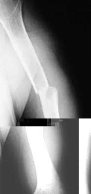
A left-handed 23-year-old man who fell 5 feet from a ladder onto his left elbow sustained the closed injury shown in Figure 26. Management should consist of


Explanation

The radiographs reveal a displaced olecranon fracture. To maximize joint congruity of this intra-articular injury, open reduction and internal fixation is the treatment of choice. A tension band plate will assist with maintenance of the reduction and may aid in early range of motion because injuries to the elbow are prone to stiffness. The oblique fracture line is particularly well suited to plate fixation. Percutaneous pin fixation is unlikely to achieve anatomic joint reduction that can be obtained with open means. External immobilization will not accomplish joint reduction and will most likely lead to a nonunion. Hotchkiss RN: Fractures and dislocations of the elbow, in Rockwood CA Jr, Green DP, Bucholz RW, Heckman JD (eds): Rockwood and Green's Fractures in Adults, ed 4. Philadelphia, PA, Lippincott-Raven, 1996, pp 929-1024. Murphy DF, Greene WB, Gilbert JA, Dameron TB Jr: Displaced olecranon fractures in adults: Biomechanical analysis of fixation methods. Clin Orthop 1987;224:210-214.

Question 2

Which of the following is a long-term complication of ankle arthrodesis for posttraumatic arthritis?


Explanation

Ankle arthrodesis for posttraumatic ankle arthrosis provides reliable pain relief. However, the long-term sequela of joint arthrodesis is the development of arthrosis in the surrounding joints. Over time, following ankle arthrodesis, the ipsilateral hindfoot and midfoot joints show signs of joint space wear, and this may be symptomatic. With a stable ankle arthrodesis, progressive limb-length discrepancy or talar osteonecrosis is not expected. Ankle arthrodesis has not been definitively linked to ipsilateral knee arthritis or contralateral ankle arthritis. Coester LM, Saltzman CL, Leupold J, Pontarelli W: Long-term results following ankle arthrodesis for post-traumatic arthritis. J Bone Joint Surg Am 2001;83:219-228.

Question 3

A 19-year-old female long-distance runner has an incomplete tension-side femoral neck stress fracture. Management should consist of


Explanation

Unlike compression-side stress fractures, tension-side stress fractures on the superior side of the femoral neck are at a very high risk of displacement, even if the patient is not bearing weight. It is highly recommended to treat these fractures like acute fractures and to proceed with internal fixation emergently. Once the fracture has displaced, the prognosis is poor in terms of returning to sports, even when reduced and internally fixed. Nonsurgical management, such as limited weight bearing and low-impact activities, works very well for other lower extremity stress fractures. A training program evaluation (shoes, tracks, schedule) is always indicated for all patients with stress fractures.

Question 4

A 7-year-old girl who sustained a type III posteromedial extension supracondylar fracture underwent a closed reduction at the time of injury. Figure 27a shows the position of the fracture fragments prior to percutaneous medial and lateral pin fixation. Following surgery, healing was uneventful and the patient regained a full painless range of motion. Fifteen months after the injury, she now reports loss of elbow motion and moderate pain with activity. A current AP radiograph is shown in Figure 27b. What is the most likely cause of her symptoms?


Explanation

27b The patient sustained a very distal supracondylar fracture of the humerus. Fractures in this area can disrupt the blood vessels supplying the lateral ossification center of the trochlea. With disturbance of the blood supply in this area, local osteonecrosis occurs and disrupts the support for the overlying articular surface, producing joint incongruity and localized degenerative arthritis. Haraldsson S: The interosseous vasculature of the distal end of the humerus with special reference to the capitellum. Acta Orthop Scand 1957;27:81-93.

Question 5

A 55-year-old man sustained an isolated closed fracture of the humerus. Initial neurologic examination reveals no active wrist or finger extension. Radiographs are shown in Figures 28a and 28b. Management should consist of


Explanation

28b The patient has an isolated closed injury involving the humeral diaphysis. The lack of wrist and finger extension indicates injury to the radial nerve. Based on these findings, ongoing observation of the nerve is warranted with delayed exploration after 3 to 4 months if there are no signs of progressive return of nerve function. Treatment of the fracture should include external immobilization and fracture bracing. An indication for nerve exploration and surgical stabilization would be an open fracture. Zuckerman JD, Kovil KJ: Fractures of the shaft of the humerus, in Rockwood CA Jr, Green DP, Bucholz RW, Heckman JD (eds): Rockwood and Green's Fractures in Adults, ed 4. Philadelphia, PA, Lippincott-Raven, 1996, pp 1025-1053.

Question 6

Examination of a 41-year-old man who was thrown from a motorcycle reveals that both legs appear externally rotated and there is bruising in the perineal area. He has a blood pressure of 80/40 mm Hg, a pulse rate of 140/min, a respiratory rate of 25/min, and he appears confused. Following administration of 4 L of saline solution and 2 units of packed red blood cells, he has a blood pressure of 80/40 mm Hg, a pulse rate of 160/min, and a respiratory rate of 25/min. The abdominal assessment for intraperitoneal blood is negative. An AP radiograph shows an anteroposterior compression injury with 7 cm of symphysis diastasis but no posterior displacement in the sacroiliac joints. What is the next most appropriate step in management?


Explanation

Because the patient has sustained a major high-energy injury to the pelvic ring, it can be assumed that there is serious bleeding or hemodynamic instability related to a pelvic vascular injury. The goal of intervention at this time is to assist in the resuscitative effort and to stop the bleeding. All attempts at providing fluid and blood are important, but without cessation of the bleeding continued loss occurs and significant problems can ensue such as coagulopathy and multiple organ failure. Noninvasive methods of stabilizating the pelvic ring should be used to stop the bleeding. These methods include wrapping a sheet around the pelvis or using commercially available belts, vacuum beanbags, or pneumatic shock garments. This will provide time to prepare for arteriography and/or external fixation. The next step is debatable but in view of negative findings for intra-abdominal blood, arteriography performed with the pelvis reduced using noninvasive methods would be ideal. Bassam D, Cephas GA, Ferguson KA, Beard LN, Young JS: A protocol for the initial management for unstable pelvic fractures. Am Surg 1998;64:862-867. Levine AM (ed): Orthopaedic Knowledge Update: Trauma. Rosemont, IL, American Academy of Orthopaedic Surgeons, 1996, pp 217-226.

Question 7

A 32-year-old man sustained a closed injury after falling 25 feet from a roof. His ankle and foot are severely swollen. Radiographs and CT scans are shown in Figures 29a through 29d. Initial management should consist of


Explanation

29b 29c 29d The patient has a severe high-energy injury from axial loading to the left ankle and distal tibia. This is a closed injury, but the soft tissues are injured and severely swollen. Initial treatment should focus on skeletal stabilization, and incisions directly over the fracture area should be avoided until soft-tissue stabilization has occurred. Immediate spanning external fixation with plans for a delayed reconstruction as needed for the joint surface is the treatment of choice. Closed reduction and application of a constrictive long leg cast may lead to increased risk of tissue necrosis. Immediate open procedures to internally fix the fracture add the risks of soft-tissue necrosis and are to be avoided. Percutaneous plating may be one of the delayed fixation options but should not be used immediately. Primary ankle arthrodesis is not indicated. Thordarson DB: Complications after treatment of tibial pilon fractures: Prevention and management strategies. J Am Acad Orthop Surg 2000;8:253-265. Marsh JL, Bonar S, Nepola JV, DeCoster TA, Hurwitz SR: Use of an articulated external fixator for fractures of the tibial plafond. J Bone Joint Surg Am 1995;77:1498-1509.

Question 8

Which of the following parameters is considered most important when assessing an acetabular fracture for surgical indications?


Explanation

The most important aspect in the decision for surgery in an acetabular fracture is the ability of the femoral head to remain concentrically reduced under the dome in AP and Judet oblique views of the pelvis. If this parameter is present, then the need for surgery is determined by other aspects such as fragmentation, age, incongruity, and displacement. If the head remains stable under the dome without traction, there is sufficient acetabular dome to provide stability, and nonsurgical treatment may be appropriate. Tile M: Assessment and management of acetabular fractures, in Tile M (ed): Pelvic and Acetabular Fractures, ed 2. Baltimore, MD, Williams and Wilkins, 1995, pp 305-354. Letournel E: Acetabular fractures: Classification and management. Clin Orthop 1980;151:81-106.

Question 9

A 57-year-old man has had right ankle pain for the past 10 months following an injury that went untreated. Radiographs are shown in Figures 30a through 30c. Management should consist of


Explanation

30b 30c The radiographs reveal a malunited distal fibular fracture with shortening. Because there appears to be an adequate cartilage space within the ankle joint, the role of reconstruction would be to prevent arthrosis and the need for ankle arthrodesis, as well as to decrease symptoms. The treatment of choice is restoration of fibular length, alignment, and rotation with osteotomy plating, and bone grafting as needed. There is no indication for ligament reconstruction of a mechanically stable ankle, and tibial shortening osteotomy will not assist in correcting the deformity. Cast immobilization may assist with improvement of symptoms but will not correct the overall process. Determination of fibular length is best done by comparing the talocrural angle of the injured side with the uninjured side. The goal is to perfectly reduce the talus in the ankle mortise. Marti RK, Raaymakers EL, Nolte PA: Malunited ankle fractures: The late results of reconstruction. J Bone Joint Surg Br 1990;72:709-713. Geissler W, Tsao A, Hughes J: Fractures and injuries of the ankle, in Rockwood CA Jr, Green DP, Bucholz RW, Heckman JD (eds): Rockwood and Green's Fractures in Adults, ed 4. Philadelphia, PA, Lippincott-Raven, 1996, pp 2201-2206.

Question 10

A 32-year-old man sustains a forceful inversion injury while playing soccer. Examination reveals tenderness in the lateral hindfoot and midfoot region with associated ecchymosis and swelling. Radiographs show proximal migration of the os peroneum. Active eversion is still present. These findings indicate disruption of the


Explanation

The os peroneum is an accessory ossicle located within the peroneus longus tendon. It is typically located at the level of the cuboid groove in the lateral hindfoot and midfoot region. Proximal migration of the os peroneum indicates disruption of the peroneus longus tendon and is an important clue to diagnosis. This unusual condition can cause chronic lateral ankle pain, and surgical repair may be indicated. Active eversion indicates that the peroneus brevis is clinically intact. Disruption of the extensor digitorum brevis, plantar fascia, or syndesmosis would have no effect on the position of the os peroneum. Thompson FM, Patterson AH: Rupture of the peroneus longus tendon: Report of three cases. J Bone Joint Surg Am 1989;71:293-295.

Question 11

A 24-year-old man sustained a grade IIIb open tibial fracture and an ipsilateral grade IIIa femoral fracture in a motorcycle accident. He is unresponsive, intubated, and has a Glasgow Coma Scale score of 8. He is resuscitated and taken to the operating room for definitive orthopaedic care. Which of the following intraoperative problems will most likely adversely affect his long-term outcome?


Explanation

Traumatic brain injury is considered to be either primary or secondary. Primary injury is direct or impact damage to the brain, and secondary injury can have intracranial or systemic causes. While treatment has little impact on primary brain injury, secondary brain injury can be avoided. There are also many causes of intracranial secondary brain injury, including intracranial hypertension or cerebral edema. There are many causes of systemic secondary brain injury, but none has a greater impact on outcome than hypotension or hypoxia. In fact, the occurrence of hypotension postinjury causes a 10- to 15-fold increase in mortality. In a series by Pietropaoli and associates, the mortality rate for head-injured patients that were normotensive during surgery was 25%, but if they were hypotensive the mortality rate was 82%. In the same series, the number of patients with a Glasgow Coma Scale score of either 4 or 5 dropped from 58% in those patients that were normotensive during surgery to 6% in those patients that became hypotensive during surgery. Efforts to avoid hypotension postinjury and especially during surgery should be of primary importance. Chesnut RM, Marshall LF, Klauber MR, et al: The role of secondary brain injury in determining outcome from severe head injury. J Trauma 1993;34:216-222. Pietropaoli JA, Rogers FB, Shackford SR, Wald SL, Schmoker JD, Zhuang J: The deleterious effects of intraoperative hypotension on outcome in patients with severe head injury. J Trauma 1992;33:403-407. Schmeling GJ, Schwab JP: Polytrauma care: The effect of head injuries and timing of skeletal fixation. Clin Orthop 1995;318:106-116.

Question 12

Figure 31 shows the radiograph of an 8-year-old boy who has a swollen forearm after falling out of a tree. Examination reveals that all three nerves are functionally intact, and there is no evidence of circulatory embarrassment. Management should consist of


Explanation

The patient has a Bado type IV Monteggia lesion. It involves dislocation of the radial head and fractures of both the radial and ulnar shafts. These fractures are very difficult to manage by closed reduction alone. The radial and ulnar shafts first have to be stabilized surgically to give a lever arm to reduce the radial head. In this age group, intramedullary pins are easy to insert percutaneously and cause less tissue trauma than plates and screws. In these types of injuries, the focus is often on the forearm fracture; the radial head dislocation may not be appreciated as was the case with this patient. Gibson WK, Timperlake RW: Operative treatment of a type IV Monteggia fracture-dislocation in a child. J Bone Joint Surg Br 1992;74:780-781.

Question 13

Figure 32 shows the radiograph of a laborer who jammed his thumb in a fall. Examination reveals pain at the base of the thumb and proximal thenar eminence region. Management should consist of


Explanation

The radiographs are classic for a Bennett's fracture, which involves a fracture of the palmar ulnar aspect of the proximal phalanx. This fracture fragment is still attached to the anterior oblique ligament. The deforming forces that cause subluxation of the base of the proximal phalanx include the pull of the abductor pollicis longus as well as the adductor pollicis. Adequate reduction can be achieved by closed reduction, percutaneous pin fixation, and casting. The fragment is too small for secure internal fixation. Stern PJ: Fractures of the metacarpals and phalanges, in Green DP, Hotchkiss RN, Pederson WC (eds): Green's Operative Hand Surgery, ed 4. Philadelphia, PA, 1999, pp 711-771.

Question 14

In displaced calcaneal fractures, what fragment is the only one that remains in its anatomic position?


Explanation

The sustentaculum tali remains in its anatomic position because of its supporting ligamentous structures. This provides the key to the reconstruction of the calcaneus. The posterior facet is reduced to the sustentaculum tali and then fixed to it for stability. All of the other components of the calcaneus are then reduced to this complex. Sanders R: Displaced intra-articular fractures of the calcaneus. J Bone Joint Surg Am 2000;82:225-250. Eastwood DM, Gregg PJ, Atkins RM: Intra-articular fractures of the calcaneum: Part I. Pathological anatomy and classification. J Bone Joint Surg Br 1993;75:183-188.

Question 15

A 46-year-old man sustains a calcaneal fracture in a fall off a scaffold. During surgical reconstruction using an extended lateral incision, the fracture is reduced and fixed with a plate and screws. One of the posterior facet screws is found to be 5 mm out of the bone on the Harris view. What structure is most likely at risk because of this finding?


Explanation

The abductor hallucis muscle is the most medial structure. The posterior tibial tendon and the flexor digitorum longus tendon lie more cephalad to the sustentaculum tali. There is a groove under the sustentaculum for the flexor hallucis longus tendon. Subchondral lag screws placed across the posterior facet exit the medial side of the calcaneus in this groove. Just medial to the flexor hallucis longus tendon is the neurovascular bundle. A screw that is out of the bone a short distance can cause triggering of the flexor hallucis longus tendon. Patients will report loss of great toe excursion in the early postoperative period. Accurate measurement of subchondral lag screw length avoids this complication. Hollinshead WH: Anatomy for Surgeons, ed 3. Philadelphia, PA, Harper and Row, 1982, pp 802-852. Rosenberg AS, Cheung Y: Diagnostic imaging of the ankle and foot, in Jahss MH (ed): Disorders of the Foot and Ankle, ed 2. Philadelphia, PA, WB Saunders, 1991, pp 109-154.

Question 16

A 23-year-old man sustained an injury to his left foot when a forklift rolled over it at work. Examination reveals marked swelling of the midfoot and forefoot, with tenderness to palpation over the medial hindfoot and dorsomedial forefoot. The distal dorsalis pedis pulse is audible on Doppler examination, and his sensation is intact to touch. Radiographs are shown in Figures 33a and 33b. Management should consist of


Explanation

33a 33b The best results after dislocations of the tarsometatarsal joints are seen with anatomic reduction; this is best achieved by open reduction and maintained with internal fixation with either pins or screws. Open reduction provides a means of debriding small bony fragments from the joint and allowing direct inspection of the reduction. Associated crush or shearing fractures of the cuboid or tarsal navicula are signs that suggest a Lisfranc injury. Because patients can function quite well despite the development of arthrosis in the Lisfranc joint, primary arthrodesis is not indicated in the management of this injury. Resch S, Stenstrom A: The treatment of tarsometatarsal injuries. Foot Ankle 1990;11:117-123. Schenck RC Jr, Heckman JD: Fractures and dislocations of the forefoot: Operative and nonoperative treatment. J Am Acad Orthop Surg 1995;3:70-78.

Question 17

A 24-year-old woman who has hypotension, a head injury, and who experienced a poor response to resuscitation has been taken to the operating room for a splenectomy. Following abdominal surgery she remains unstable with increasing pulmonary respiratory pressures and decreasing oxygen saturation. She has a transverse mid-diaphyseal fracture of the tibia with a 4-cm laceration and soil-contaminated muscle in the wound. Based on these findings, management should consist of


Explanation

Because the patient is critically ill and requires expeditious care, stabilization of the long bone fracture is required, but definitive care of the fracture should be postponed. The treatment of choice at this time is irrigation with 12 L of saline solution, followed by debridement and nondefinitive stabilization with a simple four-pin external frame to regain axial and rotational alignment. When the patient's condition is more stable, more definitive care can be performed. Bosse MJ, Kellam JF: Orthopaedic management decisions in the multiple trauma patient, in Browner BD, Jupiter JP, Levine AM, Trafton P (eds): Skeletal Trauma, ed 2. Philadelphia, PA, WB Saunders, 1998, pp 151-164. Weresh MJ, Stover MD, Bosse MJ, Jeray K, Kellam JF: Pulmonary gas exchange during intramedullary fixation of femoral shaft fractures. J Trauma 1999;46:863-868.

Question 18

A 53-year-old woman has severe neck and left shoulder pain after a rollover motor vehicle accident. Radiographs and a CT scan of the cervical spine are shown in Figures 34a through 34c. Management should consist of


Explanation

34b 34c The plain radiographs show a horizontal orientation of the C5 facet joint. The CT scan through C5 reveals an ipsilateral pedicle and lamina fracture (floating facet). This injury involves two adjacent motion segments and is extremely unstable. Lateral mass plates, with or without the purchase of the "floating facet," provide the best means of stabilization and should include the facet above (C4) and below (C6) the level of injury. Orthotic immobilization is insufficient for this particular injury. Halo vest treatment does not control the subaxial spine well and is of limited value. While simple midline (Rogers) wiring provides some tension band restoration, it is not optimal for rotational control. The use of lateral mass plates provides rotational stability. Another option would be anterior fusion and plating, which would save cervical segments. Levine AM, Mazel C, Roy-Camille R: Management of fracture separations of the articular mass using posterior cervical plating. Spine 1992;17:S447-S454. Levine AM: Facet fractures and dislocations, in Levine AM, Eismont FJ, Garfin S, Zigler JE (eds): Spine Trauma. Philadelphia, PA, WB Saunders, 1998, pp 360-362. Whitehill R, Richman JA, Glaser JA: Failure of immobilization of the cervical spine by the halo vest: A report of five cases. J Bone Joint Surg Am 1986;68:326-332.

Question 19

What is the most common clinically significant preventable complication secondary to the treatment of a displaced talar neck fracture?


Explanation

The most important consequence of a displaced talar neck fracture after closed or open treatment is malunion. Because displacement of the talar neck is associated with displacement of the subtalar joint, any malunion leads to intra-articular incongruity or malalignment of the subtalar joint. Varus malunion is common when there is comminution of the medial talar neck. This results in pain, osteoarthritis, and hindfoot deformity that requires further treatment. Because of these complications, it is imperative that all displaced talar neck fractures are reduced anatomically; fragmented fractures may require bone grafting to maintain the length and rotation of the neck. Tile M: Fractures of the talus, in Schatzker J, Tile M (eds): Rationale of Operative Fracture Care, ed 2. Berlin, Springer-Verlag, 1996, pp 563-588. Daniels TR, Smith JW, Ross TI: Varus malalignment of the talar neck: Its effect on the position of the foot and on subtalar motion. J Bone Joint Surg Am 1996;78:1559-1567.

Question 20

Examination of a carpenter who hit his thumb with a hammer reveals that the nail plate is broken but in place, and there is a 100% subungual hematoma that covers 100% of the area under the nail plate. Radiographs reveal a comminuted distal phalangeal tuft fracture. Management should consist of


Explanation

This is a classic situation for a distal phalanx tuft fracture with associated nail bed injury and subungual hematoma. In general, when the subungual hematoma is greater than 50% of the surface area under the nail plate, treatment should consist of nail plate removal, nail bed repair, oral antibiotics, and a fingertip splint. Oral antibiotics and fingertip splinting alone do not address the nail bed laceration, which will most likely lead to nail plate deformity if not repaired. Kirschner pin stabilization is not indicated because these fractures are nondisplaced and usually are inherently stable after nail bed repair. The use of IV antibiotics alone does not address the nail bed laceration surgically. Casting, followed by hydrotherapy and topical antibiotics, is not indicated because it does not address the nail bed laceration. Further, a nondisplaced distal phalangeal tuft fracture does not require cast immobilization. Stern PJ: Fractures of the metacarpals and phalanges, in Green DP, Hotchkiss RN, Pederson WC (eds): Green's Operative Hand Surgery, ed 4. Philadelphia, PA, 1999, pp 711-771.

Question 21

An olecranon fracture-dislocation of the elbow in which the fracture line exits distal to the coronoid process is best managed by open reduction and


Explanation

Fracture-dislocations of the elbow present difficult management problems. Standard olecranon fractures normally are not associated with a dislocation; however, the surgeon needs to recognize that some fractures that have a dislocation, in particular a posterior dislocation, represent a Monteggia equivalent. These injuries are not ulnar shaft fractures because they are fractured at or just distal to the coronoid; however, because of the unstable fracture-dislocation, the forces across this reduction are high. Two Kirschner wires and a tension band wire provide inadequate fixation. Therefore, the preferred method of fixation is plate osteosynthesis with a 3.5-mm low-contact dynamic compression plate or reconstruction plate. Jupiter JP, Kellam JF: Fractures of the forearm, in Browner BD, Jupiter JP, Levine AM, Trafton P (eds): Skeletal Trauma, ed 2. Philadelphia, PA, WB Saunders, 1998, pp 421-454. Quintero J: Fracture of the forearm, in Ruedi TP, Murphy WM (eds): AO Principles of Fracture Management. Stuttgart, Thieme, 2000, pp 323-337.

Question 22

A 15-year-old baseball pitcher who reports increasing pain in his right shoulder over the past 3 weeks states that the pain increases the more he pitches. Radiographs of both shoulders are shown in Figures 35a and 35b. What is the next most appropriate step in management?


Explanation

35b The patient has a rotational stress fracture of the proximal humeral physis (Little Leaguer's shoulder). The symptoms of increasing pain with activity and relief with rest are typical of a stress injury. Treatment should consist of cessation of throwing activity but rehabilitation of the shoulder girdle muscles. The pitching technique should be evaluated as well. Barnett LS: Little League shoulder syndrome: Proximal humeral epiphyseolysis in the adolescent baseball pitchers: A case report. J Bone Joint Surg Am 1985;67:495-496.

Question 23

A 36-year-old man sustains a traumatic spondylolisthesis of L5 on S1. Surgical stabilization requires pedicular fixation into the sacrum. If the screw is placed in a medial to lateral direction and penetrates the sacral ala, what nerve root is at risk?


Explanation

The L5 nerve root lies directly over the superior and anterior alae. If the screw is directed approximately 20 degrees laterally and bicortical purchase is achieved, there is the risk of injuring the L5 nerve root. If the screw is directed medially into the body of S1, there is little risk of injury. The same root is at risk during placement of an iliosacral screw. Ebraheim NA, et al: Lumbosacral nerve and dorsal screw placement. Orthopedics 2000;23:245-247. Ebraheim NA, Mermer M, Xu R, Yeasting RA: Radiological evaluation of S1 dorsal screw placement. J Spinal Disord 1996;9:527-535.

Question 24

A 25-year-old woman who fell on her outstretched hand reports chronic pain over the hypothenar eminence region and some dorsal ulnar wrist pain. She also notes difficulty playing golf and tennis. Plain radiographs of the hand and wrist are unremarkable. A CT scan is shown in Figure 36. What is the next most appropriate step in management?


Explanation

The CT scan reveals a hook of the hamate nonunion with irregular resorption at the fracture site, which is at the base of the hamate. Symptomatic relief of the pain and discomfort has been well documented after excision of the hook of the hamate. Ultrasound therapy will not provide long-term symptomatic relief or induce nonunion healing. MRI for further soft-tissue evaluation is inappropriate because this is a bony problem; the bony architecture of the wrist is best visualized by CT. Open reduction and internal fixation of the hook of the hamate does not provide the symptomatic relief that is found with excision of the hook of the hamate. In addition, the technical difficulties and relative risk of persistent nonunion after open reduction and internal fixation are not merited when hamate excision can be effected easily and causes no long-term untoward effects. Electrodiagnostic evaluation is inappropriate because there is no history of the persistent numbness and tingling that is found in peripheral compression neuropathies. Stark HH, Chao EK, Zemel NP, Rickard TA, Ashworth CR: Fracture of the hook of the hamate. J Bone Joint Surg Am 1989;71:1206-1207. Failla JM: Hook of hamate vascularity: Vulnerability to osteonecrosis and nonunion. J Hand Surg Am 1993;18:1075-1079. Carter PR, Easton RG, Littler JW: Ununited fracture of the hook of the hamate. J Bone Joint Surg Am 1977;59:583-588.

Question 25

An active 72-year-old woman sustained a mid-diaphyseal right humerus fracture 16 months ago. History reveals that she was first treated with a brace for 7 months. Additional treatment consisted of intramedullary nailing 9 months ago. Recently the rod was removed, and the patient now reports pain and gross motion at the fracture site. Current radiographs are shown in Figures 37a and 37b. What is the next most appropriate step in management?


Explanation

37b The patient has a well-established nonunion in a very porotic bone. Electrical stimulation has been found effective in treating tibial nonunions, but there is very little data on humeral nonunions, especially chronic well-established ones. Ultrasound stimulation is effective in accelerating fracture healing, but there is little data concerning the treatment of nonunions. Intramedullary nailing with bone graft is an option, but it maybe difficult to obtain a rigid construct in a very porotic bone. An Ilizarov-type external fixator would be an alternative, but there is little clinical data for the humerus and it may be poorly tolerated. A plate and screw construct with bone graft combines rigidity with the biologic advantage of the bone graft. A recent series reported on the use of a plate combined with onlay allograft for recalitrant nonunions. Cement augmentation for screw fixation either in the canal or added to the screw holes may be helpful in select cases. Hornicek FJ, Zych GA, Hutson JJ, Malinin TI: Salvage of humeral nonunions with onlay bone plate allograft augmentation. Clin Orthop 2001;386:203-209.

Question 26

A 78-year-old female presents with a highly comminuted, intra-articular distal humerus fracture after a fall. Her bone quality is osteoporotic. Which of the following surgical options is associated with the most reliable return to independent activities of daily living in this specific patient demographic?


Explanation

Total elbow arthroplasty (TEA) is the preferred treatment for elderly patients with highly comminuted distal humerus fractures and osteoporotic bone. TEA allows for immediate weight-bearing for transfers and early range of motion, providing more reliable functional outcomes than ORIF in this population.

Question 27

A 30-year-old male with a closed midshaft tibia fracture is treated with intramedullary nailing. Six hours postoperatively, he complains of pain out of proportion to the injury. His blood pressure is 110/70 mmHg. Direct measurement of the anterior compartment pressure reveals a value of 45 mmHg. What is the most appropriate next step in management?


Explanation

Compartment syndrome is diagnosed when the Delta P (diastolic blood pressure minus compartment pressure) is less than 30 mmHg. This patient has a Delta P of 25 mmHg (70 - 45), which is a definitive indication for an urgent four-compartment fasciotomy.

Question 28

A 25-year-old male arrives at the trauma bay hemodynamically unstable after a high-speed motorcycle collision. An AP pelvic radiograph demonstrates a symphysis pubis diastasis of 4 cm and widening of the sacroiliac joints consistent with an APC III injury. During initial resuscitation, a pelvic binder is applied. To maximize reduction of the pelvic volume, over which anatomical landmark should the binder be centered?


Explanation

To effectively reduce pelvic volume and tamponade venous bleeding in an open-book pelvic fracture, a pelvic binder must be centered directly over the greater trochanters. Placement over the iliac crests is incorrect and can paradoxically worsen the deformity.

Question 29

A 40-year-old construction worker falls from a height of 15 feet and sustains bilateral joint-depression calcaneus fractures. Which of the following injuries is most commonly associated with this mechanism of trauma and must be actively ruled out?


Explanation

High-energy axial loading injuries that cause bilateral calcaneus fractures have a highly established association with spinal trauma, particularly thoracolumbar burst fractures. A thorough clinical and radiographic evaluation of the spine is mandatory.

Question 30

A 28-year-old female sustains a high-energy basicervical femoral neck fracture (Pauwels III) following a motor vehicle collision. Which of the following fixation constructs is biomechanically optimal to resist the high shear forces inherent in this fracture pattern?


Explanation

Pauwels III fractures are high-angle vertical fractures characterized by significant shear instability. A sliding hip screw coupled with a derotational screw provides superior biomechanical stability and higher load to failure compared to multiple cancellous screws.

Question 31

A 35-year-old male sustains a purely ligamentous Lisfranc injury. There are no associated fractures, but weight-bearing radiographs show 3 mm of widening between the medial and middle cuneiforms. What is the recommended definitive treatment to optimize long-term functional outcomes?


Explanation

Evidence demonstrates that primary arthrodesis yields superior functional outcomes and lower reoperation rates compared to open reduction and internal fixation for purely ligamentous Lisfranc injuries. ORIF is associated with higher rates of hardware failure and post-traumatic arthritis in purely ligamentous variants.

Question 32

A 50-year-old female sustains an open tibia fracture (Gustilo-Anderson IIIB) and undergoes initial surgical debridement and intramedullary nailing. A rotational muscle flap is planned. To minimize the risk of deep infection, what is the optimal timeframe for soft-tissue coverage?


Explanation

Early soft-tissue coverage of Gustilo IIIB tibia fractures, optimally performed within 72 hours (and generally less than 5 to 7 days), significantly decreases the risk of deep infection and promotes fracture union. Delays beyond this window are associated with higher complication rates.

Question 33

A 22-year-old male presents with a closed, distal-third humeral shaft fracture. He is neurologically intact on initial evaluation in the emergency department. Following closed reduction and placement of a coaptation splint, he exhibits a complete wrist drop and inability to extend his metacarpophalangeal joints. What is the most appropriate next step in management?


Explanation

While a primary radial nerve palsy associated with a closed humeral shaft fracture is typically observed, a secondary palsy occurring after a reduction maneuver is a strong indication for immediate surgical exploration. This secondary deficit suggests possible entrapment or laceration of the nerve in the fracture site.

Question 34

A 45-year-old male presents with a high-energy pilon fracture. The limb exhibits marked soft-tissue swelling and extensive fracture blisters circumferentially. Definitive open reduction and internal fixation is planned. What is the most appropriate initial management?


Explanation

The standard of care for severe pilon fractures with massive soft-tissue compromise is the 'span-scan-plan' approach. Initial management consists of a spanning external fixator and elevation to allow the soft-tissue envelope to recover before attempting definitive fixation.

Question 35

A 60-year-old female taking alendronate for 10 years presents with 3 months of lateral thigh pain. Radiographs reveal diffuse cortical thickening of the lateral subtrochanteric femur with a transverse radiolucent 'beak' on the lateral cortex extending halfway through the bone. What is the most appropriate management?


Explanation

This patient has an impending atypical femur fracture associated with long-term bisphosphonate use. Prophylactic intramedullary nailing is indicated to relieve pain and prevent completion of the fracture, followed by medical management including cessation of the bisphosphonate.

Question 36

A 32-year-old male is evaluated 8 weeks after open reduction and internal fixation of a displaced talar neck fracture. The AP radiograph of the ankle reveals a subchondral radiolucent band in the talar dome. What does this radiographic finding indicate?


Explanation

The subchondral radiolucency is known as the Hawkins sign. It represents subchondral osteopenia secondary to hyperemia, which proves that the talar body has sufficient blood supply, making avascular necrosis highly unlikely.

Question 37

A 48-year-old male sustains a posterior hip dislocation in a motor vehicle collision. The dislocation is successfully reduced closed in the emergency department within 2 hours of the injury. What is the most significant long-term complication associated with this injury?


Explanation

Osteonecrosis (avascular necrosis) of the femoral head is a major long-term complication of posterior hip dislocations. The risk is minimized by urgent reduction within 6 hours, but remains a significant potential complication even with prompt treatment.

Question 38

A 29-year-old male sustains a terrible triad injury of the elbow (elbow dislocation, radial head fracture, and coronoid fracture). Operative management is planned. According to standard treatment algorithms, what is the typical sequence of surgical repair?


Explanation

The standard surgical algorithm for a terrible triad injury proceeds from deep to superficial. Fixation begins with the coronoid, followed by addressing the radial head (repair or replacement), and finally repairing the lateral collateral ligament complex.

Question 39

A 38-year-old male sustains an isolated, displaced, midshaft clavicle fracture with 2.5 cm of shortening. What is the primary established benefit of operative fixation over non-operative management for this specific injury pattern?


Explanation

Displaced and shortened (>2 cm) midshaft clavicle fractures have a significantly higher nonunion rate and worse functional outcomes when treated non-operatively. Operative fixation significantly decreases the risk of nonunion and symptomatic malunion.

Question 40

A 24-year-old male is brought to the trauma bay with scapulothoracic dissociation following a motorcycle accident. What is the most critical immediate life-threatening concern associated with this injury?


Explanation

Scapulothoracic dissociation involves a massive disruption of the shoulder girdle from the axial skeleton. Vascular injury, particularly massive hemorrhage from subclavian artery or vein disruption, is the most immediate life-threatening concern.

Question 41

A 55-year-old male undergoes an extensile lateral approach for open reduction and internal fixation of a joint-depression calcaneus fracture. During the development of the full-thickness subperiosteal flap, which neurological structure is at highest risk of iatrogenic injury?


Explanation

The sural nerve courses along the lateral aspect of the hindfoot and is at significant risk of injury during the incision, flap elevation, and retraction required for the extensile lateral approach to the calcaneus.

Question 42

A 26-year-old male professional soccer player sustains an acute Zone 2 (Jones) fracture of the proximal fifth metatarsal. He wishes to return to play as safely and rapidly as possible. What is the most appropriate management?


Explanation

In elite athletes, early intramedullary screw fixation of a Jones fracture is recommended. It minimizes the high risk of nonunion associated with this watershed area and allows for a more accelerated return to competitive sports compared to non-operative treatment.

Question 43

A 33-year-old male sustains an anteroposterior compression (APC) pelvic ring injury. Which of the following anatomic disruptions defines an APC III injury and differentiates it from an APC II injury?


Explanation

According to the Young-Burgess classification, an APC II injury involves disruption of the anterior sacroiliac ligaments with an intact posterior hinge. An APC III injury involves complete disruption of both the anterior and posterior sacroiliac ligaments, leading to complete global instability.

Question 44

A 65-year-old male with a native hip sustains a low-energy anterior hip dislocation after a fall. Upon initial inspection in the emergency department, what is the typical clinical posture of the affected lower extremity?


Explanation

Anterior hip dislocations typically present with the affected limb in a flexed, abducted, and externally rotated position. This contrasts with posterior hip dislocations, which classically present flexed, adducted, and internally rotated.

Question 45

A 40-year-old male sustains a low-velocity gunshot wound to the right thigh, resulting in a non-displaced midshaft femur fracture. Clinical examination reveals normal distal pulses and no neurologic deficits. What is the most appropriate initial management?


Explanation

Low-velocity gunshot wounds resulting in non-displaced fractures without vascular compromise, severe contamination, or significant soft tissue loss can safely be managed non-operatively. Treatment consists of local wound care, tetanus prophylaxis, and appropriate immobilization.

Question 46

A 25-year-old male sustains a high-energy motor vehicle collision, resulting in a displaced talar neck fracture with dislocation of both the subtalar and tibiotalar joints. According to the Hawkins classification, what is the approximate expected rate of avascular necrosis (AVN) of the talar body for this injury pattern?


Explanation

This describes a Hawkins Type III talar neck fracture. Because three distinct sources of blood supply are disrupted (artery of the tarsal canal, deltoid branches, and anterior tibial branches), the rate of AVN is exceptionally high, approaching 80-100%.

Question 47

A 35-year-old male undergoes open reduction and internal fixation of a transverse posterior wall acetabular fracture via a Kocher-Langenbeck approach. Postoperatively, the patient demonstrates weak ankle dorsiflexion and eversion, but normal plantarflexion. Which nerve was most likely injured during the procedure?


Explanation

The peroneal division of the sciatic nerve is uniquely susceptible to iatrogenic injury during a Kocher-Langenbeck approach due to its lateral and superficial position and tethering at the sciatic notch. Injury results in foot drop and weak eversion.

Question 48

A 40-year-old presents to the emergency department with a closed middle-third humeral shaft fracture and an immediate, complete radial nerve palsy upon initial presentation. What is the most appropriate initial management?


Explanation

Immediate radial nerve palsy in a closed humeral shaft fracture is typically managed nonoperatively with a functional brace, as the majority are neuropraxias that recover spontaneously. Operative exploration is reserved for open fractures, failure to recover by 3-4 months, or palsies that develop after closed reduction.

Question 49

A 28-year-old male sustains a highly vertical, displaced femoral neck fracture (Pauwels III). Which fixation construct offers the most optimal biomechanical stability for this specific, high-shear fracture pattern?


Explanation

Pauwels III fractures are characterized by a highly vertical fracture line and tremendous shear forces. A sliding hip screw with an anti-rotation screw or a fixed-angle device provides superior biomechanical stability and higher union rates compared to parallel cancellous screws alone.

Question 50

A 45-year-old farmer is struck by a tractor, sustaining a Gustilo-Anderson Type IIIA open tibial shaft fracture heavily contaminated with soil and organic debris. Which initial intravenous antibiotic regimen is most appropriate?


Explanation

For severe open fractures (Type III) with agricultural or soil contamination, standard protocols dictate coverage for Clostridium species. This requires adding high-dose penicillin to the baseline gram-positive (cephalosporin) and gram-negative (aminoglycoside) coverage.

Question 51

A 50-year-old man sustains a high-energy bicondylar tibial plateau fracture. Axial and coronal CT imaging reveals a large, separate, and distally displaced posteromedial fragment. What is the optimal surgical approach to adequately address this specific fragment?


Explanation

Posteromedial fragments in tibial plateau fractures typically displace distally and posteriorly due to the pull of the semimembranosus. A dedicated posteromedial approach with anti-glide or buttress plating is required because anterolateral locked plates cannot adequately capture or compress this coronal shear fragment.

Question 52

A 25-year-old male is brought to the trauma bay after a motorcycle crash with an Antero-Posterior Compression type III (APC-III) pelvic ring injury. He remains hypotensive despite 2 liters of crystalloid and application of a pelvic binder. A FAST exam is negative. What is the next best step in management?


Explanation

In a hemodynamically unstable patient with a pelvic ring injury and a negative FAST (which rules out major intra-abdominal hemorrhage), the bleeding is primarily retroperitoneal. Pelvic angiography with embolization or preperitoneal pelvic packing is the preferred intervention.

Question 53

A 40-year-old patient presents with a highly comminuted, displaced tibial pilon fracture resulting from a fall from height. The soft tissues are severely swollen with extensive fracture blisters spanning the ankle. What is the preferred initial management strategy?


Explanation

High-energy pilon fractures with severe soft tissue compromise must be managed in a staged fashion to minimize devastating wound complications. A spanning external fixator provides length and alignment while allowing the soft tissue envelope to recover prior to definitive delayed ORIF.

Question 54

A 60-year-old woman falls on an outstretched hand, sustaining a volar Barton's fracture of the distal radius. Which of the following best describes this fracture pattern and its optimal management?


Explanation

A volar Barton fracture is a coronal shear fracture of the volar lip of the distal radius associated with volar subluxation of the carpus. It is inherently unstable and typically requires open reduction and volar buttress plating to restore articular congruity.

Question 55

A 22-year-old athlete sustains a midfoot sprain. Weight-bearing radiographs demonstrate a 3 mm diastasis between the base of the first and second metatarsals. What is the recommended treatment approach?


Explanation

A diastasis greater than 2 mm between the first and second metatarsals on weight-bearing films indicates instability of the Lisfranc ligament complex. Operative stabilization via ORIF or primary arthrodesis is indicated to prevent midfoot collapse and post-traumatic arthritis.

Question 56

A 30-year-old male sustains a closed comminuted tibial shaft fracture. Which of the following clinical findings is the most sensitive early indicator of acute compartment syndrome?


Explanation

Pain out of proportion to the apparent injury and pain on passive stretch of the muscles in the involved compartments are the most sensitive and earliest clinical signs of acute compartment syndrome. Pulselessness, pallor, and paralysis are very late signs indicating irreversible ischemia.

Question 57

A 45-year-old sustains a distal femur fracture. Coronal CT imaging demonstrates an isolated coronal shear fracture of the lateral femoral condyle (Hoffa fracture). Which surgical approach and fixation strategy is generally considered most appropriate?


Explanation

Hoffa fractures are coronal shear fractures, most commonly of the lateral condyle. They require an anatomic reduction via a lateral or anterolateral arthrotomy, followed by rigid fixation typically utilizing anterior-to-posterior (or posterior-to-anterior) countersunk lag screws to compress the articular fragment.

Question 58

In evaluating a patient with a displaced intra-articular calcaneus fracture, which of the following demographic or socioeconomic factors is most strongly associated with a poor clinical outcome and lower return-to-work rates following operative intervention?


Explanation

Large prospective studies (such as Buckley et al.) have consistently demonstrated that patients receiving Workers' Compensation have significantly poorer functional outcomes and markedly lower return-to-work rates following operative fixation of calcaneus fractures compared to those who do not.

Question 59

A 24-year-old male falls on an outstretched hand and sustains a displaced fracture of the proximal pole of the scaphoid. He is at high risk for avascular necrosis (AVN) primarily due to which unique anatomical feature of the scaphoid's blood supply?


Explanation

The primary arterial supply to the scaphoid enters the dorsal ridge near the waist and flows in a retrograde fashion to the proximal pole. Fractures at the waist or proximal pole disrupt this retrograde supply, leading to high rates of nonunion and AVN.

Question 60

A 45-year-old man feels a 'pop' in his anterior elbow while lifting a heavy object. The Hook test is positive. If surgical repair of the distal biceps is performed utilizing a single-incision anterior approach, which nerve is at greatest risk of iatrogenic injury?


Explanation

During a single-incision anterior approach for distal biceps repair, the lateral antebrachial cutaneous nerve (LABCN) is the most commonly injured nerve due to its superficial location in the surgical field. The PIN is more classically at risk during a two-incision approach.

Question 61

During open reduction and internal fixation of a bimalleolar equivalent ankle fracture, the intraoperative Cotton test reveals syndesmotic instability. Which of the following is an accepted biomechanical principle regarding syndesmotic screw fixation?


Explanation

Because the fibula sits posterior to the tibia at the level of the syndesmosis, syndesmotic screws must be angled 20-30 degrees anteriorly from lateral to medial to accurately capture the tibia. Maximum dorsiflexion during insertion is traditionally taught, though modern evidence shows ankle position does not affect syndesmotic width.

Question 62

An 80-year-old woman with a well-fixed cementless total hip arthroplasty sustains a fall. Radiographs demonstrate a periprosthetic femur fracture extending around the stem, but the stem remains completely stable (Vancouver B1). What is the standard operative management?


Explanation

A Vancouver B1 fracture is defined as a fracture around or just below a well-fixed femoral stem. The gold standard treatment is open reduction and internal fixation using locking plates, cerclage cables, and potentially cortical strut allografts, while retaining the stable prosthesis.

Question 63

A 35-year-old male involved in a high-speed MVC sustains an ipsilateral displaced midshaft clavicle fracture and a displaced scapular neck fracture ('floating shoulder'). What is the primary rationale for performing operative fixation of the clavicle in this specific scenario?


Explanation

A 'floating shoulder' represents a double disruption of the superior suspensory shoulder complex. Plating the clavicle is often sufficient to restore stability to the entire complex, indirectly reducing and stabilizing the scapular neck without necessarily requiring scapular fixation.

Question 64

A 40-year-old male presents after a high-speed motorcycle crash. His blood pressure is 70/40 mmHg. Pelvic radiographs show an APC-III injury. A pelvic binder is applied, and 2 units of PRBCs are given. Repeat blood pressure is 75/40 mmHg. FAST exam is negative. What is the most appropriate next step in management?


Explanation

In a hemodynamically unstable patient with a pelvic ring injury and negative FAST, the source of bleeding is likely retroperitoneal. Pelvic packing or angiography with embolization is the appropriate next step to control hemorrhage.

Question 65

A 45-year-old male is involved in a high-speed motorcycle crash. Pelvic radiographs demonstrate a widened symphysis pubis of 3.5 cm and widened anterior sacroiliac joints bilaterally, but the posterior sacroiliac ligaments remain intact. What is the most appropriate definitive management for this specific injury pattern?


Explanation

This is an Anteroposterior Compression Type II (APC-II) pelvic ring injury, characterized by symphyseal diastasis and disruption of the anterior SI ligaments with intact posterior SI ligaments. Anterior symphyseal plate fixation is the standard definitive treatment to restore stability.

Question 66

A 30-year-old female falls from a significant height and sustains a Hawkins Type III talar neck fracture. Based on this classification, what is the approximate risk of developing avascular necrosis (AVN) of the talar body?


Explanation

A Hawkins Type III fracture involves a displaced talar neck fracture with both subtalar and tibiotalar dislocation. This severe disruption of the talar blood supply carries an AVN risk of approximately 75% to 90%.

Question 67

A 28-year-old male sustains a closed tibial shaft fracture and presents with out-of-proportion leg pain and tense compartments. Which of the following pressure measurements is the most reliable objective indicator for performing an emergent fasciotomy?


Explanation

A delta pressure (diastolic blood pressure minus compartment pressure) of less than 30 mmHg is the most reliable objective diagnostic criterion for acute compartment syndrome. Absolute pressures can be misleading due to variations in systemic blood pressure.

Question 68

A 55-year-old male sustains a high-energy closed tibial pilon fracture. Clinical examination reveals massive soft tissue swelling, fracture blisters over the medial ankle, and threatened skin. What is the standard of care for the initial management of this injury?


Explanation

High-energy pilon fractures with severe soft tissue compromise are best managed with a staged protocol. Initial spanning external fixation protects the soft tissues, allowing for swelling to subside before definitive delayed ORIF is safely performed.

Question 69

During the operative management of a supracondylar distal femur fracture, a coronal plane fracture of the lateral femoral condyle (Hoffa fragment) is identified. Which surgical approach and fixation strategy is most appropriate for this specific fragment?


Explanation

A Hoffa fracture is a coronal shear fracture of the femoral condyle. Fixation is biomechanically optimal using anterior-to-posterior lag screws placed perpendicular to the fracture plane, typically accessed via a lateral approach for a lateral condyle fragment.

Question 70

A 40-year-old man presents to the emergency department with a posterior hip dislocation and an associated posterior wall acetabular fracture following a dashboard injury. Which nerve is most commonly injured in this specific clinical scenario?


Explanation

The sciatic nerve, particularly its peroneal division, is the most commonly injured nerve in posterior hip dislocations and posterior wall acetabular fractures due to its anatomic proximity to the posterior acetabulum.

Question 71

A 35-year-old female sustains a "terrible triad" injury of the elbow after a fall onto an outstretched hand. Which of the following correctly describes the anatomic components of this injury pattern?


Explanation

The "terrible triad" of the elbow consists of an elbow dislocation, a radial head fracture, and a coronoid fracture. It is notoriously unstable and almost always involves disruption of the lateral collateral ligament (LCL) complex.

Question 72

A 22-year-old farmer sustains a Gustilo-Anderson Type IIIb open tibial shaft fracture heavily contaminated with soil and organic debris. According to current guidelines, what is the most appropriate initial empiric intravenous antibiotic regimen?


Explanation

For a heavily contaminated agricultural injury (Type III open fracture), broad coverage is required. A first-generation cephalosporin, an aminoglycoside, and high-dose penicillin are recommended to cover Gram-positive, Gram-negative, and anaerobic organisms (such as Clostridium).

Question 73

A 29-year-old motorcyclist presents with massive swelling over the shoulder girdle, a pulseless upper extremity, and a completely flail arm. Radiographs show extreme lateral displacement of the scapula. After the primary ATLS survey, what is the most critical initial diagnostic step?


Explanation

This presentation is highly suspicious for scapulothoracic dissociation, a severe injury frequently associated with limb-threatening subclavian or axillary artery disruption. CT angiography is urgently required to diagnose and plan intervention for potential vascular injury.

Question 74

A 45-year-old construction worker falls from scaffolding, sustaining a joint-depressed intra-articular calcaneus fracture. Bohler's angle is measured at 5 degrees. What is the primary anatomic goal of open reduction and internal fixation (ORIF) in this patient?


Explanation

The primary goals of operative treatment for intra-articular calcaneus fractures are the anatomic reduction of the posterior facet (to minimize post-traumatic arthritis) and the restoration of calcaneal height, width, and alignment.

Question 75

A 60-year-old female presents with a Schatzker Type II tibial plateau fracture. Which concomitant soft tissue injury is most commonly associated with this specific fracture pattern?


Explanation

Schatzker II fractures (split-depression of the lateral tibial plateau) are highly associated with lateral meniscal tears. These are frequently peripheral or capsular separations, occurring in up to 50% of these injuries.

Question 76

A 25-year-old male sustains a vertically oriented, displaced femoral neck fracture (Pauwels Type III) during a motor vehicle collision. What biomechanical complication is most likely to occur if this is fixed with standard multiple parallel cancellous screws alone?


Explanation

Pauwels Type III fractures have a high-angle, vertical orientation that subjects the fracture site to massive shear forces. Parallel screw fixation alone often fails to resist these forces, leading to varus collapse and potential nonunion.

Question 77

A 32-year-old polytrauma patient presents with bilateral femoral shaft fractures, a grade IV liver laceration, and a severe pulmonary contusion. His lactate is 4.5 mmol/L, and pH is 7.21. What is the most appropriate initial orthopedic management for his femur fractures?


Explanation

This patient is physiologically unstable with acidosis, elevated lactate, and severe chest trauma. Damage control orthopedics using rapid external fixation is indicated to stabilize the fractures while avoiding the "second hit" of systemic inflammation caused by early definitive intramedullary nailing.

Question 78

A 24-year-old football player sustains a hyperplantarflexion injury to his midfoot. Weight-bearing radiographs reveal a 3 mm diastasis between the base of the 1st and 2nd metatarsals. Injury to which of the following anatomic structures is the primary cause of this finding?


Explanation

The Lisfranc ligament originates on the lateral aspect of the medial cuneiform and inserts on the medial base of the second metatarsal. Rupture of this critical stabilizing structure leads to diastasis between the first and second rays.

Question 79

A 72-year-old female sustains a 4-part proximal humerus fracture. According to Hertel's radiographic criteria, which of the following findings is the strongest independent predictor of subsequent humeral head ischemia?


Explanation

According to Hertel's criteria, a disrupted medial hinge (>2 mm) and a short metaphyseal calcar extension (<8 mm) attached to the articular fragment are the most reliable anatomic predictors of humeral head ischemia and subsequent avascular necrosis.

Question 80

A 35-year-old male presents with a hemodynamically unstable pelvic ring injury after a motorcycle collision. To be most effective at reducing pelvic volume, at what anatomic level should a pelvic circumferential compression device (binder) be placed?


Explanation

Pelvic binders are most effective at reducing pelvic volume and closing the symphysis when placed at the level of the greater trochanters. Placement higher over the iliac crests is less effective and may cause paradoxical widening of the pelvic brim.

Question 81

According to classic Godina principles, what is the optimal timeframe for definitive soft tissue coverage (flap) in a Gustilo-Anderson type IIIB open tibia fracture to minimize flap failure and infection rates?


Explanation

Godina's landmark study demonstrated that early soft tissue coverage (within 72 hours) of complex extremity wounds significantly decreases flap failure rates, infection, and time to bone union.

Question 82

A 24-year-old male with bilateral femoral shaft fractures undergoes intramedullary nailing. Postoperatively, he develops a petechial rash, confusion, and hypoxia. Which of the following is the most significant risk factor for this syndrome?


Explanation

Fat embolism syndrome (FES) presents with the classic triad of hypoxemia, neurologic compromise, and petechial rash. Bilateral femoral shaft fractures carry a significantly higher risk for FES compared to unilateral fractures due to increased marrow volume embolization.

Question 83

A 65-year-old female sustains a 3-part proximal humerus fracture with anteroinferior dislocation of the humeral head. Which muscle's function must be carefully assessed during the primary evaluation due to its high risk of denervation?


Explanation

The axillary nerve is highly susceptible to injury in proximal humerus fractures with anterior or inferior dislocation. It innervates the deltoid and teres minor muscles, and its injury manifests as weakness in shoulder abduction and loss of lateral shoulder sensation.

Question 84

A 22-year-old football player presents with midfoot pain after his foot was axially loaded in plantar flexion. Radiographs show a "fleck sign" at the base of the second metatarsal. Which ligament is avulsed in this injury?


Explanation

The "fleck sign" is pathognomonic for a Lisfranc injury and represents an avulsion fracture of the Lisfranc ligament. This strong interosseous ligament connects the lateral aspect of the medial cuneiform to the medial base of the second metatarsal.

Question 85

A 25-year-old male sustains a proximal pole scaphoid fracture. Which of the following best describes the primary vascular supply to the scaphoid that makes this fracture prone to nonunion and avascular necrosis?


Explanation

The scaphoid relies on a retrograde blood supply primarily from the dorsal carpal branch of the radial artery, which enters the dorsal ridge at the distal pole and waist. Proximal pole fractures disrupt this flow, leading to high rates of avascular necrosis.

Question 86

A 30-year-old male with a comminuted tibial plateau fracture complains of pain out of proportion. Intracompartmental pressure monitoring is performed. Which value is the universally accepted threshold indicating four-compartment fasciotomy?


Explanation

A delta pressure (diastolic blood pressure minus compartment pressure) of less than 30 mm Hg is the most reliable threshold for diagnosing acute compartment syndrome and indicating fasciotomy. Absolute pressures are less accurate due to variations in patient hemodynamics.

Question 87

A 40-year-old female falls on an outstretched hand and sustains a "terrible triad" injury of the elbow. What are the three classic components of this injury pattern?


Explanation

The "terrible triad" of the elbow consists of an elbow dislocation, a radial head or neck fracture, and a fracture of the coronoid process. It results in severe instability requiring operative repair of the lateral collateral ligament, coronoid, and radial head.

Question 88

A 28-year-old man sustains a closed, high-energy vertically oriented femoral neck fracture (Pauwels Type III). Open reduction is performed via a Smith-Petersen approach to achieve an anatomic reduction. To maximize biomechanical stability and minimize shear forces across the fracture site, which of the following fixation constructs is most appropriate?


Explanation

For vertically oriented (Pauwels Type III) femoral neck fractures, shear forces at the fracture site are significantly increased, predisposing the injury to varus collapse and nonunion. Biomechanical studies consistently demonstrate that a fixed-angle device, such as a sliding hip screw combined with an anti-rotation screw, provides superior resistance to these vertical shear forces compared to parallel cannulated screws.

None

Clinic OS
Medically Verified Content by
Prof. Clinic OS
Consultant Orthopedic & Spine Surgeon
Chapter Index