العربية
Part of the Master Guide

Orthopedic Surgery Board Prep: Set 403 (100 MCQs on Fractures & Casting)

AAOS Orthopedic MCQs (Set 3): Long Bone Fractures & Joint Dislocations | 2026 Board Review

23 Apr 2026 66 min read 98 Views
Trauma 2000 MCQs - Part 3

Key Takeaway

This high-yield question set (Set 3) for AAOS/ABOS exams focuses on critical orthopedic trauma topics. It covers diagnosis, classification, and management of long bone fractures, common joint dislocations, and essential principles of polytrauma patient care. Prepare effectively for board certification or OITE with these solved MCQs.

AAOS Orthopedic MCQs (Set 3): Long Bone Fractures & Joint Dislocations | 2026 Board Review

Comprehensive 100-Question Exam


00:00

Start Quiz

Question 1

A left-handed 23-year-old man who fell 5 feet from a ladder onto his left elbow sustained the closed injury shown in Figure 26. Management should consist of


Explanation

The radiographs reveal a displaced olecranon fracture. To maximize joint congruity of this intra-articular injury, open reduction and internal fixation is the treatment of choice. A tension band plate will assist with maintenance of the reduction and may aid in early range of motion because injuries to the elbow are prone to stiffness. The oblique fracture line is particularly well suited to plate fixation. Percutaneous pin fixation is unlikely to achieve anatomic joint reduction that can be obtained with open means. External immobilization will not accomplish joint reduction and will most likely lead to a nonunion. Hotchkiss RN: Fractures and dislocations of the elbow, in Rockwood CA Jr, Green DP, Bucholz RW, Heckman JD (eds): Rockwood and Green's Fractures in Adults, ed 4. Philadelphia, PA, Lippincott-Raven, 1996, pp 929-1024. Murphy DF, Greene WB, Gilbert JA, Dameron TB Jr: Displaced olecranon fractures in adults: Biomechanical analysis of fixation methods. Clin Orthop 1987;224:210-214.

Question 2

Which of the following is a long-term complication of ankle arthrodesis for posttraumatic arthritis?


Explanation

Ankle arthrodesis for posttraumatic ankle arthrosis provides reliable pain relief. However, the long-term sequela of joint arthrodesis is the development of arthrosis in the surrounding joints. Over time, following ankle arthrodesis, the ipsilateral hindfoot and midfoot joints show signs of joint space wear, and this may be symptomatic. With a stable ankle arthrodesis, progressive limb-length discrepancy or talar osteonecrosis is not expected. Ankle arthrodesis has not been definitively linked to ipsilateral knee arthritis or contralateral ankle arthritis. Coester LM, Saltzman CL, Leupold J, Pontarelli W: Long-term results following ankle arthrodesis for post-traumatic arthritis. J Bone Joint Surg Am 2001;83:219-228.

Question 3

A 19-year-old female long-distance runner has an incomplete tension-side femoral neck stress fracture. Management should consist of


Explanation

Unlike compression-side stress fractures, tension-side stress fractures on the superior side of the femoral neck are at a very high risk of displacement, even if the patient is not bearing weight. It is highly recommended to treat these fractures like acute fractures and to proceed with internal fixation emergently. Once the fracture has displaced, the prognosis is poor in terms of returning to sports, even when reduced and internally fixed. Nonsurgical management, such as limited weight bearing and low-impact activities, works very well for other lower extremity stress fractures. A training program evaluation (shoes, tracks, schedule) is always indicated for all patients with stress fractures.

Question 4

A 7-year-old girl who sustained a type III posteromedial extension supracondylar fracture underwent a closed reduction at the time of injury. Figure 27a shows the position of the fracture fragments prior to percutaneous medial and lateral pin fixation. Following surgery, healing was uneventful and the patient regained a full painless range of motion. Fifteen months after the injury, she now reports loss of elbow motion and moderate pain with activity. A current AP radiograph is shown in Figure 27b. What is the most likely cause of her symptoms?


Explanation

The patient sustained a very distal supracondylar fracture of the humerus. Fractures in this area can disrupt the blood vessels supplying the lateral ossification center of the trochlea. With disturbance of the blood supply in this area, local osteonecrosis occurs and disrupts the support for the overlying articular surface, producing joint incongruity and localized degenerative arthritis. Haraldsson S: The interosseous vasculature of the distal end of the humerus with special reference to the capitellum. Acta Orthop Scand 1957;27:81-93.


Question 5

A 55-year-old man sustained an isolated closed fracture of the humerus. Initial neurologic examination reveals no active wrist or finger extension. Radiographs are shown in Figures 28a and 28b. Management should consist of


Explanation

The patient has an isolated closed injury involving the humeral diaphysis. The lack of wrist and finger extension indicates injury to the radial nerve. Based on these findings, ongoing observation of the nerve is warranted with delayed exploration after 3 to 4 months if there are no signs of progressive return of nerve function. Treatment of the fracture should include external immobilization and fracture bracing. An indication for nerve exploration and surgical stabilization would be an open fracture. Zuckerman JD, Kovil KJ: Fractures of the shaft of the humerus, in Rockwood CA Jr, Green DP, Bucholz RW, Heckman JD (eds): Rockwood and Green's Fractures in Adults, ed 4. Philadelphia, PA, Lippincott-Raven, 1996, pp 1025-1053.


Question 6

Examination of a 41-year-old man who was thrown from a motorcycle reveals that both legs appear externally rotated and there is bruising in the perineal area. He has a blood pressure of 80/40 mm Hg, a pulse rate of 140/min, a respiratory rate of 25/min, and he appears confused. Following administration of 4 L of saline solution and 2 units of packed red blood cells, he has a blood pressure of 80/40 mm Hg, a pulse rate of 160/min, and a respiratory rate of 25/min. The abdominal assessment for intraperitoneal blood is negative. An AP radiograph shows an anteroposterior compression injury with 7 cm of symphysis diastasis but no posterior displacement in the sacroiliac joints. What is the next most appropriate step in management?


Explanation

Because the patient has sustained a major high-energy injury to the pelvic ring, it can be assumed that there is serious bleeding or hemodynamic instability related to a pelvic vascular injury. The goal of intervention at this time is to assist in the resuscitative effort and to stop the bleeding. All attempts at providing fluid and blood are important, but without cessation of the bleeding continued loss occurs and significant problems can ensue such as coagulopathy and multiple organ failure. Noninvasive methods of stabilizating the pelvic ring should be used to stop the bleeding. These methods include wrapping a sheet around the pelvis or using commercially available belts, vacuum beanbags, or pneumatic shock garments. This will provide time to prepare for arteriography and/or external fixation. The next step is debatable but in view of negative findings for intra-abdominal blood, arteriography performed with the pelvis reduced using noninvasive methods would be ideal. Bassam D, Cephas GA, Ferguson KA, Beard LN, Young JS: A protocol for the initial management for unstable pelvic fractures. Am Surg 1998;64:862-867. Levine AM (ed): Orthopaedic Knowledge Update: Trauma. Rosemont, IL, American Academy of Orthopaedic Surgeons, 1996, pp 217-226.

Question 7

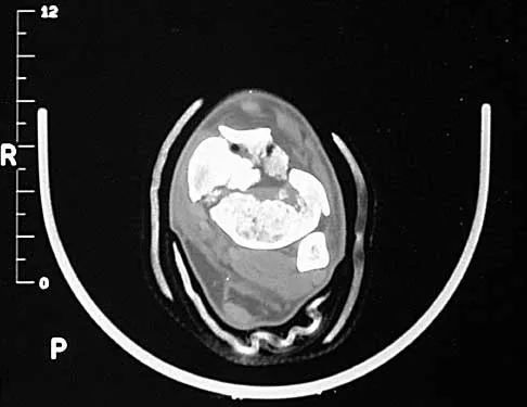
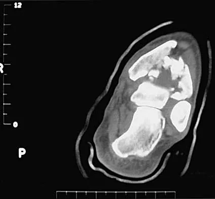
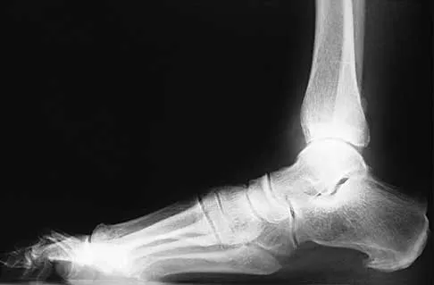
A 32-year-old man sustained a closed injury after falling 25 feet from a roof. His ankle and foot are severely swollen. Radiographs and CT scans are shown in Figures 29a through 29d. Initial management should consist of


Explanation

The patient has a severe high-energy injury from axial loading to the left ankle and distal tibia. This is a closed injury, but the soft tissues are injured and severely swollen. Initial treatment should focus on skeletal stabilization, and incisions directly over the fracture area should be avoided until soft-tissue stabilization has occurred. Immediate spanning external fixation with plans for a delayed reconstruction as needed for the joint surface is the treatment of choice. Closed reduction and application of a constrictive long leg cast may lead to increased risk of tissue necrosis. Immediate open procedures to internally fix the fracture add the risks of soft-tissue necrosis and are to be avoided. Percutaneous plating may be one of the delayed fixation options but should not be used immediately. Primary ankle arthrodesis is not indicated. Thordarson DB: Complications after treatment of tibial pilon fractures: Prevention and management strategies. J Am Acad Orthop Surg 2000;8:253-265. Marsh JL, Bonar S, Nepola JV, DeCoster TA, Hurwitz SR: Use of an articulated external fixator for fractures of the tibial plafond. J Bone Joint Surg Am 1995;77:1498-1509.


Question 8

Which of the following parameters is considered most important when assessing an acetabular fracture for surgical indications?


Explanation

The most important aspect in the decision for surgery in an acetabular fracture is the ability of the femoral head to remain concentrically reduced under the dome in AP and Judet oblique views of the pelvis. If this parameter is present, then the need for surgery is determined by other aspects such as fragmentation, age, incongruity, and displacement. If the head remains stable under the dome without traction, there is sufficient acetabular dome to provide stability, and nonsurgical treatment may be appropriate. Tile M: Assessment and management of acetabular fractures, in Tile M (ed): Pelvic and Acetabular Fractures, ed 2. Baltimore, MD, Williams and Wilkins, 1995, pp 305-354. Letournel E: Acetabular fractures: Classification and management. Clin Orthop 1980;151:81-106.

Question 9

A 57-year-old man has had right ankle pain for the past 10 months following an injury that went untreated. Radiographs are shown in Figures 30a through 30c. Management should consist of


Explanation

The radiographs reveal a malunited distal fibular fracture with shortening. Because there appears to be an adequate cartilage space within the ankle joint, the role of reconstruction would be to prevent arthrosis and the need for ankle arthrodesis, as well as to decrease symptoms. The treatment of choice is restoration of fibular length, alignment, and rotation with osteotomy plating, and bone grafting as needed. There is no indication for ligament reconstruction of a mechanically stable ankle, and tibial shortening osteotomy will not assist in correcting the deformity. Cast immobilization may assist with improvement of symptoms but will not correct the overall process. Determination of fibular length is best done by comparing the talocrural angle of the injured side with the uninjured side. The goal is to perfectly reduce the talus in the ankle mortise. Marti RK, Raaymakers EL, Nolte PA: Malunited ankle fractures: The late results of reconstruction. J Bone Joint Surg Br 1990;72:709-713. Geissler W, Tsao A, Hughes J: Fractures and injuries of the ankle, in Rockwood CA Jr, Green DP, Bucholz RW, Heckman JD (eds): Rockwood and Green's Fractures in Adults, ed 4. Philadelphia, PA, Lippincott-Raven, 1996, pp 2201-2206.


Question 10

A 32-year-old man sustains a forceful inversion injury while playing soccer. Examination reveals tenderness in the lateral hindfoot and midfoot region with associated ecchymosis and swelling. Radiographs show proximal migration of the os peroneum. Active eversion is still present. These findings indicate disruption of the


Explanation

The os peroneum is an accessory ossicle located within the peroneus longus tendon. It is typically located at the level of the cuboid groove in the lateral hindfoot and midfoot region. Proximal migration of the os peroneum indicates disruption of the peroneus longus tendon and is an important clue to diagnosis. This unusual condition can cause chronic lateral ankle pain, and surgical repair may be indicated. Active eversion indicates that the peroneus brevis is clinically intact. Disruption of the extensor digitorum brevis, plantar fascia, or syndesmosis would have no effect on the position of the os peroneum. Thompson FM, Patterson AH: Rupture of the peroneus longus tendon: Report of three cases. J Bone Joint Surg Am 1989;71:293-295.

Question 11

A 24-year-old man sustained a grade IIIb open tibial fracture and an ipsilateral grade IIIa femoral fracture in a motorcycle accident. He is unresponsive, intubated, and has a Glasgow Coma Scale score of 8. He is resuscitated and taken to the operating room for definitive orthopaedic care. Which of the following intraoperative problems will most likely adversely affect his long-term outcome?


Explanation

Traumatic brain injury is considered to be either primary or secondary. Primary injury is direct or impact damage to the brain, and secondary injury can have intracranial or systemic causes. While treatment has little impact on primary brain injury, secondary brain injury can be avoided. There are also many causes of intracranial secondary brain injury, including intracranial hypertension or cerebral edema. There are many causes of systemic secondary brain injury, but none has a greater impact on outcome than hypotension or hypoxia. In fact, the occurrence of hypotension postinjury causes a 10- to 15-fold increase in mortality. In a series by Pietropaoli and associates, the mortality rate for head-injured patients that were normotensive during surgery was 25%, but if they were hypotensive the mortality rate was 82%. In the same series, the number of patients with a Glasgow Coma Scale score of either 4 or 5 dropped from 58% in those patients that were normotensive during surgery to 6% in those patients that became hypotensive during surgery. Efforts to avoid hypotension postinjury and especially during surgery should be of primary importance. Chesnut RM, Marshall LF, Klauber MR, et al: The role of secondary brain injury in determining outcome from severe head injury. J Trauma 1993;34:216-222. Pietropaoli JA, Rogers FB, Shackford SR, Wald SL, Schmoker JD, Zhuang J: The deleterious effects of intraoperative hypotension on outcome in patients with severe head injury. J Trauma 1992;33:403-407. Schmeling GJ, Schwab JP: Polytrauma care: The effect of head injuries and timing of skeletal fixation. Clin Orthop 1995;318:106-116.

Question 12

Figure 31 shows the radiograph of an 8-year-old boy who has a swollen forearm after falling out of a tree. Examination reveals that all three nerves are functionally intact, and there is no evidence of circulatory embarrassment. Management should consist of


Explanation

The patient has a Bado type IV Monteggia lesion. It involves dislocation of the radial head and fractures of both the radial and ulnar shafts. These fractures are very difficult to manage by closed reduction alone. The radial and ulnar shafts first have to be stabilized surgically to give a lever arm to reduce the radial head. In this age group, intramedullary pins are easy to insert percutaneously and cause less tissue trauma than plates and screws. In these types of injuries, the focus is often on the forearm fracture; the radial head dislocation may not be appreciated as was the case with this patient. Gibson WK, Timperlake RW: Operative treatment of a type IV Monteggia fracture-dislocation in a child. J Bone Joint Surg Br 1992;74:780-781.


Question 13

Figure 32 shows the radiograph of a laborer who jammed his thumb in a fall. Examination reveals pain at the base of the thumb and proximal thenar eminence region. Management should consist of


Explanation

The radiographs are classic for a Bennett's fracture, which involves a fracture of the palmar ulnar aspect of the proximal phalanx. This fracture fragment is still attached to the anterior oblique ligament. The deforming forces that cause subluxation of the base of the proximal phalanx include the pull of the abductor pollicis longus as well as the adductor pollicis. Adequate reduction can be achieved by closed reduction, percutaneous pin fixation, and casting. The fragment is too small for secure internal fixation. Stern PJ: Fractures of the metacarpals and phalanges, in Green DP, Hotchkiss RN, Pederson WC (eds): Green's Operative Hand Surgery, ed 4. Philadelphia, PA, 1999, pp 711-771.


Question 14

In displaced calcaneal fractures, what fragment is the only one that remains in its anatomic position?


Explanation

The sustentaculum tali remains in its anatomic position because of its supporting ligamentous structures. This provides the key to the reconstruction of the calcaneus. The posterior facet is reduced to the sustentaculum tali and then fixed to it for stability. All of the other components of the calcaneus are then reduced to this complex. Sanders R: Displaced intra-articular fractures of the calcaneus. J Bone Joint Surg Am 2000;82:225-250. Eastwood DM, Gregg PJ, Atkins RM: Intra-articular fractures of the calcaneum: Part I. Pathological anatomy and classification. J Bone Joint Surg Br 1993;75:183-188.

Question 15

A 46-year-old man sustains a calcaneal fracture in a fall off a scaffold. During surgical reconstruction using an extended lateral incision, the fracture is reduced and fixed with a plate and screws. One of the posterior facet screws is found to be 5 mm out of the bone on the Harris view. What structure is most likely at risk because of this finding?


Explanation

The abductor hallucis muscle is the most medial structure. The posterior tibial tendon and the flexor digitorum longus tendon lie more cephalad to the sustentaculum tali. There is a groove under the sustentaculum for the flexor hallucis longus tendon. Subchondral lag screws placed across the posterior facet exit the medial side of the calcaneus in this groove. Just medial to the flexor hallucis longus tendon is the neurovascular bundle. A screw that is out of the bone a short distance can cause triggering of the flexor hallucis longus tendon. Patients will report loss of great toe excursion in the early postoperative period. Accurate measurement of subchondral lag screw length avoids this complication. Hollinshead WH: Anatomy for Surgeons, ed 3. Philadelphia, PA, Harper and Row, 1982, pp 802-852. Rosenberg AS, Cheung Y: Diagnostic imaging of the ankle and foot, in Jahss MH (ed): Disorders of the Foot and Ankle, ed 2. Philadelphia, PA, WB Saunders, 1991, pp 109-154.

Question 16

A 23-year-old man sustained an injury to his left foot when a forklift rolled over it at work. Examination reveals marked swelling of the midfoot and forefoot, with tenderness to palpation over the medial hindfoot and dorsomedial forefoot. The distal dorsalis pedis pulse is audible on Doppler examination, and his sensation is intact to touch. Radiographs are shown in Figures 33a and 33b. Management should consist of


Explanation

The best results after dislocations of the tarsometatarsal joints are seen with anatomic reduction; this is best achieved by open reduction and maintained with internal fixation with either pins or screws. Open reduction provides a means of debriding small bony fragments from the joint and allowing direct inspection of the reduction. Associated crush or shearing fractures of the cuboid or tarsal navicula are signs that suggest a Lisfranc injury. Because patients can function quite well despite the development of arthrosis in the Lisfranc joint, primary arthrodesis is not indicated in the management of this injury. Resch S, Stenstrom A: The treatment of tarsometatarsal injuries. Foot Ankle 1990;11:117-123. Schenck RC Jr, Heckman JD: Fractures and dislocations of the forefoot: Operative and nonoperative treatment. J Am Acad Orthop Surg 1995;3:70-78.


Question 17

A 24-year-old woman who has hypotension, a head injury, and who experienced a poor response to resuscitation has been taken to the operating room for a splenectomy. Following abdominal surgery she remains unstable with increasing pulmonary respiratory pressures and decreasing oxygen saturation. She has a transverse mid-diaphyseal fracture of the tibia with a 4-cm laceration and soil-contaminated muscle in the wound. Based on these findings, management should consist of


Explanation

Because the patient is critically ill and requires expeditious care, stabilization of the long bone fracture is required, but definitive care of the fracture should be postponed. The treatment of choice at this time is irrigation with 12 L of saline solution, followed by debridement and nondefinitive stabilization with a simple four-pin external frame to regain axial and rotational alignment. When the patient's condition is more stable, more definitive care can be performed. Bosse MJ, Kellam JF: Orthopaedic management decisions in the multiple trauma patient, in Browner BD, Jupiter JP, Levine AM, Trafton P (eds): Skeletal Trauma, ed 2. Philadelphia, PA, WB Saunders, 1998, pp 151-164. Weresh MJ, Stover MD, Bosse MJ, Jeray K, Kellam JF: Pulmonary gas exchange during intramedullary fixation of femoral shaft fractures. J Trauma 1999;46:863-868.

Question 18

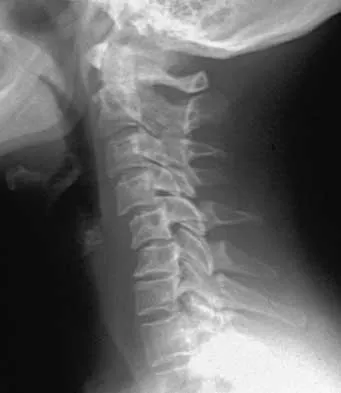
A 53-year-old woman has severe neck and left shoulder pain after a rollover motor vehicle accident. Radiographs and a CT scan of the cervical spine are shown in Figures 34a through 34c. Management should consist of


Explanation

The plain radiographs show a horizontal orientation of the C5 facet joint. The CT scan through C5 reveals an ipsilateral pedicle and lamina fracture (floating facet). This injury involves two adjacent motion segments and is extremely unstable. Lateral mass plates, with or without the purchase of the "floating facet," provide the best means of stabilization and should include the facet above (C4) and below (C6) the level of injury. Orthotic immobilization is insufficient for this particular injury. Halo vest treatment does not control the subaxial spine well and is of limited value. While simple midline (Rogers) wiring provides some tension band restoration, it is not optimal for rotational control. The use of lateral mass plates provides rotational stability. Another option would be anterior fusion and plating, which would save cervical segments. Levine AM, Mazel C, Roy-Camille R: Management of fracture separations of the articular mass using posterior cervical plating. Spine 1992;17:S447-S454. Levine AM: Facet fractures and dislocations, in Levine AM, Eismont FJ, Garfin S, Zigler JE (eds): Spine Trauma. Philadelphia, PA, WB Saunders, 1998, pp 360-362. Whitehill R, Richman JA, Glaser JA: Failure of immobilization of the cervical spine by the halo vest: A report of five cases. J Bone Joint Surg Am 1986;68:326-332.


Question 19

What is the most common clinically significant preventable complication secondary to the treatment of a displaced talar neck fracture?


Explanation

The most important consequence of a displaced talar neck fracture after closed or open treatment is malunion. Because displacement of the talar neck is associated with displacement of the subtalar joint, any malunion leads to intra-articular incongruity or malalignment of the subtalar joint. Varus malunion is common when there is comminution of the medial talar neck. This results in pain, osteoarthritis, and hindfoot deformity that requires further treatment. Because of these complications, it is imperative that all displaced talar neck fractures are reduced anatomically; fragmented fractures may require bone grafting to maintain the length and rotation of the neck. Tile M: Fractures of the talus, in Schatzker J, Tile M (eds): Rationale of Operative Fracture Care, ed 2. Berlin, Springer-Verlag, 1996, pp 563-588. Daniels TR, Smith JW, Ross TI: Varus malalignment of the talar neck: Its effect on the position of the foot and on subtalar motion. J Bone Joint Surg Am 1996;78:1559-1567.

Question 20

Examination of a carpenter who hit his thumb with a hammer reveals that the nail plate is broken but in place, and there is a 100% subungual hematoma that covers 100% of the area under the nail plate. Radiographs reveal a comminuted distal phalangeal tuft fracture. Management should consist of


Explanation

This is a classic situation for a distal phalanx tuft fracture with associated nail bed injury and subungual hematoma. In general, when the subungual hematoma is greater than 50% of the surface area under the nail plate, treatment should consist of nail plate removal, nail bed repair, oral antibiotics, and a fingertip splint. Oral antibiotics and fingertip splinting alone do not address the nail bed laceration, which will most likely lead to nail plate deformity if not repaired. Kirschner pin stabilization is not indicated because these fractures are nondisplaced and usually are inherently stable after nail bed repair. The use of IV antibiotics alone does not address the nail bed laceration surgically. Casting, followed by hydrotherapy and topical antibiotics, is not indicated because it does not address the nail bed laceration. Further, a nondisplaced distal phalangeal tuft fracture does not require cast immobilization. Stern PJ: Fractures of the metacarpals and phalanges, in Green DP, Hotchkiss RN, Pederson WC (eds): Green's Operative Hand Surgery, ed 4. Philadelphia, PA, 1999, pp 711-771.

Question 21

An olecranon fracture-dislocation of the elbow in which the fracture line exits distal to the coronoid process is best managed by open reduction and


Explanation

Fracture-dislocations of the elbow present difficult management problems. Standard olecranon fractures normally are not associated with a dislocation; however, the surgeon needs to recognize that some fractures that have a dislocation, in particular a posterior dislocation, represent a Monteggia equivalent. These injuries are not ulnar shaft fractures because they are fractured at or just distal to the coronoid; however, because of the unstable fracture-dislocation, the forces across this reduction are high. Two Kirschner wires and a tension band wire provide inadequate fixation. Therefore, the preferred method of fixation is plate osteosynthesis with a 3.5-mm low-contact dynamic compression plate or reconstruction plate. Jupiter JP, Kellam JF: Fractures of the forearm, in Browner BD, Jupiter JP, Levine AM, Trafton P (eds): Skeletal Trauma, ed 2. Philadelphia, PA, WB Saunders, 1998, pp 421-454. Quintero J: Fracture of the forearm, in Ruedi TP, Murphy WM (eds): AO Principles of Fracture Management. Stuttgart, Thieme, 2000, pp 323-337.

Question 22

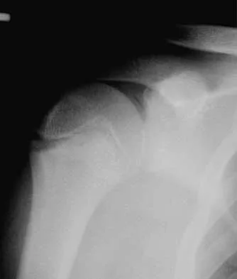
A 15-year-old baseball pitcher who reports increasing pain in his right shoulder over the past 3 weeks states that the pain increases the more he pitches. Radiographs of both shoulders are shown in Figures 35a and 35b. What is the next most appropriate step in management?


Explanation

The patient has a rotational stress fracture of the proximal humeral physis (Little Leaguer's shoulder). The symptoms of increasing pain with activity and relief with rest are typical of a stress injury. Treatment should consist of cessation of throwing activity but rehabilitation of the shoulder girdle muscles. The pitching technique should be evaluated as well. Barnett LS: Little League shoulder syndrome: Proximal humeral epiphyseolysis in the adolescent baseball pitchers: A case report. J Bone Joint Surg Am 1985;67:495-496.


Question 23

A 36-year-old man sustains a traumatic spondylolisthesis of L5 on S1. Surgical stabilization requires pedicular fixation into the sacrum. If the screw is placed in a medial to lateral direction and penetrates the sacral ala, what nerve root is at risk?


Explanation

The L5 nerve root lies directly over the superior and anterior alae. If the screw is directed approximately 20 degrees laterally and bicortical purchase is achieved, there is the risk of injuring the L5 nerve root. If the screw is directed medially into the body of S1, there is little risk of injury. The same root is at risk during placement of an iliosacral screw. Ebraheim NA, et al: Lumbosacral nerve and dorsal screw placement. Orthopedics 2000;23:245-247. Ebraheim NA, Mermer M, Xu R, Yeasting RA: Radiological evaluation of S1 dorsal screw placement. J Spinal Disord 1996;9:527-535.

Question 24

A 25-year-old woman who fell on her outstretched hand reports chronic pain over the hypothenar eminence region and some dorsal ulnar wrist pain. She also notes difficulty playing golf and tennis. Plain radiographs of the hand and wrist are unremarkable. A CT scan is shown in Figure 36. What is the next most appropriate step in management?


Explanation

The CT scan reveals a hook of the hamate nonunion with irregular resorption at the fracture site, which is at the base of the hamate. Symptomatic relief of the pain and discomfort has been well documented after excision of the hook of the hamate. Ultrasound therapy will not provide long-term symptomatic relief or induce nonunion healing. MRI for further soft-tissue evaluation is inappropriate because this is a bony problem; the bony architecture of the wrist is best visualized by CT. Open reduction and internal fixation of the hook of the hamate does not provide the symptomatic relief that is found with excision of the hook of the hamate. In addition, the technical difficulties and relative risk of persistent nonunion after open reduction and internal fixation are not merited when hamate excision can be effected easily and causes no long-term untoward effects. Electrodiagnostic evaluation is inappropriate because there is no history of the persistent numbness and tingling that is found in peripheral compression neuropathies. Stark HH, Chao EK, Zemel NP, Rickard TA, Ashworth CR: Fracture of the hook of the hamate. J Bone Joint Surg Am 1989;71:1206-1207. Failla JM: Hook of hamate vascularity: Vulnerability to osteonecrosis and nonunion. J Hand Surg Am 1993;18:1075-1079. Carter PR, Easton RG, Littler JW: Ununited fracture of the hook of the hamate. J Bone Joint Surg Am 1977;59:583-588.


Question 25

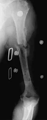
An active 72-year-old woman sustained a mid-diaphyseal right humerus fracture 16 months ago. History reveals that she was first treated with a brace for 7 months. Additional treatment consisted of intramedullary nailing 9 months ago. Recently the rod was removed, and the patient now reports pain and gross motion at the fracture site. Current radiographs are shown in Figures 37a and 37b. What is the next most appropriate step in management?


Explanation

The patient has a well-established nonunion in a very porotic bone. Electrical stimulation has been found effective in treating tibial nonunions, but there is very little data on humeral nonunions, especially chronic well-established ones. Ultrasound stimulation is effective in accelerating fracture healing, but there is little data concerning the treatment of nonunions. Intramedullary nailing with bone graft is an option, but it maybe difficult to obtain a rigid construct in a very porotic bone. An Ilizarov-type external fixator would be an alternative, but there is little clinical data for the humerus and it may be poorly tolerated. A plate and screw construct with bone graft combines rigidity with the biologic advantage of the bone graft. A recent series reported on the use of a plate combined with onlay allograft for recalitrant nonunions. Cement augmentation for screw fixation either in the canal or added to the screw holes may be helpful in select cases. Hornicek FJ, Zych GA, Hutson JJ, Malinin TI: Salvage of humeral nonunions with onlay bone plate allograft augmentation. Clin Orthop 2001;386:203-209.


Question 26

A 24-year-old man presents with a closed femoral shaft fracture, multiple rib fractures, pulmonary contusions, and an initial Glasgow Coma Scale (GCS) of 6 following a high-speed motor vehicle collision. He is hypotensive in the emergency department and requires vasopressor support. What is the most appropriate management of his femur fracture?


Explanation

In a hemodynamically unstable polytrauma patient (borderline or in extremis), damage control orthopedics (DCO) with rapid external fixation is indicated. Early reamed intramedullary nailing in this setting significantly increases the risk of ARDS and secondary brain injury.

Question 27

A 28-year-old man sustains a closed spiral fracture of the distal third of the humerus (Holstein-Lewis fracture). Physical examination reveals an inability to actively extend the wrist or fingers. What is the most appropriate initial management?


Explanation

The presence of a primary radial nerve palsy in a closed humeral shaft fracture is not an absolute indication for surgery. Initial management consists of coaptation splinting and observation, as the majority of these palsies spontaneously resolve.

Question 28

A 32-year-old man sustains a posterior hip dislocation. Following emergent closed reduction, a CT scan reveals a concentric joint space, no intra-articular loose bodies, and a posterior wall fracture involving 15% of the acetabular articular surface. What is the most appropriate definitive management?


Explanation

Posterior wall fractures involving less than 20% of the articular surface are generally stable. Non-operative management with protected weight-bearing is indicated when the hip is concentrically reduced and clinically stable on dynamic stress testing.

Question 29

A 28-year-old man sustains a closed midshaft humeral fracture. He presents with a wrist drop and inability to extend his fingers. Radiographs show a transverse midshaft fracture. What is the most appropriate initial management of the nerve injury?


Explanation

Radial nerve palsy associated with closed humeral shaft fractures typically represents a neuropraxia. The standard initial management is functional bracing or splinting and observation for 3 to 4 months before considering nerve exploration.

Question 30

A 45-year-old polytrauma patient presents with a closed left femoral shaft fracture, multiple rib fractures, and bilateral pulmonary contusions. His serum lactate is 4.5 mmol/L and pH is 7.15. Which of the following is the most appropriate management of his femur fracture?


Explanation

This patient is in a borderline/unstable physiologic state as evidenced by elevated lactate and acidosis. Damage control orthopedics (DCO) with temporary external fixation is indicated to minimize the inflammatory "second hit" associated with definitive intramedullary nailing.

Question 31

A 24-year-old unrestrained driver is involved in a motor vehicle collision. He presents with his right lower extremity positioned in flexion, adduction, and internal rotation.

Which of the following structures is at greatest risk of injury?


Explanation

The clinical posture described is classic for a posterior hip dislocation. This injury places the sciatic nerve, specifically the common peroneal division, at greatest risk due to stretching or direct compression by the displaced femoral head.

Question 32

A 65-year-old woman presents with an anterior shoulder dislocation and an associated greater tuberosity fracture. Following closed reduction, radiographs reveal that the greater tuberosity fragment is displaced superiorly by 8 mm. What is the most appropriate next step in management?


Explanation

In greater tuberosity fractures associated with shoulder dislocations, displacement greater than 5 mm increases the risk of subacromial impingement and altered rotator cuff mechanics. Surgical fixation is highly recommended for functional recovery.

Question 33

A 32-year-old man undergoes reamed intramedullary nailing for a closed tibial shaft fracture. Postoperatively, he complains of severe leg pain out of proportion to the injury, exacerbated by passive toe stretch. His blood pressure is 130/80 mmHg. What intracompartmental pressure finding definitively confirms acute compartment syndrome?


Explanation

The diagnosis of acute compartment syndrome is confirmed when the differential (delta) pressure between the diastolic blood pressure and the measured intracompartmental pressure falls below 30 mmHg. This indicates critical ischemia requiring immediate fasciotomy.

Question 34

A 70-year-old woman on long-term alendronate therapy presents with vague thigh pain. Radiographs demonstrate an incomplete transverse fracture of the lateral cortex of the subtrochanteric femur with localized periosteal thickening.

What is the recommended prophylactic surgical treatment?


Explanation

Atypical femur fractures related to prolonged bisphosphonate use feature characteristic lateral cortical thickening and a transverse fracture pattern. Prophylactic full-length cephalomedullary nailing is recommended to prevent completion of the fracture.

Question 35

A 25-year-old cyclist falls directly onto his shoulder. Radiographs reveal a midshaft clavicle fracture with 2.5 cm of shortening and complete displacement. Which of the following is an expected outcome if this is treated nonoperatively rather than surgically?


Explanation

Completely displaced midshaft clavicle fractures with greater than 2 cm of shortening treated nonoperatively have a significantly higher rate of symptomatic malunion and nonunion. This can lead to objectively decreased shoulder strength and rapid fatigability.

Question 36

A 19-year-old athlete sustains a high-energy traumatic knee dislocation that is spontaneously reduced in the emergency department. His ankle-brachial index (ABI) is measured at 0.8. What is the most appropriate next step in management?


Explanation

An ABI of less than 0.9 following a knee dislocation is highly suspicious for a popliteal artery injury. A CT angiogram is the gold standard next step to pinpoint the location and extent of vascular injury prior to operative intervention.

Question 37

A 7-year-old boy falls on an outstretched hand and sustains a plastic deformation fracture of the proximal ulna with an associated radial head dislocation. Which nerve is most commonly injured in this specific fracture-dislocation pattern?


Explanation

This injury pattern is a Monteggia fracture-dislocation. The posterior interosseous nerve (PIN), a deep branch of the radial nerve, wraps around the radial neck and is the most frequently injured nerve during radial head dislocation.

Question 38

A 34-year-old man sustains an isolated, closed, diaphyseal fracture of both the radius and ulna.

What is the preferred definitive management to maximize the restoration of forearm pronation and supination?


Explanation

In adults, diaphyseal both-bone forearm fractures are treated as articular injuries because perfect alignment is required for normal pronation and supination. The standard of care is anatomic open reduction and internal fixation (ORIF) with compression plating.

Question 39

A 40-year-old man presents after a high-energy motor vehicle crash with a distal femur fracture. CT scan reveals a coronal plane fracture of the lateral femoral condyle. What is the optimal fixation strategy for this specific fracture fragment?


Explanation

A coronal shear fracture of the femoral condyle is known as a Hoffa fracture. It requires anatomic reduction and absolute stability, best achieved with lag screws directed anterior-to-posterior (AP) or posterior-to-anterior (PA).

Question 40

In a subtrochanteric femur fracture, the proximal fragment is typically deformed by specific muscle forces. Which of the following describes the typical position of the proximal fragment and the primary muscle responsible for its flexion?


Explanation

In subtrochanteric fractures, the short proximal fragment is pulled into a characteristic position: flexion by the iliopsoas, abduction by the gluteus medius and minimus, and external rotation by the short external rotators.

Question 41

A 10-year-old boy presents with knee pain and inability to actively extend his knee after a jumping injury. Radiographs reveal a high-riding patella and a small bony avulsion from the inferior pole of the patella. What is the most appropriate management?


Explanation

This clinical picture represents a patellar sleeve fracture, a severe pediatric injury involving a large cartilaginous avulsion from the patella that disrupts the extensor mechanism. It mandates surgical repair (ORIF) to restore active extension.

Question 42

An 80-year-old woman with a cemented total hip arthroplasty sustains a fall. Radiographs show a periprosthetic fracture around the stem tip. The stem appears loose within the cement mantle, but there is adequate distal bone stock. According to the Vancouver classification, what is the appropriate treatment?


Explanation

A Vancouver B2 periprosthetic femur fracture is characterized by a fracture around a loose stem in the presence of adequate bone stock. The definitive treatment is revision arthroplasty with a long stem that bypasses the fracture site.

Question 43

A 28-year-old polytrauma patient sustains a highly comminuted, closed femoral shaft fracture in a motor vehicle collision. On arrival, he is tachycardic, has an SpO2 of 88% on room air, and exhibits petechiae across his axillae and chest. Which of the following is the most appropriate initial management for his femur fracture?


Explanation

This patient exhibits clinical signs of fat embolism syndrome and pulmonary compromise. In a physiologically unstable or borderline polytrauma patient, damage control orthopedics using temporary external fixation is favored to avoid the additional physiological 'hit' of reamed intramedullary nailing.

Question 44

A 34-year-old man sustains a transverse midshaft humerus fracture during an arm wrestling match. In the emergency department, he demonstrates an inability to actively extend his wrist or digits. What is the most appropriate initial management?


Explanation

A primary radial nerve palsy associated with a closed humeral shaft fracture typically represents a neuropraxia or axonotmesis. Observation with coaptation splinting or a functional brace is the standard of care, as spontaneous nerve recovery occurs in the vast majority of cases.

Question 45

A 45-year-old male presents to the emergency department after a seizure with his left arm locked in internal rotation. Radiographs reveal a posterior shoulder dislocation. A subsequent CT scan demonstrates an anteromedial humeral head impression defect involving 45% of the articular surface. What is the most appropriate definitive management?


Explanation

Posterior shoulder dislocations often result in a reverse Hill-Sachs lesion. When the articular defect exceeds 40%, joint-preserving procedures such as the McLaughlin procedure are prone to failure, and arthroplasty (hemiarthroplasty or total shoulder) is the recommended treatment.

Question 46

The deforming muscular forces acting on a subtrochanteric femur fracture characteristically result in what position of the proximal fragment?


Explanation

In subtrochanteric femur fractures, the proximal fragment is acted upon by specific muscular deforming forces. It is flexed by the iliopsoas, abducted by the gluteus medius and minimus, and externally rotated by the short external rotators.

Question 47

A 25-year-old man sustains a posterior hip dislocation following a dashboard injury. A closed reduction is performed within 4 hours. Post-reduction CT imaging shows a concentric joint reduction, no intra-articular fragments, and a non-displaced posterior wall fracture involving 10% of the articular surface. What is the most appropriate next step in management?


Explanation

Small (<20%), non-displaced posterior wall fractures associated with a stable, concentrically reduced hip joint can be managed nonoperatively. Protected weight-bearing and close radiographic follow-up is the standard of care.

Question 48

A 30-year-old man is brought to the trauma bay after a high-velocity knee injury. Examination reveals a severe posterolateral dislocation with a prominent transverse groove or 'dimple sign' at the medial joint line. Attempted closed reduction is unsuccessful. What is the most likely anatomic reason for the irreducibility?


Explanation

A 'dimple sign' on the medial aspect of the knee indicates a posterolateral knee dislocation where the medial femoral condyle has buttonholed through the medial capsule and medial collateral ligament. This is an irreducible injury that requires emergent open reduction.

Question 49

A 55-year-old woman presents with severe thigh pain and a low-energy, transverse diaphyseal femur fracture with focal lateral cortical thickening. She has been taking alendronate for 8 years. Which of the following statements regarding her management is most accurate?


Explanation

This patient has an atypical femur fracture (AFF) associated with long-term bisphosphonate use. Teriparatide (recombinant PTH) is an anabolic agent that stimulates bone formation and has been shown to aid in the healing of these challenging fractures.

Question 50

A 22-year-old male sustains a high-energy closed tibial diaphysis fracture. Four hours later, his leg is tense, and he complains of pain out of proportion to the injury. Passive stretch of his hallux elicits excruciating pain. His diastolic blood pressure is 65 mmHg, and his anterior compartment pressure measures 45 mmHg. What is the most appropriate management?


Explanation

Acute compartment syndrome is a surgical emergency. A delta pressure (Diastolic BP minus Compartment Pressure) of less than 30 mmHg (in this case, 65 - 45 = 20 mmHg) is a definitive indication for immediate four-compartment fasciotomy.

Question 51

In a Galeazzi fracture-dislocation, which of the following stabilizing structures is most commonly disrupted, leading to instability of the distal radioulnar joint (DRUJ)?


Explanation

A Galeazzi fracture involves a fracture of the distal third of the radial shaft with associated disruption of the DRUJ. The Triangular Fibrocartilage Complex (TFCC) is the primary stabilizer of the DRUJ and is typically torn or avulsed in this injury pattern.

Question 52

A 60-year-old patient sustains a Vancouver type B2 periprosthetic femur fracture around a cemented, polished taper-slip stem. Radiographs demonstrate a loose femoral component but adequate proximal bone stock. What is the gold standard surgical treatment?


Explanation

A Vancouver B2 periprosthetic fracture is characterized by a fracture around a loose stem in the presence of good proximal bone stock. The standard of care is revision to a long, cementless, extensively porous-coated or fluted tapered stem that bypasses the fracture site.

Question 53

A 66-year-old woman presents with atraumatic thigh pain. She has been taking oral alendronate for 9 years. Radiographs reveal a transverse fracture of the femoral shaft with lateral cortical thickening and a medial cortical spike. What is the most appropriate management of this condition?


Explanation

This is an atypical femur fracture associated with long-term bisphosphonate use, characterized by severely suppressed bone turnover. Treatment requires intramedullary nailing of the affected side and critical evaluation of the contralateral femur, which is highly at risk for a synchronous lesion.

Question 54

A 24-year-old man sustains a closed spiral fracture of the distal third of the humerus (Holstein-Lewis fracture). Initial examination in the emergency department documents normal wrist and finger extension. Following closed reduction and application of a coaptation splint, the patient immediately develops an inability to extend his wrist and digits. What is the most appropriate next step in management?


Explanation

A radial nerve palsy that develops after a closed reduction attempt indicates potential iatrogenic entrapment of the nerve within the fracture site. Immediate surgical exploration and fracture stabilization are required.

Question 55

A 30-year-old male sustains an anterior knee dislocation during a sporting event. The joint is reduced in the emergency department. Palpation reveals symmetric pedal pulses, but the measured Ankle-Brachial Index (ABI) is 0.85. What is the most appropriate next step in the management of this patient?


Explanation

Following a knee dislocation, an ABI of less than 0.90, even with palpable pulses, is highly suspicious for a clinically significant intimal tear or vascular injury. CT angiography is indicated to evaluate the popliteal artery definitively.

Question 56

A 45-year-old woman falls on an outstretched hand and sustains an elbow injury. Imaging confirms a posterior elbow dislocation, a type II coronoid fracture, and a comminuted radial head fracture (the "terrible triad"). During surgical reconstruction, what is the standard recommended sequence of repair?


Explanation

The standard surgical sequence for a terrible triad injury begins deep and moves superficial, working from inside to outside. Coronoid fixation restores anterior stability, followed by radial head repair/replacement to restore the anterior column, and finally LCL repair to restore posterolateral stability.

Question 57

A 25-year-old man sustains a subtrochanteric femur fracture. To achieve an anatomic reduction, the surgeon must overcome the deforming forces acting on the proximal fragment. The proximal fragment is typically pulled into which of the following positions, and by which corresponding muscles?


Explanation

In subtrochanteric fractures, the proximal fragment is classically deformed into flexion by the iliopsoas, abduction by the gluteus medius and minimus, and external rotation by the short external rotators.

Question 58

A 32-year-old man undergoes reamed intramedullary nailing for a closed tibial shaft fracture. Twelve hours postoperatively, he complains of severe leg pain requiring rapidly escalating doses of narcotics. Which of the following is the most objective and definitive threshold indicating the need for emergent fasciotomy?


Explanation

A delta pressure (diastolic blood pressure minus compartment pressure) of less than 30 mm Hg is an absolute indication for fasciotomy in suspected acute compartment syndrome. Clinical signs like pain with passive stretch are sensitive early indicators but are subjective, while loss of pulses is a very late and unreliable finding.

Question 59

A 22-year-old athlete sustains a midfoot injury with severe midfoot pain and plantar ecchymosis. Radiographs reveal widening of the interval between the first and second metatarsal bases. The primary stabilizing ligament disrupted in this classic Lisfranc injury connects which two osseous structures?


Explanation

The Lisfranc ligament is an intra-articular ligament that connects the lateral aspect of the medial cuneiform to the medial base of the second metatarsal. It provides critical stability to the tarsometatarsal joint complex.

Question 60

A 28-year-old driver is involved in a motor vehicle collision and sustains a posterior hip dislocation. He presents with a foot drop and decreased sensation over the dorsum of the foot. Following emergent, uneventful closed reduction within 2 hours of injury, the neurologic deficit persists. What is the most appropriate management of this persistent deficit?


Explanation

Sciatic nerve palsy (specifically the peroneal division) complicates 10-15% of posterior hip dislocations. If the hip is concentrically reduced, the standard management is observation and supportive care (e.g., AFO), as the majority of these neuropraxias recover spontaneously over months.

Question 61

While most middle-third clavicle fractures can be managed nonoperatively, certain clinical and radiographic criteria strictly dictate surgical intervention. Which of the following is an absolute indication for operative fixation of an acute clavicle fracture?


Explanation

Absolute indications for clavicle fracture fixation include open fractures, neurovascular compromise, and impending skin breakdown (severe tenting with blanching). Displacement and shortening are relative indications based on patient activity level and shared decision-making.

Question 62

A 22-year-old man presents with a closed right femoral shaft fracture and a severe closed head injury with a Glasgow Coma Scale (GCS) score of 6. He is hemodynamically labile. According to the principles of Damage Control Orthopedics (DCO), what is the most appropriate initial management of the femur fracture?


Explanation

In severely polytraumatized patients (e.g., severe head injury, hemodynamic instability), prolonged surgeries and the systemic inflammatory hit from reamed nailing can be fatal (second hit phenomenon). Damage Control Orthopedics dictates rapid, temporary stabilization with external fixation.

Question 63

A 35-year-old man undergoes open reduction and internal fixation of a Hawkins Type II talar neck fracture. At 8 weeks postoperatively, an anteroposterior mortise radiograph reveals a subchondral radiolucent band in the dome of the talus (Hawkins sign). What does this radiographic finding indicate?


Explanation

The Hawkins sign represents subchondral osteopenia (resorption of bone) seen 6 to 8 weeks after injury. Its presence requires an intact blood supply to the talus, indicating that avascular necrosis is highly unlikely.

Question 64

A 55-year-old woman sustained a nondisplaced distal radius fracture treated in a short arm cast. Two weeks after the cast was removed (6 weeks post-injury), she notes a sudden inability to actively extend the interphalangeal joint of her thumb. Radiographs confirm the distal radius fracture is healing well in anatomic alignment. What is the most appropriate surgical treatment?


Explanation

Delayed rupture of the EPL tendon occurs due to mechanical attrition or ischemia at Lister's tubercle, especially in nondisplaced distal radius fractures. Because the tendon ends are typically retracted and degenerated, direct repair is usually impossible, making an EIP to EPL transfer the gold standard.

Question 65

A 40-year-old man sustains a high-energy trauma resulting in a distal femur fracture. CT imaging reveals a coronal plane fracture of the lateral femoral condyle (Hoffa fracture). What is the optimal surgical approach and fixation strategy for this specific fragment?


Explanation

A Hoffa fracture is a coronal plane fracture of the femoral condyle. It requires anatomic articular reduction and fixation with lag screws oriented perpendicular to the fracture plane, typically placed in an anterior-to-posterior (AP) or posterior-to-anterior (PA) direction.

Question 66

A 21-year-old collegiate rugby player with a history of recurrent anterior shoulder instability undergoes preoperative imaging, which reveals 25% anterior glenoid bone loss and a large, engaging Hill-Sachs lesion. Which of the following procedures is most appropriate to restore stability and prevent recurrence?


Explanation

In the presence of critical glenoid bone loss (typically > 20-25%) and an engaging Hill-Sachs lesion, isolated soft tissue repairs (like a Bankart repair) have unacceptably high failure rates. A bony augmentation procedure, such as the Latarjet procedure, is required to restore the glenoid arc and confer stability.

Question 67

A 29-year-old motorcyclist sustains a completely displaced, closed midshaft femur fracture and a closed midshaft tibia fracture on the same limb (ipsilateral floating knee). Both fractures are amenable to antegrade intramedullary nailing. Which of the following describes the most universally accepted sequence of definitive stabilization?


Explanation

In a floating knee injury, the femur is generally stabilized first. Stabilizing the femur restores limb alignment and allows the knee to be safely flexed, which is necessary to gain the proper starting trajectory for subsequent tibial intramedullary nailing.

Question 68

A 25-year-old man with a severe closed head injury (GCS 7) and a closed femoral shaft fracture is admitted to the trauma bay. Initial head CT shows cerebral edema with mass effect. Which of the following is the most appropriate initial management of his femur fracture?


Explanation

In a polytrauma patient with a severe head injury and elevated intracranial pressure, early total care (IM nailing) risks a 'second hit' phenomenon, exacerbating brain injury due to embolization and hypotension. Damage control orthopedics with temporary external fixation is the safest initial management.

Question 69

A 34-year-old man sustains an open humeral shaft fracture (Gustilo-Anderson Type II) after a motorcycle collision. Examination reveals a complete inability to extend the wrist and fingers, which was noted immediately after the injury. What is the most appropriate management of the nerve injury?


Explanation

While radial nerve palsy in a closed humeral shaft fracture is typically observed initially, an open fracture with an associated radial nerve palsy is an absolute indication for immediate nerve exploration during the mandatory surgical debridement and fracture fixation.

Question 70

During surgical reconstruction for a 'terrible triad' injury of the elbow, what is the recommended sequence of repair to best restore elbow stability?


Explanation

The standard surgical sequence for a terrible triad injury addresses the structures from deep to superficial: first fixing the coronoid, then repairing or replacing the radial head, and finally repairing the lateral collateral ligament (LCL) complex.

Question 71

A 68-year-old woman on long-term alendronate therapy presents with a low-energy, transverse subtrochanteric femur fracture. Radiographs show lateral cortical thickening and a medial spike. What is the most crucial next step in the workup prior to surgical intervention?


Explanation

Bisphosphonate-associated atypical femur fractures are frequently bilateral. It is critical to obtain radiographs of the contralateral femur to evaluate for a prodromal or complete atypical fracture, which may require prophylactic fixation.

Question 72

A 22-year-old football player sustains a high-energy knee injury resulting in a multi-ligamentous knee dislocation. Upon reduction in the emergency department, his pedal pulses are palpable, but his ankle-brachial index (ABI) is 0.8. What is the most appropriate next step in management?


Explanation

An ankle-brachial index (ABI) less than 0.9 in the setting of a knee dislocation is highly suspicious for a vascular injury, even if pulses are palpable. CT angiography is the gold standard next step to rapidly delineate a popliteal artery intimal tear or occlusion.

Question 73

A 24-year-old male presents with recurrent anterior shoulder instability. Advanced imaging reveals a 30 percent anterior glenoid bone loss and an engaging Hill-Sachs lesion. Which of the following surgical procedures provides the lowest rate of recurrent instability for this patient?


Explanation

In the setting of critical glenoid bone loss (>25%), soft tissue stabilization (Bankart repair) has an unacceptably high failure rate. A bone block procedure, such as the Latarjet (coracoid transfer), is required to restore the glenoid articular arc and provide a 'sling' effect.

Question 74

A 28-year-old man sustains a completely displaced, high-shear vertical femoral neck fracture (Pauwels Type III). Which of the following internal fixation constructs provides the highest biomechanical stability for this fracture pattern?


Explanation

Pauwels Type III fractures experience extremely high vertical shear forces, making standard parallel cannulated screws prone to failure. A fixed-angle device, such as a sliding hip screw combined with a derotational screw, provides superior biomechanical resistance to vertical shear.

Question 75

A 40-year-old man sustains a Gustilo-Anderson Type IIIB open tibial shaft fracture. Following initial aggressive debridement and stabilization, within what timeframe should definitive soft-tissue coverage ideally be performed to minimize the risk of deep infection?


Explanation

Current evidence suggests that definitive soft-tissue coverage of Type IIIB open tibia fractures within 3 to 7 days (optimally within 5 days) significantly reduces the rate of deep infection and flap failure compared to delayed coverage.

Question 76

A 35-year-old male sustained a closed tibial shaft fracture treated with a reamed intramedullary nail 9 months ago. He presents with persistent weight-bearing pain. Radiographs demonstrate an oligotrophic nonunion at the fracture site without hardware failure. Which of the following is the most appropriate definitive management?


Explanation

Exchange nailing with a larger reamed nail is the gold standard for aseptic oligotrophic or hypertrophic nonunion of the tibia treated initially with an IM nail. It provides improved mechanical stability and stimulates a biological healing response via reaming.

Question 77

A 25-year-old male is brought to the emergency department after a high-speed motorcycle collision. He is hemodynamically unstable, intubated for severe bilateral pulmonary contusions, and has bilateral closed femoral shaft fractures. What is the most appropriate initial orthopedic management?


Explanation

In a polytrauma patient who is hemodynamically unstable ('in extremis') with pulmonary compromise, damage control orthopedics using external fixation is indicated. This minimizes the 'second hit' inflammatory cascade associated with prolonged surgery and intramedullary reaming.

Question 78

A 22-year-old professional rugby player presents with recurrent anterior shoulder instability. A 3D CT scan reveals 28% anteroinferior glenoid bone loss. Which of the following is the most appropriate surgical intervention to restore stability?


Explanation

The Latarjet procedure is indicated for recurrent anterior shoulder instability in the setting of critical glenoid bone loss (typically >20-25%). Isolated soft-tissue Bankart repairs in this setting have unacceptably high failure rates.

Question 79

A 40-year-old female sustains a high-energy knee dislocation. Following closed reduction, her foot is warm, but the Ankle-Brachial Index (ABI) is calculated to be 0.8. What is the most appropriate next step in management?


Explanation

An ABI less than 0.9 in the setting of a knee dislocation is highly suspicious for a popliteal artery intimal tear or occlusion. It warrants immediate advanced vascular imaging, most commonly CT angiography, to map the injury prior to surgical intervention.

Question 80

A 30-year-old male sustains a closed midshaft humerus fracture and presents with an immediate, complete radial nerve palsy. The fracture is acceptably reduced and placed in a coaptation splint. If there is no clinical sign of nerve recovery, at what post-injury timeframe is an EMG/NCS most indicated to evaluate for subclinical reinnervation?


Explanation

Primary radial nerve palsies associated with closed humerus fractures are usually managed observationally, as the majority are neurapraxias. If no clinical signs of recovery are present by 6 to 12 weeks, an EMG/NCS should be obtained to assess for denervation and subclinical reinnervation.

Question 81

A 35-year-old restrained driver in a motor vehicle collision sustains a traumatic posterior hip dislocation. Following successful closed reduction, the patient exhibits weak ankle dorsiflexion and decreased sensation over the dorsum of the foot. Which nerve division is most commonly injured in this injury pattern?


Explanation

The peroneal (fibular) division of the sciatic nerve is most commonly injured during posterior hip dislocations. Its lateral position and relatively fixed tethering at the sciatic notch make it more vulnerable to stretch than the tibial division.

Question 82

A 28-year-old male falls on his outstretched hand and presents with a displaced distal third radial shaft fracture.

After anatomic rigid plate fixation of the radius, how should the distal radioulnar joint (DRUJ) be managed?


Explanation

This describes a Galeazzi fracture-dislocation. Following anatomic fixation of the radius, DRUJ stability must be assessed; if it is unstable, it is most stable in supination and should be pinned in this position for 4 to 6 weeks.

Question 83

A 72-year-old female on alendronate for 8 years presents with a transverse, minimally displaced subtrochanteric femur fracture after a ground-level fall. Radiographs of the contralateral asymptomatic femur show lateral cortical thickening and a 'beaked' appearance. What is the most appropriate management?


Explanation

Atypical femur fractures from prolonged bisphosphonate use are frequently bilateral. Prophylactic fixation of the contralateral side is strongly recommended when there is radiographic evidence of a stress reaction (lateral cortical thickening) and impending failure.

Question 84

A 31-year-old pedestrian struck by a vehicle sustains ipsilateral fractures of the femoral and tibial shafts (floating knee). What systemic complication is statistically most associated with this specific injury pattern compared to isolated long bone fractures?


Explanation

The 'floating knee' involves significant marrow-replacing fractures of two major long bones. These highly energetic injuries carry a significantly elevated risk of fat embolism syndrome due to the large volume of intramedullary fat released into the venous circulation.

Question 85

An 18-year-old male undergoes closed reduction of a both-bone forearm fracture. He develops severe pain out of proportion to the injury and tense compartments, prompting a volar fasciotomy. To adequately decompress the deep volar compartment, which specific fascial structure MUST be released?


Explanation

Adequate decompression of the deep volar compartment of the forearm requires incising the fascia overlying the deep muscles, specifically the pronator quadratus, flexor pollicis longus, and flexor digitorum profundus.

Question 86

A 65-year-old female sustains a severely displaced 4-part proximal humerus fracture. According to recent quantitative anatomical studies, preservation of which artery is most critical for the primary blood supply to the humeral head?


Explanation

While classic teaching (Laing) emphasized the anterior humeral circumflex artery, recent cadaveric studies (Hettrich et al.) demonstrate that the posterior humeral circumflex artery provides the overwhelming majority (up to 64%) of the blood supply to the humeral head.

Question 87

A 19-year-old football player complains of dyspnea, dysphagia, and right-sided neck pain after being tackled. Examination reveals a palpable depression at the right medial clavicle.

What is the most appropriate management plan?


Explanation

Posterior sternoclavicular dislocations are orthopedic emergencies due to potential compression of the trachea, esophagus, and great vessels. Because of the high risk of catastrophic vascular injury during reduction, it must be performed in the OR with cardiothoracic surgery available.

Question 88

A 24-year-old warehouse worker drops a heavy crate on his midfoot. AP and oblique radiographs are obtained to evaluate for a Lisfranc injury. On a normal anteroposterior (AP) radiograph of the foot, what is the key radiographic parameter that confirms structural integrity of the Lisfranc complex?


Explanation

On a standard AP radiograph of the foot, the critical landmark for Lisfranc joint integrity is the precise alignment of the medial border of the second metatarsal with the medial border of the middle cuneiform.

None

Clinic OS
Medically Verified Content by
Prof. Clinic OS
Consultant Orthopedic & Spine Surgeon
Chapter Index