ABOS Foot & Ankle MCQs (Set 4): Ankle Fractures & Diabetic Foot | OITE & SMLE Review
Key Takeaway
This high-yield MCQ set for the AAOS, ABOS, and OITE exams focuses on crucial Foot & Ankle topics. It covers the diagnosis and management of common ankle fractures, complexities of diabetic foot care, and surgical considerations for various forefoot deformities, ensuring comprehensive board preparation.
ABOS Foot & Ankle MCQs (Set 4): Ankle Fractures & Diabetic Foot | OITE & SMLE Review
Comprehensive 100-Question Exam
00:00
Start Quiz
Question 1
A 50-year-old woman who underwent a joint replacement of the hallux metatarsophalangeal joint 6 months ago now has pain and swelling about the great toe. Radiographs are shown in Figures 39a and 39b. What is the next most appropriate step in management?
Explanation
Question 2
What is the most common foot and ankle deformity in patients with arthrogryposis?
Explanation
Question 3
A 16-year-old girl has had pain and swelling along the medial arch of her left foot for the past 3 months. She also reports pain from shoe wear and while running. Nonsteroidal anti-inflammatory drugs have failed to provide relief. Radiographs are shown in Figures 40a through 40c. What is the next most appropriate step in management?
Explanation
Question 4
A 28-year-old man was shot in the foot with a .22 caliber handgun approximately 2 hours ago. Examination reveals an entrance wound dorsally and a plantar exit wound. The foot is neurovascularly intact. Radiographs reveal a nondisplaced fracture of the third metatarsal. Soft-tissue management for this injury should consist of
Explanation
Question 5
The photomicrograph seen in Figure 41 shows which of the following conditions?
Explanation
Question 6
A 33-year-old man had his foot run over by a forklift 1 hour ago. Examination reveals that the head of the fifth metatarsal is extruded through the plantar aspect of the foot. The foot is severely swollen and pale, there is no sensation in the toes, and the pulses are not palpable. Radiographs are shown in Figures 42a and 42b. Emergent management should consist of
Explanation
Question 7
A 2-year-old boy has been referred for musculoskeletal evaluation. Examination reveals shortened proximal limbs, hip and knee flexion contractures, an abducted thumb, and ear abnormalities. His parents are concerned about his deformed feet. What is the most common foot deformity associated with this patient's diagnosis?
Explanation
Question 8
A 31-year-old woman has a history of a painful ankle that has failed to respond to conservative management. She has associated night pain that is relieved with nonsteroidal anti-inflammatory drugs. MRI and technetium Tc 99m scans are consistent with an osteoid osteoma. Management should now consist of
Explanation
Question 9
A 13-year-old girl has had pain in her ankle and difficulty with sporting activities for the past 6 months. Nonsteroidal anti-inflammatory drugs and use of a short leg cast have provided minimal relief. A radiograph and MRI scan are shown in Figures 43a and 43b. What is the next most appropriate step in treatment?
Explanation
Question 10
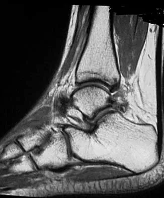
A 16-year-old female dancer has persistent posterior ankle pain, particularly after a vigorous dancing schedule. Examination reveals tenderness both posteromedially and posterolaterally. MRI scans are seen in Figures 44a and 44b. What is the most likely diagnosis?
Explanation
Question 11
Which of the following nerves is most commonly injured during ankle arthroscopy?
Explanation
Question 12
An obese 56-year-old woman with hypertension has had posterior heel pain for the past 6 months. She also notes some enlargement over the posterior aspect of the heel. Examination reproduces pain with palpation at the insertion of the Achilles tendon. A lateral radiograph is shown in Figure 45. What is the most likely diagnosis?
Explanation
Question 13
A 42-year-old woman has a history of nontraumatic ankle swelling with tenderness over the Achilles tendon and plantar fascia. She reports that while vacationing in Connecticut 2 months ago she noted the presence of a "red bull's eye" rash. Management should consist of
Explanation
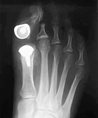
Question 14
A 50-year-old woman has a painful hallux valgus and a painful callus beneath the second metatarsal head. A radiograph is shown in Figure 46. To correct these problems, treatment of the great toe deformity should consist of
Explanation
Question 15
The lower extremity motor dysfunction in Charcot-Marie-Tooth disease most commonly involves which of the following muscles?
Explanation
Question 16
Fixed hyperextension of the metatarsophalangeal joint is associated with
Explanation
Question 17
The orthosis shown in Figure 47 is commonly used for
Explanation
Question 18
A 14-year-old boy has medial ankle pain, progressive unilateral flatfoot deformity, and pain with most activities of daily living. He denies any recent injury. His parents recall that at age 7 years he sustained an injury that was treated as a sprain. Examination reveals valgus deformity with painless, unrestricted passive motion of the ankle. He has grossly equal limb lengths. A radiograph of the affected ankle is shown in Figure 48a, and the contralateral ankle is shown in Figure 48b. Management should consist of
Explanation
Question 19
What is the most common organism found following a nail puncture wound through tennis shoes in a host without immunocompromise?
Explanation
Question 20
Examination of a 28-year-old woman reveals a moderate hallux valgus deformity and a prominence of the medial eminence. She reports that she can participate in all activities, wear 3-inch heels with minimal discomfort, and walk in a 1-inch heel with no pain. However, she is concerned that the deformity will get worse and requests recommendations regarding surgical correction. What is the best course of action?
Explanation
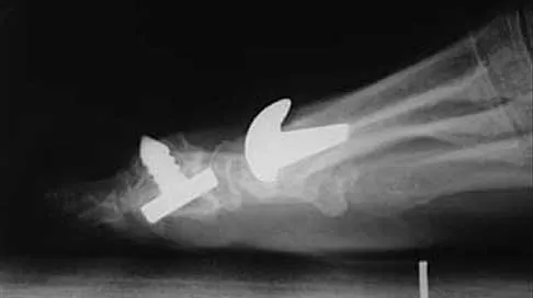
Question 21
A 25-year-old man has ankle instability and a lateral foot callosity. Radiographs are shown in Figures 49a through 49c. Management options are best determined by the
Explanation
Question 22
A 17-year-old boy underwent open reduction and internal fixation of a navicular fracture 5 days ago. A follow-up examination now reveals a tensely swollen foot with erythema and multiple skin bullae. The patient is febrile and has marked pain with palpation of the entire forefoot and hindfoot. What is the next step in management?
Explanation
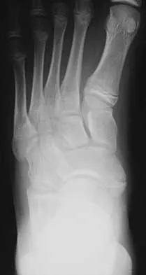
Question 23
A 14-year-old girl has had mild pain and nail deformity of the great toe for the past 4 months. A radiograph is shown in Figure 50. What is the most likely etiology of the lesion?
Explanation
Question 24
The third plantar intrinsic muscle layer of the foot consists of which of the following structures?
Explanation
Question 25
Which of the following results cannot be achieved with an in-shoe orthosis?
Explanation
Question 26
A 55-year-old poorly controlled diabetic presents with a swollen, erythematous, warm, and painless right foot. Pulses are palpable and bounding. Radiographs are unremarkable. MRI demonstrates diffuse marrow edema in the midfoot without focal fluid collections. What is the most appropriate initial management?
Explanation
Question 27
According to the classic biomechanical study by Ramsey and Hamilton, a 1-mm lateral displacement of the talus within the ankle mortise following an ankle fracture reduces the tibiotalar contact area by approximately what percentage?
Explanation
Question 28
A 62-year-old diabetic patient presents with a deep plantar neuropathic ulcer under the first metatarsal head. On examination, a sterile metal probe smoothly passes through the ulcer base and strikes hard, gritty bone. Which of the following is true regarding this clinical finding?
Explanation
Question 29
In a Supination-External Rotation (SER) stage IV ankle fracture, which of the following structures is injured last (Stage IV) according to the Lauge-Hansen classification?
Explanation
Question 30
A 60-year-old diabetic woman with a history of recurrent forefoot ulcers and osteomyelitis presents for preoperative evaluation for a planned Syme amputation. Which of the following noninvasive vascular parameters is the most reliable predictor of successful wound healing at this specific amputation level?
Explanation
Question 31
A 45-year-old man sustains a pronation-external rotation (PER) ankle fracture. Intraoperatively, after rigid internal fixation of the medial and lateral malleoli, an intraoperative Hook test demonstrates 3 mm of lateral syndesmotic widening. Which of the following represents the most appropriate next step?
Explanation
Question 32
A 50-year-old diabetic male undergoes total contact casting for Eichenholtz Stage I Charcot arthropathy of the midfoot. Which of the following radiographic findings marks the transition to Eichenholtz Stage II (Coalescence)?
Explanation
Question 33
A 30-year-old male presents with a severely displaced ankle fracture-dislocation. Closed reduction under conscious sedation in the ED is mechanically blocked. A true lateral radiograph shows the proximal segment of the fibula locked posterior to the tibia. Behind which specific anatomic structure is the fibula typically entrapped in this rare injury pattern?
Explanation
Question 34
A 65-year-old poorly controlled diabetic sustains an unstable bimalleolar ankle fracture. Operative fixation is planned. Compared to a non-diabetic patient, what is the most appropriate modification to the surgical technique and postoperative protocol?
Explanation
Question 35
A 55-year-old patient with long-standing diabetes presents with a unilateral warm, erythematous, and swollen foot and ankle. There are no open wounds. Radiographs show soft tissue swelling without bony abnormalities. Which of the following physical examination findings is most useful to differentiate acute Charcot arthropathy from cellulitis?
Explanation
Question 36
A 42-year-old man sustains an ankle injury. Radiographs reveal an ankle fracture-dislocation that is irreducible in the emergency department. A CT scan demonstrates the proximal fibular shaft fragment is locked behind the posterior tubercle of the tibia. Which of the following is the most likely diagnosis?
Explanation
Question 37
The most common anatomic location for the development of Charcot neuroarthropathy in the diabetic foot is:
Explanation
Question 38
A 35-year-old woman sustains a trimalleolar ankle fracture. The posterior malleolus fracture involves 30% of the articular surface. Which of the following is the primary biomechanical advantage of open reduction and internal fixation of the posterior malleolus compared to placing a trans-syndesmotic screw?
Explanation
Question 39
A 60-year-old diabetic patient presents with a chronic, recurrent plantar neuropathic ulcer under the first metatarsal head despite the use of total contact casting and accommodative footwear. Ankle dorsiflexion is limited to 5 degrees past neutral with the knee extended, but improves to 15 degrees with the knee flexed. What is the most appropriate surgical intervention to promote healing and prevent recurrence?
Explanation
Question 40
According to the Lauge-Hansen classification, what is the initial ligamentous injury in a supination-external rotation (SER) type ankle fracture?
Explanation
Question 41
A 58-year-old patient with poorly controlled type 2 diabetes presents with an ulcerated, swollen midfoot. The clinician is concerned about osteomyelitis versus acute Charcot arthropathy. Which of the following MRI findings is most specific for diagnosing osteomyelitis over Charcot arthropathy?
Explanation
Question 42
A 60-year-old diabetic patient presents with a swollen, erythematous foot without an open ulcer. Radiographs show periarticular fragmentation and subluxation at the tarsometatarsal joint. Skin temperature is elevated compared to the contralateral side. What is the most appropriate initial management?
Explanation
Question 43
A 45-year-old man sustains an ankle injury. Radiographs show an isolated lateral malleolus fracture at the level of the syndesmosis. A gravity stress view shows 6 mm of medial clear space widening. What Lauge-Hansen classification does this injury represent?
Explanation
Question 44
In a patient with long-standing diabetes mellitus and a plantar forefoot ulcer that has failed to heal despite total contact casting, what surgical intervention is most likely to promote healing and prevent recurrence?
Explanation
Question 45
Which of the following clinical tests has the highest positive predictive value for diagnosing osteomyelitis beneath a diabetic foot ulcer?
Explanation
Question 46
A 55-year-old poorly controlled diabetic patient undergoes open reduction and internal fixation of a bimalleolar ankle fracture. What modification to the standard postoperative protocol is most strongly recommended for this patient?
Explanation
Question 47
A patient sustains a pronation-external rotation (PER) ankle injury. According to the Lauge-Hansen classification, what is the first structure injured in this sequential failure pattern?
Explanation
Question 48
What is the pathomechanical consequence of a 1-mm lateral shift of the talus within the ankle mortise following a malreduced ankle fracture?
Explanation
Question 49
A 35-year-old man presents with an ankle fracture-dislocation. Radiographs show a posterior dislocation of the proximal fibular fragment behind the lateral ridge of the distal tibia, which is irreducible by closed means. What is this specific injury pattern named?
Explanation
Question 50
In the treatment of acute Charcot neuroarthropathy (Eichenholtz stage I), when is the transition from a total contact cast to a Charcot Restraint Orthotic Walker (CROW) or custom therapeutic footwear most appropriate?
Explanation
Question 51
A 50-year-old diabetic woman with peripheral neuropathy undergoes ORIF for an unstable ankle fracture. To enhance construct stability and reduce the high risk of catastrophic failure, which of the following techniques is most appropriate?
Explanation
Question 52
A 65-year-old diabetic patient presents with a deep, non-healing plantar midfoot ulcer. Radiographs show a bony prominence causing the ulcer, but MRI is equivocal for osteomyelitis. What is the gold standard for diagnosing osteomyelitis in this setting?
Explanation
Question 53
What is the most appropriate indication for repairing the deltoid ligament during open reduction and internal fixation of a supination-external rotation stage IV ankle fracture?
Explanation
Question 54
A 40-year-old patient presents with a trimalleolar ankle fracture. The posterior malleolus fracture involves 35% of the articular surface and is displaced. What is the primary biomechanical rationale for surgical fixation of the posterior malleolus in this case?
Explanation
Question 55
In diabetic patients, measuring tissue oxygenation is critical for determining the healing potential of an ulcer or a planned surgical incision. What is the minimum transcutaneous oxygen tension (TcPO2) generally considered necessary to support wound healing?
Explanation
Question 56
A 55-year-old patient with long-standing, poorly controlled diabetes presents with a red, hot, swollen left foot. The patient denies any trauma. Pedal pulses are bounding. Inflammatory markers are within normal limits. Radiographs show soft tissue swelling but no fractures or joint subluxation. What is the most appropriate next step in management?
Explanation
Question 57
When treating a trimalleolar ankle fracture, which of the following is the most widely accepted absolute indication for operative fixation of the posterior malleolus?
Explanation
Question 58
A 60-year-old poorly controlled diabetic patient has a chronic plantar foot ulcer beneath the first metatarsal head. Which of the following clinical findings has the highest positive predictive value for underlying osteomyelitis?
Explanation
Question 59
A 32-year-old man presents to the emergency department after a skiing fall. He complains of severe medial ankle pain and proximal lateral leg pain. Ankle radiographs show an isolated widening of the medial clear space. Knee radiographs reveal a proximal third fibula fracture. According to the Lauge-Hansen classification, what is the mechanism of this injury?
Explanation
Question 60
A 65-year-old diabetic patient presents with a recurrent neuropathic ulcer beneath the first metatarsal head despite compliant use of a total contact cast and custom orthotics. Examination reveals that ankle dorsiflexion is limited to 5 degrees of plantarflexion when the knee is fully extended, and remains limited to 5 degrees of plantarflexion when the knee is flexed to 90 degrees. What is the most appropriate surgical intervention?
Explanation
Question 61
A 40-year-old male sustains a severe ankle fracture-dislocation. Closed reduction in the emergency department is unsuccessful despite adequate sedation. A CT scan reveals that the proximal fragment of the fibula is locked behind the posterior tubercle of the distal tibia. What is the eponymous name for this specific injury pattern?
Explanation
Question 62
Operative reconstruction (e.g., corrective arthrodesis) for a patient with midfoot Charcot neuroarthropathy is most commonly indicated and safely performed during which Eichenholtz stage?
Explanation
Question 63
When treating a Weber B (supination-external rotation) distal fibula fracture, what is the primary biomechanical advantage of utilizing a posterior antiglide plate compared to a lateral neutralization plate?
Explanation
Question 64
A diabetic patient is undergoing evaluation for a major lower extremity amputation due to gangrene. Compared to a healthy individual, what is the approximate percentage increase in energy expenditure required for ambulation following a unilateral transtibial (below-knee) amputation?
Explanation
Question 65
Ankle radiographs of a 28-year-old male reveal a vertical shear fracture of the medial malleolus and a transverse fracture of the lateral malleolus at the level of the joint line. According to the Lauge-Hansen classification, what is the most likely mechanism of injury?
Explanation
Question 66
According to the Wagner classification system for diabetic foot ulcers, a lesion described as a deep ulcer with localized gangrene isolated to the great toe and forefoot is classified as:
Explanation
Question 67
A 35-year-old woman is 4 months postoperative from open reduction and internal fixation of an ankle fracture, which included placement of two 3.5mm trans-syndesmotic screws. She is completely asymptomatic and asks if the screws must be removed. Based on current orthopedic literature, what is the recommendation regarding routine removal of syndesmotic screws?
Explanation
Question 68
Differentiating acute Charcot neuroarthropathy from osteomyelitis in the diabetic foot is challenging. On magnetic resonance imaging (MRI), which of the following findings is most specific for osteomyelitis rather than acute Charcot arthropathy?
Explanation
Question 69
A 14-year-old adolescent sustains a twisting ankle injury. Radiographs demonstrate a Salter-Harris III fracture of the anterolateral aspect of the distal tibial epiphysis. What anatomical structure is responsible for the avulsion of this fracture fragment?
Explanation
Question 70
During the radiographic evaluation of a suspected syndesmotic injury, external rotation stress views are obtained. Which of the following radiographic measurements on an AP or mortise view is the most reliable indicator of deep deltoid ligament incompetence and syndesmotic instability?
Explanation
Question 71
A 62-year-old patient with poorly controlled type 2 diabetes and profound peripheral neuropathy sustains a displaced bimalleolar ankle fracture. Which of the following modifications to standard internal fixation is most strongly recommended to minimize catastrophic complications?
Explanation
Question 72
A 55-year-old man with a 15-year history of diabetes presents with a red, swollen, and warm right foot. He denies trauma or fevers. Radiographs show periarticular debris, fragmentation of the tarsometatarsal joints, and subluxation. What is the most appropriate initial management?
Explanation
Question 73
A 65-year-old poorly controlled diabetic with severe peripheral neuropathy sustains a closed, displaced bimalleolar equivalent ankle fracture. He undergoes stable open reduction and internal fixation. What is the most appropriate postoperative weight-bearing protocol for this specific patient?
Explanation
Question 74
A 58-year-old male with long-standing diabetes presents with a red, hot, swollen unilateral foot without open wounds. Radiographs show periarticular fragmentation and subluxation at the tarsometatarsal joints. What is the most appropriate initial management?
Explanation
Question 75
A 32-year-old male sustains a pronation-external rotation (PER) ankle fracture. Intraoperatively, after medial and lateral malleolar fixation, the Cotton test is positive indicating syndesmotic instability. When placing a syndesmotic screw, what is the optimal ankle position and screw technique?
Explanation
Question 76
A 60-year-old diabetic male has a chronic plantar ulcer under the first metatarsal head. On examination, a sterile metal probe easily advances through the ulcer base and palpably taps against a hard, gritty surface. What is the approximate positive predictive value of this clinical finding for underlying osteomyelitis?
Explanation
Question 77
A 45-year-old female sustains a trimalleolar ankle fracture. The posterior malleolar fragment involves 35% of the articular surface and remains displaced 3 mm after fibular fixation. Which of the following is the primary biomechanical advantage of directly fixing this posterior malleolar fragment?
Explanation
Question 78
A 28-year-old male presents with a severe ankle injury following a fall. Closed reduction in the emergency department is unsuccessful. Radiographs show a distinct fracture-dislocation of the ankle.
Which of the following pathoanatomic features most likely prevents closed reduction in this specific injury pattern?
Explanation
Question 79
A 72-year-old obese female with advanced diabetic neuropathy and a history of a contralateral Charcot midfoot presents with a closed, unstable, displaced bimalleolar ankle fracture. In addition to standard open reduction and internal fixation, what supplemental surgical strategy is increasingly favored to minimize the high risk of catastrophic failure?
Explanation
Question 80
A 55-year-old diabetic man presents with a plantar midfoot ulceration measuring 2 cm in diameter. Which of the following is an absolute contraindication to the use of a total contact cast for offloading this patient's ulcer?
Explanation
Question 81
A 24-year-old athlete sustains a twisting injury to the ankle. Anteroposterior (AP) and mortise radiographs are obtained to evaluate for a syndesmotic injury. Which of the following radiographic parameters is considered abnormal and highly suggestive of a syndesmotic disruption on a standard mortise view?
Explanation
Question 82
A 40-year-old male sustains an ankle fracture. Radiographs reveal a transverse fracture of the medial malleolus and a high spiral fracture of the fibula above the syndesmosis (Weber C).
According to the Lauge-Hansen classification, what was the mechanism of injury?
Explanation
None
