العربية
Part of the Master Guide

AAOS & ABOS Foot & Ankle Board Review MCQs (Set 2): Ankle Fractures, Lisfranc, Diabetic Foot

AAOS Foot & Ankle MCQs (Set 4): Ankle Fractures & Hindfoot Deformities | Board Review

23 Apr 2026 59 min read 87 Views
Foot & Ankle 2006 MCQs - Part 4

Key Takeaway

This high-yield question set for the AAOS/ABOS exams focuses on critical Foot & Ankle topics. It covers the diagnosis, classification, and management of various ankle fractures, complex hindfoot deformities, and principles of diabetic foot care. Ideal for board preparation and OITE revision.

AAOS Foot & Ankle MCQs (Set 4): Ankle Fractures & Hindfoot Deformities | Board Review

Comprehensive 100-Question Exam


00:00

Start Quiz

Question 1

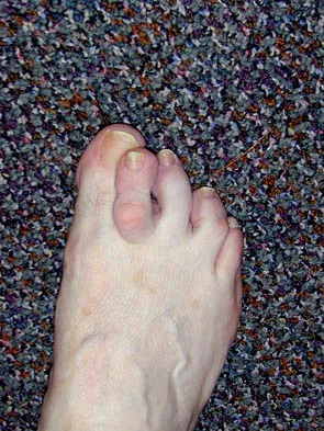
Figures 34a and 34b show the clinical photographs of a 46-year-old woman who has a painful deformity of the second toe. Surgical treatment consisting of metatarsophalangeal capsulotomy and proximal interphalangeal joint resection arthroplasty resulted in satisfactory correction, but the toe remains unstable at the metatarsophalangeal joint. What is the next most appropriate step?


Explanation

Crossover second toes are attributed to attenuation or rupture of the plantar plate and lateral collateral ligament and are associated with varying degrees of instability. Flexor-to-extensor transfer (Girdlestone/Taylor procedure) can provide intrinsic stability to the toe. Although plantar metatarsal head condylectomy can increase stability by resulting in scarring of the plantar plate, excision of the entire second metatarsal head carries a high risk of transfer metatarsalgia. Removal of the base of the proximal phalanx destabilizes the toe and should be reserved as a salvage procedure. Simple flexor tenotomy alone will not improve stability, and arthrodesis of the second metatarsophalangeal joint will limit motion and impair function. Coughlin MJ: Crossover second toe deformity. Foot Ankle 1987;8:29-39.

Question 2

A 40-year-old man fell 10 feet from a tree and sustained the closed isolated injury shown in Figures 35a and 35b. Management consists of splinting. At his 2-week follow-up visit, he clinically passes the wrinkle test. He agrees to open reduction and internal fixation. What is the best surgical approach to obtain anatomic reduction and limit wound dehiscence?


Explanation

The approach to the calcaneus has evolved from several different patterns, driven by a high wound complication rate of 10%. The current extensile lateral approach was described by Zwipp and associates in 1988. The surgical exposure uses an L-shaped incision, with the vertical component positioned one half a finger's breath anterior to the Achilles tendon and extending distally to the junction of the lateral skin and the plantar skin. Borrelli and Lashgari mapped the angiosome of the lateral calcaneal flap and found that the major arterial blood supply to this flap consisted of three arteries: the lateral calcaneal artery, the lateral malleolar artery, and the lateral tarsal artery. The lateral calcaneal artery appeared to be responsible for most of the blood supply to the corner of the flap. This was found 1.5 cm anterior to the Achilles tendon. Division of this artery with inaccurate placement of the vertical limb of the incision can cause ischemia of the lateral skin flap. Borrelli J Jr, Lashgari C: Vascularity of the lateral calcaneal flap: A cadaveric injection study. J Orthop Trauma 1999;13:73-77. Freeman BJC, Duff S, Allen PE, et al: The extended lateral approach to the hindfoot: An anatomical basis and surgical implications. J Bone Joint Surg Br 1998;80:139-142.


Question 3

In the treatment of all magnitudes of bunionette deformities, what is the most common complication associated with lateral condylectomy of the fifth metatarsal head?


Explanation

When a lateral condylectomy alone is performed for all bunionette deformities, a high recurrence rate is expected. Lateral condylectomy should be used alone when the primary deformity is an enlarged lateral condyle of the fifth metatarsal head. In cases with significant divergence of the fifth metatarsal shaft in relationship to the fourth metatarsal shaft or with lateral bowing of the distal fifth metatarsal shaft, the lateral fifth metatarsal prominence will not be effectively reduced and recurrent symptoms and deformity are expected. Transfer metatarsalgia and/or dislocation of the metatarsophalangeal joint can infrequently occur with excessive metatarsal head excision. Arthrosis of the metatarsophalangeal joint has not been frequently reported. Coughlin MJ, Mann RA: Keratotic disorders of the plantar skin, in Coughlin MJ, Mann RA (eds): Surgery of the Foot and Ankle, ed 6. St Louis, MO, Mosby-Year Book, 1993, pp 413-465. Kelikian H: Deformities of the lesser toe, in Kelikian H (ed): Hallux Valgus, Allied Deformities of the Forefoot and Metatarsalgia. Philadelphia, PA, WB Saunders, 1965, pp 327-330.

Question 4

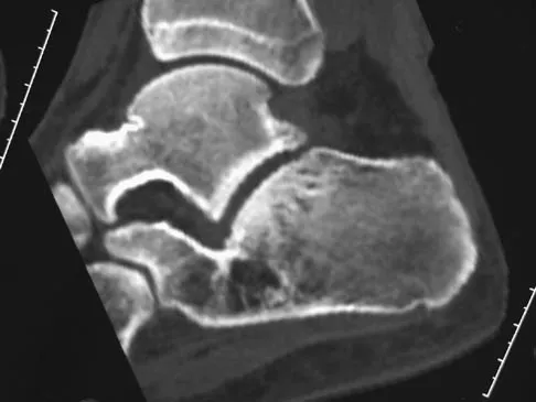
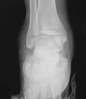
A 17-year-old patient sustained a closed calcaneal fracture when he jumped off of a roof 2 years ago, and he underwent nonsurgical management at the time of injury. The patient now reports lateral hindfoot pain that is worse with weight-bearing activities. Anti-inflammatory drugs and orthoses have failed to provide relief. Coronal and sagittal CT scans are shown in Figures 36a and 36b. What is the best course of action?


Explanation

The CT scans show evidence of a lateral wall blowout and malunion without significant arthrosis of the subtalar joint. In a young patient, it is preferable to avoid a fusion and allow residual motion by performing an exostectomy that decompresses the lateral subtalar joint and peroneal tendons. Chandler JT, Bonar SK, Anderson RB, et al: Results of in situ subtalar arthrodesis for late sequelae of calcaneus fractures. Foot Ankle Int 1999;20:18-24.


Question 5

A 52-year-old woman with diabetes mellitus has had a plantar foot ulcer under the second metatarsal head for the past week. The patient had a similar ulcer 2 months ago, and total contact casting resulted in healing. Examination reveals no signs of infection. What procedure will best prevent recurrence of the ulcer?


Explanation

The contracted Achilles tendon leads to increased forefoot pressure, thus increasing the risk for ulceration in neuropathic patients. Several studies have shown the benefit of Achilles tendon lengthening to heal and prevent forefoot ulceration in these patients. The flexor hallucis longus transfer is used for chronically torn/deficient Achilles tendons, not a contracted Achilles tendon. The Jones procedure works well for the first ray but does not help to alleviate pressure under the second ray. Peripheral bypass surgery is unnecessary because the ulcer healed during the initial treatment, indicating that the patient has adequate circulation. The posterior tibial tendon transfer is used for foot drop or other neuromuscular conditions to correct deformity and increase function. It is not used for forefoot ulcers in patients with diabetes mellitus. Armstrong DG, Stacpoole-Shea S, Nguyen H, et al: Lengthening of the Achilles tendon in diabetic patients who are at high risk for ulceration of the foot. J Bone Joint Surg Am 1999;81:535-538.

Question 6

Figures 37a and 37b show the clinical photographs of a 43-year-old patient with type I diabetes mellitus who has a stump ulcer after undergoing successful transtibial amputation 1 year ago. Which of the following is considered the most predictable method of healing the ulcer and preventing recurrent ulceration?


Explanation

The ulcer occurred as the result of a mismatch between the shape of the residual limb and the prosthetic socket. With the mismatch, the residual limb pistoned and the tissue failed because of the applied shear forces. The most predictable short- and long-term solution is reconstruction of the residual limb. Refraining from use of the prosthesis will prevent the patient from walking for months. It is unlikely that prosthetic socket modification will allow resolution of this large ulcer.


Question 7

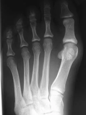
A 15-year-old girl who plays high school basketball has had worsening forefoot pain and swelling that is aggravated by activity for the past 5 weeks. She denies any history of an injury. Examination reveals no deformities. A radiograph is shown in Figure 38. Initial management should consist of


Explanation

Freiberg's infraction is believed to be an osteochondrosis of the second metatarsal head. It is the only osteochondrosis that has a predilection for females. The typical patient is an athletically active adolescent female. The radiograph shows stage II disease wherein reossification is occurring; it is at this time that the second metatarsal head is most susceptible to deformation. Therefore, initial management should consist of a short leg walking cast.


Question 8

A 56-year-old woman has a painful mass on the bottom of her left foot, and orthotic management has failed to provide relief. Examination reveals that the mass is contiguous with the plantar fascia. An MRI scan shows a homogenous nodule within the plantar fascia. Resection of the tumor is shown in the clinical photograph in Figure 39. What type of cell is most likely responsible for the formation of this tumor?


Explanation

The history, examination, and surgical findings are most consistent with plantar fibromatosis. Plantar fibromatosis is a benign tumor of the plantar fascia that consists chiefly of fibromyoblasts. These cells produce excessive collagen and are similar to the cells found in the palmar fascia of patients with Dupuytren's contracture of the hand. The myocyte, synovial cell, and osteocyte all produce their respective individual tissue types but do not contribute to the formation of a plantar fibromatosis. The T-cell is an important immunologic cell that is most affected in patients with HIV.


Question 9

A 34-year-old man underwent a transtibial amputation as the result of a work-related injury. The amputation was performed at the inferior level of the tibial tubercle. The residual limb has a soft-tissue envelope composed of gastrocnemius muscle that is used as soft-tissue cushioning for the distal tibia. Despite undergoing several prosthetic fittings, he continues to report pain and instability. Examination reveals that the prosthesis appears to fit well with no apparent pressure points or areas of skin breakdown. He is not willing to have any further surgery. Which of the following modifications will most likely provide relief?


Explanation

While transtibial amputees can be fitted with a prosthesis with a residual limb as short as 5 cm, or with retention of the insertion of the patellar tendon, this patient has an unstable gait because of the limited ability of the prosthetic socket to maintain a snug and stable fit. While cumbersome and bulky, double metal uprights and a corset is the only predictable method of gaining stability. The other methods attempt to add an element of stability; however, they are unlikely to be successful. Bowker JH, Goldberg B, Poonekar PD: Transtibial amputation: Surgical procedures and postsurgical management, in Bowker JH, Michael JW (eds): Atlas of Limb Prosthetics. St Louis, MO, Mosby Year Book, 1992, pp 429-452.

Question 10

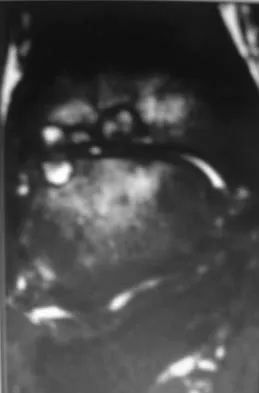
A 30-year-old man has had intermittent swelling of his right ankle for the past 6 months. He denies any history of trauma. Radiographs reveal osteolytic changes on both sides of the joint. An axial CT scan and a T2-weighted MRI scan are shown in Figures 40a and 40b. He undergoes surgical excision. An intraoperative photograph and a biopsy specimen are shown in Figures 40c and 40d. What is the most likely diagnosis?


Explanation

Pigmented villonodular synovitis often presents with intermittent swelling and minimal pain. It often occurs around joints but may be found around tendon sheaths and bursal linings. Periarticular erosions involving both sides of joints are typical, and multiple joint involvement has been described. Portions of low-signal intensity on T1- and T2-weighted images are characteristic of hemosiderin-laden processes. High-signal content is suggestive of high water content. The combination of low-signal intensity areas in intra-articular lesions with or without osseous destruction is diagnostic of pigmented villonodular synovitis. Aspiration reveals bloody or brownish fluid. The treatment of choice is synovectomy performed arthroscopically or open. Recurrence is common. Walling AK: Soft tissue and bone tumors, in Coughlin MJ, Mann RA (eds): Surgery of the Foot and Ankle, ed 7. St Louis, MO, Mosby, 1999, pp 1007-1032.


Question 11

The Coleman block test is used to evaluate the cavovarus foot. What is the most important information obtained from this test?


Explanation

Coleman block testing, performed by placing an elevation under the lateral border of the foot, is used to determine if the forefoot and/or plantar flexed first ray is causing a compensatory varus in the hindfoot. The block is placed under the lateral border of the foot, and therefore does not have any relation to the Achilles tendon and suppleness of the hindfoot. Holmes JR, Hansen ST Jr: Foot and ankle manifestations of Charcot-Marie-Tooth disease. Foot Ankle 1993;14:476-486.

Question 12

Figures 41a and 41b show the radiographs of a 22-year-old woman who has a bunion on her left foot. She denies pain in the foot, but she reports increasing difficulty with shoe wear. Management should consist of


Explanation

Surgery is not indicated in a patient who has a mild deformity and no pain. Shoe wear modifications should be recommended.


Question 13

A 35-year-old woman has had significant pain and swelling in the left medial ankle inferior to the medial malleolus for the past 8 months. Physical therapy, brace and orthotic management, and immobilization have failed to provide relief. She is now requesting a more aggressive option to assist in pain relief. Clinical photographs and radiographs are seen in Figures 42a through 42f. Following exposure, a complete rupture of the posterior tibial tendon is visible. What is the most appropriate surgical reconstruction?


Explanation

The patient has a complete rupture of the posterior tibial tendon with minimal hindfoot valgus deformity. The deformity is supple, and there is no arthritis in the subtalar, talonavicular, or calcaneocuboid joints; therefore, joint-sparing procedures are appropriate in this patient (avoidance of arthrodeses). The treatment of choice is flexor digitorum longus tendon transfer, medial slide calcaneal osteotomy, and spring ligament repair. Primary repair of an incompetent posterior tibial tendon can lead to failure and recurrence of pain and deformity. Talonavicular arthrodesis corrects the forefoot abduction and elevates a plantar flexed talus; however, the patient does not have this deformity; therefore, the procedure is not indicated. Myerson MS, Corrigan J, Thompson F, et al: Tendon transfer combined with calcaneal osteotomy for treatment of posterior tibial tendon insufficiency: A radiological investigation. Foot Ankle Int 1995;16:712-718. Trnka HJ, Easley ME, Myerson MS: The role of calcaneal osteotomies for correction of adult flat foot. Clin Orthop 1999;365:50-64. Jahss MH: Spontaneous rupture of the tibialis posterior tendon: Clinical findings, tenographic studies, and a new technique for repair. Foot Ankle 1982;3:158-166.


Question 14

A 48-year-old man reports localized plantar forefoot pain. Examination reveals a discrete callus (intractable plantar keratosis) with well-localized tenderness beneath the second metatarsal head. The callus most likely lies beneath what structure?


Explanation

A discrete or focal callus is a response to excessive weight-bearing stress beneath the lateral (fibular) condyle of a lesser metatarsal head (most commonly second). The other structures generally have not been associated with a discrete callus. Coughlin MJ, Mann RA: Keratotic disorders of the plantar skin, in Coughlin MJ, Mann RA (eds): Surgery of the Foot and Ankle, ed 6. St Louis, MO, Mosby-Year Book, 1993, pp 413-465.

Question 15

A 65-year-old woman with a history of diabetes mellitus and plantar ulcers has an erythematous and swollen right foot and ankle. Despite IV antibiotics, the erythema spreads to her lower calf within 24 hours. She has a systolic blood pressure of 80/55 mm Hg and a pulse rate of 120. Laboratory studies show a creatinine level of 1.5 mg. Initial management should consist of


Explanation

Necrotizing fasciitis is an aggressive and rapidly spreading soft-tissue infection, usually caused by group A beta-hemolytic Streptococcus pyogenes. Presentation is typical of a rapidly ascending cellulitis, recalcitrant to antibiotic treatment. Differentiation between cellulitis and impetigo is difficult, and success depends on a high level of suspicion. The skin and subcutaneous tissues are affected, with sparing of the muscles. Septic shock and multi-organ system failure can be fatal. Treatment is aggressive surgical debridement with broad-spectrum antibiotics. Repeat irrigation and debridement may be necessary. Hyperbaric oxygen studies have shown inconsistent results. Richardson EG (ed): Orthopaedic Knowledge Update: Foot and Ankle 3. Rosemont, IL, American Academy of Orthopaedic Surgeons, 2003, pp 199-205.

Question 16

A 15-year-old boy has hindfoot pain and very limited subtalar motion. A CT scan reveals a talocalcaneal coalition involving 40% of the middle facet. He has no degeneration of the posterior subtalar facet. Following failure of nonsurgical management, treatment should consist of


Explanation

The CT scan is an important test to help determine the extent of involvement of the talocalcaneal facet in a talocalcaneal coalition. In a young patient with no arthritis and joint involvement of less than 50%, resection of the coalition and fat pad interposition has been shown to be successful. A calcaneal osteotomy does not address the coalition. Subtalar arthroereisis has been used for treatment of a flexible flatfoot; tarsal coalition patients have a rigid-type flatfoot deformity. Sullivan JA: The child's foot, in Morrissy RT, Weinstein SL (eds): Lovell and Winter's Pediatric Orthopaedics, ed 4. Philadelphia, PA, Lippincott-Raven, 1996, vol 2, pp 1077-1135.

Question 17

A 55-year-old patient is seeking a surgical consultation for a painful flatfoot deformity that has failed to respond to nonsteroidal anti-inflammatory drugs, shoe and activity modifications, and orthoses. The patient is of medium build, a nonsmoker, and has no history of diabetes mellitus. Radiographs are shown in Figures 43a through 43c. Based on these findings, treatment should consist of


Explanation

The patient has a degenerative collapse of the midfoot through the tarsometatarsal joints with significant forefoot abduction; therefore, a midfoot arthrodesis is required to address the arthritic joints and deformity at the tarsometatarsal articulation. All of the other procedures correct hindfoot deformities and therefore would not be appropriate treatment. Brage M: Degenerative joint disease of the midfoot. Foot Ankle Clin 1999;4:355-367.


Question 18

A 32-year-old construction worker reports a persistent burning, tingling sensation on the dorsum of his right foot and significant sensitivity on the plantar surface after a 500-lb steel beam dropped on it 8 weeks ago. Initial radiographs revealed no fractures, and the skin remained intact at the time of injury. Physical therapy, anti-inflammatory drugs, and a serotonin reuptake inhibitor have failed to provide relief. What is the next most appropriate step in management?


Explanation

Following failure of physical therapy and pharmacologic management in a patient with complex regional pain syndrome, the management of choice is sympathetic blocks. While continued physical therapy would be assistive, sympathetic blocks allow a more rapid relief of symptoms. Neurostimulation is not appropriate at this stage because of its invasive nature. Cepeda MS, Lau J, Carr DB: Defining the therapeutic role of local anesthetic sympathetic blockade in complex regional pain syndrome: A narrative and systematic review. Clin J Pain 2002;18:216-233. Perez RS, Kwakkel G, Zuurmond WW, et al: Treatment of reflex sympathetic dystrophy (CRPS type 1): A research synthesis of 21 randomized clinical trials. J Pain Symptom Manage 2001;21:511-526. Tran KM, Frank SM, Raja SN, et al: Lumbar sympathetic block for sympathetically maintained pain changes in cutaneous temperatures and pain perception. Anesth Analg 2000;90:1396-1401.

Question 19

What nerve is most likely to develop a traumatic neuroma following open reducation and internal fixation of a talar neck fracture via a posterolateral approach?


Explanation

The preferred approach is posterolateral, placing the sural nerve most at risk. The dorsal intermediate cutaneous nerve is anterolateral to the ankle, and the medial and lateral plantar branches are medial and inferior to the surgical site. The saphenous nerve is anteromedial and away from the surgical approach. Swanson TV, Bray TJ, Holmes GB Jr: Fractures of the talar neck: A mechanical study of fixation. J Bone Joint Surg Am 1992;74:544-551.

Question 20

Patients with tarsal tunnel syndrome are most likely to obtain a favorable outcome from decompression of the posterior tibial nerve if which of the following conditions is present?


Explanation

In one series, only 44% of patients demonstrated good results from tarsal tunnel decompression if they did not have a mass in the tarsal tunnel. Conditions that produce a traction neuropathy of the posterior tibial nerve are unlikely to respond to neurolysis. The most favorable condition associated with a good response to decompression is when a compressing anatomic structure can be removed from the tarsal tunnel. Frey C, Kerr R: Magnetic resonance imaging and the evaluation of tarsal tunnel syndrome. Foot Ankle 1993;14:159-164. Garrett AL: Poliomyelitis, in Nickel VL (ed): Orthopaedic Rehabilitation. New York, NY, Churchill Livingston, 1982, pp 449-458.

Question 21

A 30-year-old man has chronic pain, joint stiffness, and symmetrical polyarthropathy but no significant synovitis. Examination reveals enlargement of the second and third metatarsal heads. Radiographs show chondrocalcinosis of the ankles and bony enlargement of the midfoot; no marginal erosions are evident at the metatarsophalangeal level. What is the most likely diagnosis?


Explanation

The patient's clinical picture is considered the classic presentation for hemochromatosis. Osteoarthritis and pseudogout more commonly affect an older age group. Rheumatoid arthritis is more common in women and is not commonly associated with chondrocalcinosis. The radiographic appearance of the forefoot in Reiter's syndrome is one of a pencil in cup deformity of the metatarsophalangeal joint, not enlargement. Stevens FM, Edwards C: Recognizing and managing hemochromatosis and hemochromatosis arthropathy. J Musculoskeletal Med 2004;4:212-225.

Question 22

The strongest biomechanical construct for open reduction and internal fixation of a talar neck fracture uses what interval and entry point?


Explanation

The strongest biomechanical construct is posterior to anterior fixation with the entry point being at the level of the posterolateral tubercle of the talus. This uses the interval between the peroneus brevis and the flexor hallucis longus. The interval between the flexor digitorum longus and the flexor hallucis longus with entry at the posteromedial tubercle of the talus is not an accepted approach for fixation of talar neck fractures. All of the other options use screw placements from anterior to posterior. Swanson TV, Bray TJ, Homes GB Jr: Fractures of the talar neck: A mechanical study of fixation. J Bone Joint Surg Am 1992;74:544-551.

Question 23

A 14-year-old boy with a history of cerebral palsy has a clawed hallux, cavus foot deformity, and associated pain. Examination reveals pain under the first metatarsal head and a rigid first tarsometatarsal joint. Treatment should consist of


Explanation

In a retrospective study for clawing of the hallux with associated cavus foot deformities, the modified Robert Jones tendon transfer was shown to be effective in relieving symptoms related to clawing of the hallux in 90% of patients but was not reliable in relieving pain under the first metatarsal head, with success in only 43% of patients. In three patients, pain under the metatarsal head was relieved initially but recurred by 18 months. The return of symptoms in these cases is the result of stretching of the muscle and tendon of the extensor hallucis longus by the more powerful peroneus longus. Basal dorsal wedge osteotomy at the time of the modified Robert Jones procedure is recommended. Mizel MS, Miller RA, Scioli MW (eds): Orthopaedic Knowledge Update: Foot and Ankle 2. Rosemont, IL, American Academy of Orthopaedic Surgeons, 1998, pp 79-100.

Question 24

A 45-year-old woman with stage II posterior tibial tendinitis has failed to respond to nonsurgical management. Recommended treatment now includes posterior tibial tendon debridement and medial calcaneal displacement osteotomy along with transfer of what tendon?


Explanation

The flexor digitorium longus is the commonly accepted tendon transfer for posterior tibial tendon insufficiency. The flexor hallucis longus has to be carefully rerouted to avoid crossing the neurovascular bundle and has not been shown clinically to provide superior results to flexor digitorum longus transfer. Use of the peroneus longus results in loss of plantar flexion strength of the first metatarsal, contributing to the flatfoot deformity. The anterior tibial tendon is in the anterior compartment and fires out of phase with the posterior tibial tendon. Sitler DF, Bell SJ: Soft tissue procedures. Foot Ankle Clin 2003;8:503-520.

Question 25

A 38-year-old marathon runner has had Achilles tendon pain for the past 2 months. Examination reveals that the tendon is thickened and tender proximal to the calcaneal insertion. The tendon sheath is not thickened or tender. The pathophysiology of the tendon is best described as


Explanation

Atraumatic Achilles tendon disease can be differentiated into Achilles tendinosis and peritendinitis. Thickening and tenderness of the Achilles tendon are present in both, but thickening and tenderness of the tendon sheath indicates peritendinitis. Histologic examination of Achilles tendinosis reveals an absence of acute and chronic inflammatory cells. Radiologists often diagnose partial tendon rupture by MRI and there may be microscopic longitudinal tears present, but there is no mechanical compromise as would be implied by a partial rupture. The thickening typically occurs in the portion of the tendon with the poorest blood supply, and biochemical analysis detects high levels of lactate and other products of anaerobic glycolysis. Astrom M, Rausing A: Chronic Achilles tendinopathy: A survey of surgical and histopathologic findings. Clin Orthop 1995;316:151-164. Ohberg L, Lorentzon R, Alfredson H: Neovascularisation in Achilles tendons with painful tendinosis but not in normal tendons: An ultrasonographic investigation. Knee Surg Sports Traumatol Arthrosc 2001;9:233-238.

Question 26

When utilizing an anterolateral approach to the distal tibia for open reduction and internal fixation of a pilon fracture, which neurologic structure is at greatest risk of iatrogenic injury during the superficial surgical dissection?


Explanation

The superficial peroneal nerve crosses the surgical field as it transitions from the lateral compartment to the anterior aspect of the leg. It must be identified and protected during the anterolateral approach to the distal tibia.

Question 27

Which of the following parameters measured on a standard anteroposterior (AP) or mortise radiograph is considered the most reliable indicator of syndesmotic widening?


Explanation

The tibiofibular clear space, measured 1 cm above the joint line on an AP or mortise radiograph, should be less than 6 mm. It is the most reliable radiographic parameter for assessing syndesmotic integrity, as it is unaffected by tibial rotation.

Question 28

A 55-year-old woman presents with a painful, flexible flatfoot deformity and an inability to perform a single-leg heel raise.

Clinical examination reveals severe forefoot abduction with 'too many toes' sign. Which of the following procedures is most appropriate to specifically address the forefoot abduction component of her deformity?


Explanation

Lateral column lengthening (Evans osteotomy) effectively corrects the forefoot abduction associated with Stage IIb adult acquired flatfoot deformity. A medial displacement calcaneal osteotomy primarily addresses hindfoot valgus but does not reliably correct severe forefoot abduction.

Question 29

In the Sanders classification of intra-articular calcaneal fractures, the primary prognostic variable that determines the classification type is the number and location of fracture lines through which of the following structures?


Explanation

The Sanders classification is based on coronal CT scan images and categorizes calcaneal fractures based on the number of primary fracture lines through the posterior articular facet. The severity correlates with the degree of comminution of this specific facet.

Question 30

A 35-year-old man sustains a severe ankle injury. Radiographs reveal a fracture-dislocation where the proximal fibular fragment is irreducible by closed means.

Intraoperative exploration demonstrates entrapment of the fibula behind the posterior tubercle of the distal tibia. What is the diagnosis?


Explanation

A Bosworth fracture is a rare fracture-dislocation of the ankle where the proximal fibular fragment becomes locked behind the posterior tubercle of the distal tibia. This entrapment makes closed reduction impossible, necessitating open reduction.

Question 31

A 24-year-old man presents with a symptomatic cavovarus foot deformity. A Coleman block test is performed, during which the first ray is allowed to drop off the block, and the hindfoot corrects to a neutral alignment. This finding indicates that the hindfoot varus is primarily driven by which of the following?


Explanation

The Coleman block test distinguishes between rigid and flexible hindfoot varus. If the hindfoot corrects to neutral when the first ray is allowed to drop, the deformity is flexible and driven by a rigidly plantarflexed first ray.

Question 32

A 14-year-old boy presents with frequent ankle sprains and a rigid, flat foot. CT imaging confirms a middle facet talocalcaneal coalition. If nonoperative management fails, what is the generally accepted threshold of joint involvement above which a subtalar or triple arthrodesis is recommended over surgical resection?


Explanation

Resection of a talocalcaneal coalition is generally contraindicated if the coalition involves more than 50% of the posterior facet area or if there are advanced degenerative changes. In these cases, arthrodesis is the preferred surgical treatment.

Question 33

According to the Lauge-Hansen classification, which of the following represents the correct sequential order of structural injury in a Supination-External Rotation (SER) ankle fracture?


Explanation

In the SER pattern, the force creates sequential injury starting anterolaterally: AITFL (Stage 1), spiral fracture of the fibula (Stage 2), PITFL or posterior malleolus (Stage 3), and finally the medial malleolus or deltoid ligament (Stage 4).

Question 34

A 45-year-old man sustains an ankle fracture. Radiographs and CT scan demonstrate a lateral malleolus fracture and a posterior malleolus fracture involving 35% of the tibial articular surface with step-off. According to current biomechanical and clinical guidelines, what is the most appropriate management of the posterior malleolus?


Explanation

Current literature supports open reduction and internal fixation of posterior malleolus fractures involving >25% of the articular surface or those associated with syndesmotic instability. Fixation restores the articular surface and improves syndesmotic stability better than syndesmotic screws alone.

Question 35

During open reduction and internal fixation of a Weber C ankle fracture, the surgeon needs to evaluate the syndesmosis. Which intraoperative method is considered the most reliable indicator of syndesmotic instability requiring fixation?


Explanation

The intraoperative fluoroscopic Cotton test (lateral pull on the fibula using a bone hook) is the most reliable clinical test to assess dynamic syndesmotic instability. Widening of the tibiofibular clear space under stress dictates the need for syndesmotic fixation.

Question 36

A 65-year-old patient with long-standing, poorly controlled diabetes mellitus and severe peripheral neuropathy presents with a displaced bimalleolar ankle fracture. To minimize the high risk of complications, which modification to standard operative management is most appropriate?


Explanation

Diabetic patients with severe neuropathy are at high risk for fixation failure and Charcot arthropathy. Enhanced fixation techniques, such as multiple trans-syndesmotic screws or stronger locking constructs, combined with prolonged non-weight-bearing, are recommended.

Question 37

A 30-year-old man presents with a high-energy closed ankle injury. The ankle is grossly deformed and irreducible in the emergency department. Radiographs demonstrate the proximal fragment of the fibula entrapped posterior to the lateral tubercle of the distal tibia. What is the correct diagnosis?


Explanation

A Bosworth fracture-dislocation is a rare, severe injury where the proximal fibular fragment dislocates posterior to the incisura fibularis of the tibia. It is characteristically irreducible by closed means and requires prompt open reduction.

Question 38

A 55-year-old woman presents with a painful, unilateral flatfoot deformity. She is unable to perform a single-leg heel rise, but manual testing reveals that her subtalar joint remains mobile and reducible. What is the most appropriate surgical treatment?


Explanation

This patient has a Stage II adult acquired flatfoot deformity (flexible flatfoot). The gold standard surgical management for a flexible deformity involves an FDL transfer to replace the diseased posterior tibial tendon, combined with a medial displacement calcaneal osteotomy.

Question 39

In a patient with Charcot-Marie-Tooth disease who presents with a progressive, flexible cavovarus foot deformity, the plantarflexed first ray is primarily driven by the relative overpull of which specific muscle?


Explanation

In Charcot-Marie-Tooth disease, muscle imbalances lead to the classic cavovarus deformity. The peroneus longus remains strong and overpulls the weak tibialis anterior, driving the first metatarsal into rigid plantarflexion.

Question 40

A 40-year-old man presents with severe lateral hindfoot pain 2 years after nonoperative management of a displaced intra-articular calcaneus fracture. Examination reveals subtalar stiffness and subfibular impingement. Radiographs show a healed calcaneal malunion with loss of Bohler's angle and advanced subtalar arthritis. What is the best surgical option?


Explanation

For a painful calcaneal malunion with subtalar arthritis and subfibular impingement, an in situ subtalar arthrodesis combined with a lateral wall exostectomy reliably relieves pain and restores lateral hindfoot anatomy.

Question 41

According to the Lauge-Hansen classification, what is the correct sequential order of tissue injury in a supination-external rotation (SER) ankle fracture?


Explanation

The SER injury progresses in four stages: Stage 1 is the AITFL; Stage 2 is the short oblique/spiral fracture of the fibula; Stage 3 is the PITFL or posterior malleolus; Stage 4 is the deltoid ligament or medial malleolus.

Question 42

A 14-year-old boy presents with a rigid flatfoot and a history of recurrent ankle sprains. Clinical suspicion is high for a calcaneonavicular coalition. Which radiographic view is most sensitive and specific for demonstrating this specific coalition?


Explanation

The 45-degree internal oblique view of the foot elongates the calcaneonavicular interval, making it the best standard radiograph to diagnose a calcaneonavicular coalition. The Harris axial view is best for talocalcaneal coalitions.

Question 43

A 15-year-old girl is evaluated for a painful, rigid flatfoot. A lateral weight-bearing radiograph demonstrates a continuous, dense osseous outline extending from the talar dome down to the sustentaculum tali (the "C-sign"). This radiographic finding strongly suggests which underlying condition?


Explanation

The "C-sign" on a lateral radiograph represents a continuous bony bridge between the talar dome and the sustentaculum tali. It is a highly reliable indicator of a talocalcaneal coalition, particularly involving the middle facet.

Question 44

A 35-year-old man sustains a high-energy, closed tibial pilon fracture. At presentation, there is massive soft tissue swelling and multiple clear fracture blisters over the medial ankle. What is the most appropriate initial orthopaedic management?


Explanation

High-energy pilon fractures are frequently associated with severe soft tissue compromise. Standard of care involves initial application of a spanning external fixator, delaying definitive ORIF until soft tissues recover and the "wrinkle sign" appears.

Question 45

When utilizing lag screws for the internal fixation of a transverse medial malleolus fracture, what is the optimal trajectory of the screws to maximize interfragmentary compression and avoid articular penetration?


Explanation

According to AO principles of fracture management, lag screws provide maximum interfragmentary compression when inserted exactly perpendicular to the fracture plane. Directing them parallel to the joint runs the risk of poor compression or joint penetration depending on the fracture angle.

Question 46

A 22-year-old woman is being evaluated for a symptomatic cavovarus foot deformity. A Coleman block test is performed by having the patient stand with her lateral heel and foot on a wooden block, allowing the first metatarsal to hang freely. During this maneuver, her hindfoot varus completely corrects to neutral. What does this indicate?


Explanation

The Coleman block test evaluates hindfoot flexibility. If the hindfoot varus corrects to neutral when the first metatarsal is allowed to drop, the hindfoot is flexible, and the deformity is primarily driven by the rigid, plantarflexed first ray.

Question 47

A 62-year-old woman has severe, long-standing, medial and lateral ankle pain with a progressive flatfoot deformity. Examination shows a fixed, rigid planovalgus deformity, and radiographs demonstrate significant joint space narrowing and subchondral sclerosis in the subtalar and talonavicular joints. What is the most definitive surgical intervention?


Explanation

This patient has a Stage III adult acquired flatfoot deformity, characterized by a rigid deformity and established hindfoot/midfoot arthritis. The standard definitive surgical treatment is a triple arthrodesis to realign and fuse the painful, arthritic joints.

Question 48

A 40-year-old patient presents with a severe ankle injury. Radiographs show a transverse fracture of the medial malleolus and a comminuted, bending-type fracture of the fibula located above the level of the syndesmosis. Which Lauge-Hansen classification does this pattern most accurately fit?


Explanation

The Pronation-Abduction injury pattern begins with tension medially (transverse medial malleolus fracture or deltoid rupture), followed by syndesmotic rupture, and concludes with a comminuted bending fracture of the fibula above the joint level.

Question 49

When counseling a patient on the choice between operative and nonoperative management for an acute Achilles tendon rupture using modern, early-functional rehabilitation protocols, operative treatment is statistically associated with a higher risk of which of the following?


Explanation

Modern randomized controlled trials show that early functional rehabilitation protocols make the re-rupture rates between operative and nonoperative management statistically similar. However, operative management consistently carries a higher risk of wound complications and infection.

Question 50

A 28-year-old woman reports persistent lateral foot pain 4 weeks after an inversion injury initially diagnosed as an ankle sprain. She has maximal point tenderness about 2 cm anterior and inferior to the lateral malleolus. Radiographs reveal a minimally displaced fracture of the anterior process of the calcaneus. This fracture usually occurs via avulsion of which ligament?


Explanation

Fractures of the anterior process of the calcaneus are often misdiagnosed as simple lateral ankle sprains. They typically result from an avulsion of the bifurcate ligament (calcaneocuboid and calcaneonavicular bands) during forceful plantarflexion and inversion.

Question 51

A 35-year-old man sustains an ankle fracture. Radiographs show a posterior malleolar fragment involving 30% of the articular surface with posterior subluxation of the talus. What is the most appropriate surgical approach for direct visualization and isolated fixation of this fragment?


Explanation

The posterolateral approach allows direct visualization and stable fixation of the posterior malleolus, which is indicated for large fragments (>25%) with posterior subluxation. It provides excellent access while avoiding the posteromedial neurovascular bundle.

Question 52

A 42-year-old roofer falls from a ladder and sustains a displaced intra-articular calcaneus fracture

. According to the Sanders classification, which of the following radiographic views is primary for determining the grade of the fracture?


Explanation

The Sanders classification of calcaneus fractures is based on the number and location of articular fracture lines through the posterior facet. This is best visualized and graded on the coronal CT image.

Question 53

A 50-year-old male presents with a high-energy closed pilon fracture accompanied by severe soft tissue swelling, fracture blisters, and skin tenting. What is the most appropriate initial management step?


Explanation

High-energy pilon fractures with severe soft tissue compromise require staged management. Temporary spanning external fixation allows soft tissues to recover before definitive internal fixation is performed, typically 10 to 21 days later.

Question 54

A 55-year-old woman presents with medial ankle pain and a progressive flatfoot deformity. She has a flexible hindfoot valgus and is unable to perform a single-leg heel raise. Nonoperative management has failed. What is the most appropriate surgical intervention?


Explanation

The patient has Stage II posterior tibial tendon dysfunction (flexible deformity). The gold standard surgical treatment involves FDL transfer to the navicular and a medializing calcaneal osteotomy to restore the medial arch and correct hindfoot valgus.

Question 55

A 62-year-old female presents with severe, rigid planovalgus deformity and lateral hindfoot pain secondary to subfibular impingement. Radiographs show significant subtalar and talonavicular arthritis. What is the most appropriate definitive treatment?


Explanation

Stage III adult acquired flatfoot deformity is characterized by a rigid deformity with associated hindfoot arthritis. Triple arthrodesis (subtalar, talonavicular, and calcaneocuboid) provides deformity correction, stability, and definitive pain relief.

Question 56

During open reduction and internal fixation of a Weber C ankle fracture, the surgeon suspects a syndesmotic injury. Which of the following intraoperative tests is considered the most reliable for evaluating syndesmotic instability?


Explanation

The Cotton test involves placing a bone hook around the fibula and applying a lateral force to assess for diastasis of the syndesmosis. It is highly reliable for intraoperative evaluation of syndesmotic integrity.

Question 57

A patient with a history of a conservatively managed calcaneus fracture presents 1 year later with persistent lateral midfoot pain exacerbated by walking on uneven ground. Examination reveals tenderness localized to the lateral column. Which joint is most likely responsible for these symptoms?


Explanation

The calcaneocuboid joint can be involved in intra-articular calcaneus fractures or affected by altered biomechanics. Post-traumatic arthritis of this joint typically presents with lateral column pain exacerbated by uneven terrain.

Question 58

A 28-year-old male undergoes ORIF for a displaced talar neck fracture. At 8 weeks postoperatively, an AP radiograph shows a subchondral radiolucent band in the talar dome. What does this finding indicate?


Explanation

Hawkins sign is a subchondral radiolucent band in the talar dome seen 6-8 weeks post-injury. It represents subchondral atrophy from disuse in the presence of an intact blood supply, effectively ruling out avascular necrosis.

Question 59

A 16-year-old boy presents with progressive bilateral cavovarus foot deformities. Neurological examination reveals weakness in foot dorsiflexion and eversion. In Charcot-Marie-Tooth disease, which muscle imbalance is the primary driver of this forefoot-driven hindfoot varus deformity?


Explanation

In Charcot-Marie-Tooth disease, the overpull of a strong peroneus longus against a weak tibialis anterior forces the first ray into plantarflexion. This creates a rigidly plantarflexed first ray, which drives the hindfoot into a secondary varus alignment.

Question 60

A patient presents with a severe cavovarus deformity. A Coleman block test is performed, and the hindfoot completely corrects to a neutral alignment when the first metatarsal is allowed to drop off the block. What does this indicate about the deformity?


Explanation

The Coleman block test assesses hindfoot flexibility in a cavovarus foot. If the hindfoot varus corrects when the plantarflexed first ray is allowed to drop, it confirms that the hindfoot is flexible and the deformity is primarily forefoot-driven.

Question 61

A 30-year-old male presents with a severely deformed ankle following a twisting injury. Closed reduction in the emergency department is unsuccessful. Radiographs show a posterior fracture-dislocation of the fibula behind the posterior tubercle of the distal tibia. What is the most likely diagnosis?


Explanation

A Bosworth fracture-dislocation occurs when the proximal fragment of the fibula becomes irreducibly entrapped behind the posterior tubercle of the distal tibia. It strictly requires open reduction.

Question 62

A 60-year-old patient with poorly controlled diabetes mellitus and severe peripheral neuropathy sustains a bimalleolar ankle fracture. To prevent fixation failure and Charcot arthropathy, what modification to standard surgical technique is recommended?


Explanation

Diabetic patients with neuropathy are at high risk for hardware failure, malunion, and Charcot arthropathy. Maximizing fixation strength (using multiple syndesmotic screws or stronger plates) and extending the non-weight-bearing period are critical.

Question 63

A 14-year-old boy sustains a Salter-Harris III fracture of the anterolateral distal tibial epiphysis

. Which of the following ligaments is responsible for avulsing this fragment?


Explanation

A juvenile Tillaux fracture is an avulsion of the anterolateral distal tibial epiphysis. It is caused by the strong pull of the anterior inferior tibiofibular ligament (AITFL) occurring after the central and medial physis has already closed.

Question 64

A 45-year-old female presents with an isolated lateral malleolus fracture and medial joint line tenderness. A gravity stress radiograph demonstrates 6 mm of medial clear space widening. Which of the following statements regarding the medial injury is most accurate?


Explanation

The deep deltoid ligament is the primary medial stabilizer against lateral talar shift. In a bimalleolar equivalent fracture, anatomic fixation of the fibula and syndesmosis restores stability; routine repair of the deltoid ligament is generally not necessary.

Question 65

A 38-year-old male complains of anterior ankle impingement and difficulty fitting into standard shoes 2 years after conservative treatment of a calcaneus fracture. Which of the following best describes the typical components of a calcaneal malunion?


Explanation

Calcaneal malunions characteristically present with a loss of heel height (causing anterior tibiotalar impingement), increased heel width (causing subfibular impingement), and varus deformity of the tuberosity.

Question 66

A 12-year-old boy presents with frequent ankle sprains and a rigid flatfoot. Radiographs show a "C sign", and CT confirms a middle facet talocalcaneal coalition. If nonoperative management fails and there is no significant arthritis, what is the best surgical option?


Explanation

For symptomatic talocalcaneal coalitions without degenerative changes, resection with interposition of a fat graft or FHL tendon is indicated to prevent recurrence. Extensor digitorum brevis interposition is typically utilized for calcaneonavicular coalitions.

Question 67

A 25-year-old athlete sustains a lateral subtalar dislocation. Closed reduction in the emergency department is unsuccessful. Which of the following anatomic structures is most commonly interposed and blocking reduction in this specific injury pattern?


Explanation

In a lateral subtalar dislocation, the posterior tibial tendon (PTT) frequently becomes entrapped between the talus and the navicular, blocking closed reduction and necessitating surgical intervention.

Question 68

A 45-year-old man undergoes ORIF for a Weber C ankle fracture with syndesmotic instability. Two cortical screws are placed across four cortices. Regarding syndesmotic screw fixation, what does the current orthopedic literature recommend?


Explanation

Current evidence shows no significant functional difference between retained and removed syndesmotic screws. Furthermore, retained screws that eventually break do not negatively impact long-term patient outcomes, making routine removal unnecessary.

Question 69

When treating a trimalleolar ankle fracture, which of the following is the strongest absolute indication for operative fixation of the posterior malleolus?


Explanation

While indications for posterior malleolus fixation have evolved to include smaller fragments if they contribute to syndesmotic instability, the absolute strongest indication remains persistent posterior subluxation of the talus. Restoring joint congruity and stability is paramount.

Question 70

A 65-year-old poorly controlled diabetic patient with peripheral neuropathy sustains a bimalleolar equivalent ankle fracture. What modification to the standard surgical protocol is most strongly recommended?


Explanation

Diabetic patients with neuropathy have a substantially higher risk of Charcot arthropathy, hardware failure, and nonunion. Enhanced rigid fixation (such as locking plates or TTC nails) and a prolonged non-weight-bearing period (often double the normal duration) are strongly recommended.

Question 71

A 55-year-old woman presents with a flexible flatfoot deformity, unable to perform a single-leg heel raise. She has pain along the medial hindfoot. Imaging shows uncovering of the talonavicular joint but no arthritis. Conservative management has failed. Which of the following surgical procedures is most appropriate?


Explanation

Stage II posterior tibial tendon dysfunction (PTTD) presents as a flexible deformity without arthritis. It is typically treated with joint-sparing procedures such as an FDL transfer combined with a medializing calcaneal osteotomy to correct the mechanical axis.

Question 72

A 24-year-old man with Charcot-Marie-Tooth disease presents with a rigid bilateral cavovarus foot deformity and recurrent lateral ankle sprains. The Coleman block test demonstrates a flexible hindfoot that corrects to neutral when the first ray is off the block. What is the primary driver of his hindfoot varus deformity?


Explanation

In Charcot-Marie-Tooth disease, the primary deforming force is typically a plantarflexed first ray driven by an overactive peroneus longus outpulling a weak tibialis anterior. The Coleman block test proves the hindfoot varus is flexible and secondary to this forefoot pronation.

Question 73

A 38-year-old construction worker falls from a ladder, sustaining an intra-articular calcaneus fracture. Radiographs reveal a decreased Böhler's angle and an increased angle of Gissane.

What is the primary anatomic goal of open reduction and internal fixation in this setting?


Explanation

The primary goals of ORIF for displaced intra-articular calcaneus fractures are the restoration of the posterior facet congruity, restoration of calcaneal height, and reduction of calcaneal width to decompress the subfibular space.

Question 74

A 45-year-old man is 2 years status-post nonoperative management of a displaced intra-articular calcaneus fracture. He now presents with lateral hindfoot pain and difficulty walking on uneven ground. Physical examination reveals impingement of the fibula against the lateral calcaneal wall. What is the most appropriate definitive surgical intervention?


Explanation

Late complications of nonoperatively managed calcaneus fractures often include subtalar arthritis and lateral impingement due to widening of the calcaneus. The standard treatment for this combined pathology is a subtalar distraction arthrodesis coupled with a lateral wall exostectomy.

Question 75

A 45-year-old man sustains a bimalleolar equivalent ankle fracture. Radiographs show a posterior malleolus fragment involving 15% of the articular surface. Intraoperatively, after fixing the lateral malleolus, the syndesmosis remains unstable. What is the most appropriate next step in management?


Explanation

Recent literature indicates that ORIF of the posterior malleolus, even for smaller fragments, restores syndesmotic stability better than trans-syndesmotic screws. Anatomic reduction of the posterior malleolus reconstructs the posterior inferior tibiofibular ligament (PITFL) footprint.

Question 76

A 32-year-old male presents to the emergency department after a twisting injury to his ankle. The ankle is grossly deformed and irreducible in the trauma bay under procedural sedation. Radiographs demonstrate a fracture-dislocation. What is the most likely anatomic block to reduction in a classic Bosworth injury?


Explanation

A Bosworth fracture-dislocation involves the proximal fragment of the fibula becoming locked behind the posterolateral tubercle of the tibia. This classically results in an irreducible deformity requiring urgent open reduction.

Question 77

A 60-year-old female with long-standing, poorly controlled type 2 diabetes mellitus and profound peripheral neuropathy sustains a displaced bimalleolar ankle fracture. Which of the following surgical strategies is most appropriate to minimize the risk of post-operative failure?


Explanation

Diabetic patients with neuropathy are at high risk for Charcot arthropathy and hardware failure following ankle fractures. Maximized rigid fixation, often including augmented syndesmotic fixation and prolonged non-weight-bearing (double the standard time), is recommended.

Question 78

A 24-year-old man with Charcot-Marie-Tooth disease presents with a progressive, bilateral cavovarus foot deformity. The Coleman block test demonstrates that the hindfoot varus corrects to neutral when the first ray is allowed to plantarflex off the block. What is the primary muscle imbalance driving this patient's forefoot deformity?


Explanation

In Charcot-Marie-Tooth disease, a strong peroneus longus overpowers a weak tibialis anterior, causing plantarflexion of the first ray and a forefoot-driven cavovarus deformity. The hindfoot varus is initially flexible (corrects on Coleman block test) but can become rigid over time.

Question 79

A 35-year-old roofer falls and sustains an isolated vertical fracture of the medial malleolus. Which of the following Lauge-Hansen injury mechanisms is most likely responsible for this specific fracture pattern?


Explanation

A vertical fracture of the medial malleolus is the hallmark of a Supination-Adduction (SAD) stage 2 injury. The talus impacts the medial malleolus, causing a vertical shear fracture, which is best treated biomechanically with an anti-glide plate.

Question 80

A 55-year-old woman presents with severe medial ankle pain and a progressively flattening arch. On examination, she is unable to perform a single-leg heel rise. Radiographs demonstrate >40% uncoverage of the talonavicular joint and a flexible hindfoot valgus deformity. What is the most appropriate surgical management?


Explanation

This patient has a Stage IIb adult-acquired flatfoot deformity, characterized by a flexible deformity with significant forefoot abduction (>40% TN uncoverage). Treatment requires a soft tissue reconstruction (FDL transfer) combined with a medial osteotomy and lateral column lengthening to correct the forefoot abduction.

Question 81

A 14-year-old boy presents with frequent ankle sprains and rigid, flat arches. Examination reveals significant restriction of subtalar motion. Lateral radiographs demonstrate a continuous osseous connection between the talus and calcaneus forming a "C-sign". Which facet of the subtalar joint is most commonly involved in this condition?


Explanation

The "C-sign" on a lateral radiograph is indicative of a talocalcaneal coalition. This coalition most commonly occurs at the middle facet of the subtalar joint.

Question 82

When evaluating an ankle mortise radiograph for a suspected syndesmotic injury, the medial clear space (MCS) is a critical parameter. Which of the following conditions definitively describes an abnormal medial clear space indicative of deep deltoid ligament disruption?


Explanation

A medial clear space of greater than 4 mm, or an MCS that is wider than the superior clear space between the talar dome and tibial plafond, is considered abnormal and highly suggestive of a deep deltoid ligament tear and syndesmotic instability.

Question 83

A 28-year-old male sustains a pronation-external rotation (PER) ankle fracture. Operative fixation of the fibula and medial malleolus is performed. Intraoperative stress testing reveals widening of the medial clear space, and a syndesmotic screw is planned. At what distance above the tibial plafond should the syndesmotic screw ideally be placed to biomechanically optimize stabilization without violating the joint capsule?


Explanation

Syndesmotic screws are typically placed 2 to 3 cm proximal to the tibial plafond. This level avoids the distal tibiofibular joint capsule while providing optimal biomechanical stability to the healing syndesmosis.

Question 84

A 55-year-old female presents with Stage IIB posterior tibial tendon dysfunction (flexible pes planovalgus with >30% uncoverage of the talar head). She has failed conservative management. Surgical reconstruction is planned, including a flexor digitorum longus (FDL) transfer and medial displacement calcaneal osteotomy (MDCO). What additional procedure is most critical to correct the forefoot abduction deformity?


Explanation

In Stage IIB PTTD with significant forefoot abduction (indicated by talar head uncoverage), a medial displacement calcaneal osteotomy alone is insufficient. A lateral column lengthening (e.g., Evans osteotomy of the calcaneus) is required to restore talonavicular joint alignment and correct the abduction.

Question 85

A 35-year-old male sustains an acute ankle injury after a fall. Radiographs demonstrate a displaced distal fibula fracture with fixed posterior subluxation of the talus. Closed reduction in the emergency department under conscious sedation is unsuccessful. What anatomic structure is most likely impeding the reduction of the fibula?


Explanation

A Bosworth fracture-dislocation involves the proximal fibular fragment becoming locked behind the posterior tubercle of the distal tibia (Volkmann's incisura). Closed reduction is typically impossible, necessitating urgent open reduction and internal fixation to prevent skin necrosis.

Question 86

A 24-year-old male with Charcot-Marie-Tooth disease presents with a progressive bilateral cavovarus foot deformity. The Coleman block test demonstrates that the hindfoot varus is flexible and completely corrects when the first metatarsal is allowed to drop off the block. What is the primary driving force for the hindfoot varus in this patient?


Explanation

In CMT-associated cavovarus foot, the relatively spared peroneus longus overpowers the weak tibialis anterior, leading to a plantarflexed first ray. This rigid plantarflexed first ray acts as a 'kickstand,' forcing the flexible hindfoot into a compensatory varus alignment during stance.

Question 87

A 14-year-old boy sustains an ankle injury while skateboarding. Radiographs show a Salter-Harris III fracture of the anterolateral distal tibia. Which ligament is responsible for the avulsion of this specific fracture fragment?


Explanation

A Tillaux fracture is a Salter-Harris III fracture of the anterolateral distal tibia that occurs as the medial physis closes before the lateral physis. The anterior inferior tibiofibular ligament (AITFL) avulses the anterolateral fragment due to an external rotation force.

Question 88

A 42-year-old man presents with chronic lateral hindfoot pain and difficulty walking on uneven ground, 18 months after nonoperative treatment of a displaced intra-articular calcaneus fracture. Examination reveals restricted subtalar motion and subfibular impingement. Radiographs show a healed calcaneus with loss of Bohler's angle, lateral wall blow-out, and subtalar arthritis. What is the most appropriate surgical management?


Explanation

A calcaneal malunion typically presents with loss of height, lateral wall exostosis causing subfibular impingement, and subtalar arthritis. Distraction bone block subtalar arthrodesis restores calcaneal height and talar declination, while lateral wall exostectomy relieves the subfibular peroneal impingement.

None

Clinic OS
Medically Verified Content by
Prof. Clinic OS
Consultant Orthopedic & Spine Surgeon
Chapter Index