Orthopedic Board Review: Mock Exam Set 574 - 100 High-Yield MCQs (Foot & Ankle Focus)
Key Takeaway
This page provides 100 randomized, high-yield orthopedic surgery multiple-choice questions for ABOS, OITE, and FRCS board exam preparation. Surgeons can test their knowledge across various subspecialties with Mock Exam Set 574, designed to reinforce critical concepts and enhance exam readiness for successful certification.
Orthopedic Board Review: Mock Exam Set 574 - 100 High-Yield MCQs (Foot & Ankle Focus)
Comprehensive 100-Question Exam
00:00
Start Quiz
Question 1
A 25-year-old athlete presents with midfoot pain and plantar ecchymosis. Weight-bearing radiographs demonstrate a 3 mm diastasis between the first and second metatarsal bases. In a purely ligamentous injury of this type, which of the following treatments provides the most predictable long-term outcome?
Explanation
Question 2
A 55-year-old female presents with a progressive, painful flatfoot deformity. Examination reveals inability to perform a single-leg heel rise and excessive forefoot abduction. Radiographs demonstrate greater than 40% talonavicular uncoverage but flexible hindfoot and forefoot joints. What is the most appropriate surgical intervention?
Explanation
Question 3
A 25-year-old athlete presents with midfoot pain after a twisting injury. Non-weight-bearing radiographs are normal. What is the most appropriate next step to diagnose a subtle Lisfranc injury?
Explanation
Question 4
A 32-year-old male sustains a Hawkins Type III talar neck fracture. Which of the following best describes the disruption of blood supply associated with this injury?
Explanation
Question 5
A 55-year-old diabetic patient presents with a red, hot, swollen foot without an ulcer. Radiographs show fragmentation and subluxation of the midfoot. What is the most appropriate initial management?
Explanation
Question 6
During a minimally invasive repair of an acute Achilles tendon rupture, the surgeon places percutaneous sutures in the proximal stump. Which neurological structure is at greatest risk during this step?
Explanation
Question 7
A 45-year-old female presents with a flexible flatfoot deformity, unable to perform a single-leg heel rise, and >40% uncovering of the talonavicular joint on weight-bearing AP radiographs. What surgical procedure is specifically indicated to correct the forefoot abduction?
Explanation
Question 8
A 40-year-old female presents with a painful bunion. Weight-bearing radiographs reveal a Hallux Valgus Angle (HVA) of 42 degrees and an Intermetatarsal Angle (IMA) of 18 degrees. Which of the following is the most appropriate surgical option?
Explanation
Question 9
A 22-year-old professional soccer player sustains an acute fracture at the metaphyseal-diaphyseal junction of the fifth metatarsal. Which of the following is the recommended treatment to minimize the risk of nonunion and allow early return to play?
Explanation
Question 10
A 35-year-old male undergoes open reduction and internal fixation of a displaced intra-articular calcaneus fracture via a lateral extensile approach. What is the most common complication associated with this specific surgical approach?
Explanation
Question 11
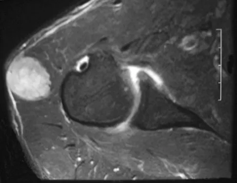
An MRI of a 28-year-old male with chronic ankle pain reveals a deep, cup-shaped osteochondral lesion on the posteromedial aspect of the talar dome. What is the typical mechanism of injury leading to this specific lesion?
Explanation
Question 12
A patient with Charcot-Marie-Tooth disease presents with a bilateral cavovarus foot deformity. A Coleman block test is performed, and the hindfoot corrects to neutral. What does this physical examination finding indicate?
Explanation
Question 13
A 45-year-old female complains of burning pain in the third web space of her foot. Non-operative management has failed. During surgical excision through a dorsal approach, which structure must be transected to adequately access the neuroma?
Explanation
Question 14
A 52-year-old male presents with dorsal foot pain and restricted first MTP joint dorsiflexion. Radiographs show dorsal osteophytes with joint space preservation on the plantar half of the first MTP joint. Which treatment is most appropriate after failed conservative care?
Explanation
Question 15
A 35-year-old male sustains a Hawkins type III talar neck fracture. Which of the following describes the anatomical displacement and the associated rate of avascular necrosis (AVN)?
Explanation
Question 16
Recent randomized controlled trials comparing operative versus non-operative management of acute Achilles tendon ruptures using early functional rehabilitation protocols have demonstrated which of the following?
Explanation
Question 17
A 55-year-old diabetic female presents with a warm, swollen, erythematous left foot. Radiographs show periarticular fragmentation, subluxation, and joint debris at the tarsometatarsal joints. According to the Eichenholtz classification, what is the appropriate initial management?
Explanation
Question 18
A 45-year-old female presents with symptomatic hallux valgus. Radiographs demonstrate a hallux valgus angle (HVA) of 45 degrees, an intermetatarsal angle (IMA) of 16 degrees, and hypermobility of the first tarsometatarsal (TMT) joint. Which surgical procedure is most appropriate?
Explanation
Question 19
A 22-year-old elite collegiate basketball player sustains an acute Zone 2 fracture of the proximal fifth metatarsal. What is the recommended treatment to minimize the risk of nonunion and expedite return to play?
Explanation
Question 20
A 60-year-old female presents with a flexible flatfoot deformity, unable to perform a single-leg heel raise. Radiographs show a talonavicular uncoverage of 40% but no arthritic changes in the subtalar, talonavicular, or calcaneocuboid joints. What is the most appropriate surgical intervention after failed conservative care?
Explanation
Question 21
Which of the following radiographic findings is most sensitive for diagnosing a subtle Lisfranc injury on a weight-bearing AP radiograph of the foot?
Explanation
Question 22
A 40-year-old male roofer falls from a height and sustains a closed, displaced intra-articular calcaneus fracture. CT scan shows a Sanders Type III fracture. Which of the following factors is most strongly associated with poor clinical outcomes following open reduction and internal fixation (ORIF)?
Explanation
Question 23
A 25-year-old female presents with persistent deep ankle pain following an inversion sprain 6 months ago. MRI reveals a 12 mm x 10 mm anterolateral osteochondral lesion of the talar dome with intact overlying cartilage. What is the most appropriate initial surgical management after failed conservative therapy?
Explanation
Question 24
A 14-year-old male presents with recurrent ankle sprains and rigid flatfeet. A "C-sign" is noted on the lateral radiograph. Which of the following physical exam findings is most characteristic of this condition?
Explanation
Question 25
Which of the following is considered an absolute contraindication to a total ankle arthroplasty in a patient with end-stage tibiotalar osteoarthritis?
Explanation
Question 26
A 28-year-old skier presents with lateral ankle pain and a snapping sensation posterior to the fibula after acute forced dorsiflexion and inversion. Physical examination reveals subluxation of the peroneal tendons with resisted eversion. Which anatomic structure is primarily compromised?
Explanation
Question 27
A professional football player sustains a hyperextension injury to his first metatarsophalangeal (MTP) joint. MRI shows complete rupture of the plantar plate and proximal migration of the sesamoids. What is the most appropriate management?
Explanation
Question 28
A 45-year-old male sustains a high-energy closed tibial pilon fracture with severe soft tissue swelling and fracture blisters. What is the most appropriate initial management strategy?
Explanation
Question 29
A 20-year-old track athlete complains of vague, aching dorsal midfoot pain that worsens with running. Radiographs are normal, but a CT scan reveals an incomplete, non-displaced stress fracture of the tarsal navicular in the sagittal plane. What is the most appropriate initial treatment?
Explanation
Question 30
A 32-year-old recreational athlete sustains an acute Achilles tendon rupture. He is considering operative versus non-operative management. According to recent high-level evidence utilizing early functional rehabilitation, what is the most accurate statement regarding outcomes?
Explanation
Question 31
A 45-year-old female presents with midfoot pain after a fall from a horse. Weight-bearing radiographs and subsequent MRI demonstrate a purely ligamentous Lisfranc injury with 3 mm of diastasis between the first and second metatarsals. What is the most appropriate surgical management?
Explanation
Question 32
A 55-year-old female presents with severe bunion pain. Radiographs reveal a hallux valgus angle (HVA) of 45 degrees, an intermetatarsal angle (IMA) of 19 degrees, and clinical hypermobility of the first ray in the sagittal plane. Which procedure is most appropriate?
Explanation
Question 33
A 50-year-old male with long-standing, poorly controlled diabetes presents with a warm, swollen, and erythematous right foot. He denies fevers or systemic symptoms. Radiographs reveal fragmentation and subluxation of the midfoot joints without osteomyelitis. Which of the following is the most appropriate initial treatment?
Explanation
Question 34
A 58-year-old female presents with adult-acquired flatfoot deformity. Examination shows a flexible hindfoot valgus, inability to perform a single-leg heel raise, and forefoot abduction with >40% talonavicular uncoverage on radiographs. What is the most appropriate surgical intervention?
Explanation
Question 35
A 21-year-old collegiate basketball player sustains a fracture at the metaphyseal-diaphyseal junction of the fifth metatarsal (Zone 2). To ensure the highest chance of union and fastest return to play, what is the best treatment option?
Explanation
Question 36
A 30-year-old male is recovering from an operatively treated Hawkins Type II talar neck fracture. At 8 weeks post-operation, an AP radiograph of the ankle demonstrates a subchondral radiolucent band in the talar dome. What does this finding signify?
Explanation
Question 37
A 45-year-old male sustains a displaced intra-articular calcaneus fracture. The surgeon is planning an extensile lateral approach for open reduction and internal fixation. Which of the following patient factors is the strongest independent predictor of postoperative wound complications?
Explanation
Question 38
In a patient with Charcot-Marie-Tooth disease presenting with a classic cavovarus foot deformity, which of the following muscle imbalances is the primary driver of the plantarflexed first ray?
Explanation
Question 39
A 60-year-old male presents with severe pain and stiffness in the first metatarsophalangeal (MTP) joint. Examination reveals pain throughout the entire arc of motion and less than 10 degrees of dorsiflexion. Radiographs show complete loss of the joint space, subchondral cysts, and large dorsal osteophytes (Grade 3 hallux rigidus). What is the gold standard surgical treatment?
Explanation
Question 40
A 45-year-old runner presents with chronic medial heel pain that is worse at the end of the day and non-responsive to plantar fasciitis treatments. Tinel's sign is positive over the medial heel, and MRI shows fatty atrophy of the abductor digiti minimi. Where is the most likely anatomic site of entrapment for the involved nerve?
Explanation
Question 41
During open reduction and internal fixation of a pronation-external rotation (Weber C) ankle fracture, what is a critical technical prerequisite before utilizing a clamp to anatomically reduce the syndesmosis?
Explanation
Question 42
A professional football player sustains a forced hyperextension injury to his great toe. MRI reveals a complete rupture of the plantar plate and joint capsule with proximal retraction of the sesamoids (Grade 3 Turf Toe). What is the recommended management?
Explanation
Question 43
A 20-year-old elite track athlete complains of insidious onset midfoot pain. A CT scan reveals a non-displaced, incomplete stress fracture involving the dorsal cortex of the tarsal navicular. What is the most appropriate initial management?
Explanation
Question 44
A 25-year-old male presents with lateral ankle pain and a snapping sensation behind the lateral malleolus when the ankle is actively dorsiflexed and everted. Radiographs demonstrate a small bony "fleck" lateral to the distal fibula. What is the anatomic basis of this pathology?
Explanation
Question 45
A diabetic patient with peripheral neuropathy presents with a chronic, recurrent plantar ulcer directly beneath the first metatarsal head. He has failed total contact casting and orthotic management. Physical examination reveals an inability to dorsiflex the ankle past neutral with the knee extended, but 15 degrees of dorsiflexion is achieved when the knee is flexed. What surgical adjunct is most likely to heal the ulcer and prevent recurrence?
Explanation
Question 46
A 55-year-old female presents with pain at the plantar aspect of the second metatarsophalangeal (MTP) joint and a new onset of the second toe crossing over the hallux. A positive dorsal drawer test of the second MTP joint is elicited. What is the most likely diagnosis?
Explanation
Question 47
A 65-year-old patient with end-stage ankle osteoarthritis is evaluated for a total ankle replacement (TAR). Which of the following is considered an absolute contraindication to modern total ankle arthroplasty?
Explanation
Question 48
A 40-year-old male sustains a high-energy, severely displaced pilon fracture (OTA 43-C3) with massive soft tissue swelling and fracture blisters. A spanning external fixator is placed on the day of injury. What is the primary reason to delay definitive open reduction and internal fixation?
Explanation
Question 49
A 15-year-old female dancer complains of insidious pain and swelling isolated to the dorsal aspect of the second metatarsal head. Radiographs demonstrate flattening, sclerosis, and fragmentation of the second metatarsal head. What is the underlying pathology?
Explanation
Question 50
A 24-year-old professional football player sustains a hyperdorsiflexion injury to his first metatarsophalangeal (MTP) joint. Exam reveals profound ecchymosis and a lack of push-off strength. MRI confirms a complete tear of the plantar plate with proximal retraction of the sesamoids. What is the most appropriate management?
Explanation
Question 51
A 45-year-old female presents with progressive flattening of her left arch. She cannot perform a single-leg heel raise. Radiographs demonstrate an uncovered talar head of 45% and a talonavicular uncoverage angle of 25 degrees. She has pain over the lateral hindfoot but a passively correctable hindfoot valgus. What is the most appropriate surgical treatment?
Explanation
Question 52
Which of the following is an absolute contraindication to a total ankle arthroplasty (TAA)?
Explanation
Question 53
A 19-year-old collegiate runner presents with midfoot pain. MRI confirms a stress fracture of the navicular involving the central third with a 1 mm gap and no sclerosis. What is the recommended initial management?
Explanation
Question 54
A 32-year-old male sustains a closed, highly comminuted, displaced tibial pilon fracture (OTA/AO 43-C3) with severe soft tissue swelling and fracture blisters. What is the most appropriate initial management?
Explanation
Question 55
A 28-year-old female presents with a painful bunion deformity. Examination reveals a hallux valgus angle of 45 degrees, an intermetatarsal angle of 18 degrees, and clinical hypermobility of the 1st tarsometatarsal (TMT) joint. What is the most appropriate surgical procedure?
Explanation
Question 56
In the Sanders classification for intra-articular calcaneal fractures, the primary coronal CT image used to determine the classification is located at which anatomic landmark?
Explanation
Question 57
A 40-year-old male sustains a Hawkins Type III talar neck fracture. Which of the following best describes this injury and its associated risk of avascular necrosis (AVN)?
Explanation
Question 58
A 22-year-old basketball player lands awkwardly on another player's foot and complains of lateral foot pain. Radiographs demonstrate a fracture at the diaphyseal-metaphyseal junction of the fifth metatarsal. He wishes to return to play as soon as possible. What is the most appropriate treatment?
Explanation
Question 59
A 55-year-old male with diabetes presents with a red, hot, swollen foot without systemic signs of infection. Radiographs reveal fragmentation and periarticular debris at the midfoot. Which of the following is the gold standard initial treatment?
Explanation
Question 60
A 42-year-old female complains of persistent dorsal midfoot pain 6 months after a seemingly minor twisting injury. Weight-bearing radiographs demonstrate a 3 mm diastasis between the base of the 1st and 2nd metatarsals. What is the most likely diagnosis?
Explanation
Question 61
A 60-year-old female undergoes surgical release of the plantar fascia for recalcitrant plantar fasciitis. Postoperatively, she develops new-onset lateral midfoot pain. What is the most likely cause of this complication?
Explanation
Question 62
A 15-year-old boy presents with a rigid flatfoot and frequent ankle sprains. Radiographs reveal an elongated anterior process of the calcaneus resembling an "anteater's nose" on the lateral view. Which of the following is the most likely diagnosis?
Explanation
Question 63
During evaluation of a patient with an acute ankle sprain, a positive external rotation stress test is elicited. On the AP radiograph, what is the normal expected tibiofibular overlap measured 1 cm proximal to the joint line?
Explanation
Question 64
A 35-year-old recreational athlete sustains an acute complete rupture of the Achilles tendon. He opts for nonoperative management. Which of the following is true regarding nonoperative versus operative management for this injury?
Explanation
Question 65
A 50-year-old female presents with a painful bunionette deformity. Radiographs show a normal 4-5 intermetatarsal angle but a lateral bowing of the 5th metatarsal shaft. What is the most appropriate surgical management?
Explanation
Question 66
A 29-year-old male sustains an inversion ankle injury resulting in a "popping" sensation behind the lateral malleolus. Examination reveals retromalleolar pain and snapping of the tendons with resisted dorsiflexion and eversion. Which of the following structures is most likely injured?
Explanation
Question 67
Which of the following physical exam findings is most specific for a complete rupture of the anterior tibial tendon?
Explanation
Question 68
A 55-year-old female presents with pain in her forefoot. She notes a feeling of "walking on a marble." Compression of the forefoot reproduces a painful click. Which interdigital space is most commonly affected by this condition?
Explanation
Question 69
A 40-year-old male sustains an acute, closed Achilles tendon rupture. He is treated non-operatively with an early functional rehabilitation protocol. Compared to surgical repair, which of the following outcomes is most closely associated with this management strategy?
Explanation
Question 70
A 24-year-old soccer player sustains an inversion ankle injury. Weight-bearing radiographs reveal a 5 mm medial clear space, which increases to 8 mm on external rotation stress views. The fibula is intact. What is the most appropriate definitive management?
Explanation
Question 71
A 58-year-old poorly controlled diabetic presents with a swollen, erythematous, and warm but painless right foot. Radiographs demonstrate periarticular debris, subchondral cyst formation, and fragmentation of the midfoot. According to the Eichenholtz classification, what stage does this represent?
Explanation
Question 72
A 30-year-old male sustains a talar neck fracture with subluxation of both the subtalar and tibiotalar joints following a motor vehicle accident. What is his estimated risk of developing avascular necrosis (AVN) of the talar body?
Explanation
Question 73
During an extensile lateral approach for open reduction and internal fixation of a displaced intra-articular calcaneus fracture, the full-thickness flap is elevated. Which of the following structures is contained within this flap and must be protected?
Explanation
Question 74
A 45-year-old female presents with a painful bunion. Weight-bearing radiographs show a hallux valgus angle (HVA) of 45 degrees, an intermetatarsal angle (IMA) of 18 degrees, and hypermobility of the first tarsometatarsal (TMT) joint. Which of the following surgical options is most appropriate?
Explanation
Question 75
A professional football player hyperextends his great toe on artificial turf. MRI confirms a complete tear of the plantar plate with proximal retraction of the medial sesamoid. What is the recommended management?
Explanation
Question 76
A 22-year-old collegiate basketball player complains of lateral foot pain after an awkward landing. Radiographs reveal a transverse fracture at the metaphyseal-diaphyseal junction of the fifth metatarsal. Which of the following is the most appropriate management to optimize his return to sport?
Explanation
Question 77
A 65-year-old male with end-stage post-traumatic ankle osteoarthritis is being evaluated for a total ankle arthroplasty. Which of the following is considered an absolute contraindication to this procedure?
Explanation
Question 78
A 35-year-old male sustains a high-energy closed tibial pilon fracture. The soft tissues are severely swollen with fracture blisters present. Which of the following defines the optimal timing for definitive open reduction and internal fixation?
Explanation
Question 79
A 28-year-old skier presents with posterolateral ankle pain and a snapping sensation behind the lateral malleolus. Physical examination reveals subluxation of the peroneal tendons with resisted eversion. Pathology involves disruption of which of the following structures?
Explanation
Question 80
A 29-year-old male suffers a severe crush injury to his foot. Compartment syndrome is suspected. How many distinct fascial compartments are generally recognized in the foot for the purpose of surgical decompression?
Explanation
Question 81
A 14-year-old boy presents with a rigid, painful flatfoot and a history of recurrent ankle sprains. Radiographs show an elongated anterior process of the calcaneus resembling an anteater's nose on the lateral view. What is the most likely diagnosis?
Explanation
Question 82
A 30-year-old male sustains a Hawkins Type III talar neck fracture. Six weeks post-operatively, an AP radiograph of the ankle reveals a subchondral radiolucent band in the talar dome. What does this finding indicate?
Explanation
Question 83
A 45-year-old female presents with a painful bunion. Clinical exam reveals a hypermobile first tarsometatarsal (TMT) joint. Radiographs show a hallux valgus angle of 35 degrees and an intermetatarsal angle of 16 degrees. Which of the following procedures is most appropriate?
Explanation
Question 84
A 65-year-old male with end-stage ankle osteoarthritis is evaluated for a total ankle arthroplasty (TAA). Which of the following is considered an absolute contraindication to TAA?
Explanation
Question 85
A 24-year-old football player presents with lateral ankle pain and a snapping sensation behind the lateral malleolus when circumducting the foot. Radiographs demonstrate a small bony avulsion off the posterolateral aspect of the distal fibula. What is the most appropriate surgical treatment?
Explanation
Question 86
A 40-year-old roofer falls from a height and sustains a severely comminuted, displaced intra-articular calcaneus fracture (Sanders Type III). A lateral extensile approach is planned for open reduction and internal fixation. Which nerve is at greatest risk of iatrogenic injury during the surgical approach?
Explanation
Question 87
A 38-year-old male sustains an acute, closed mid-substance Achilles tendon rupture. After discussing treatment options, he elects for nonoperative management with a functional rehabilitation protocol. Compared to operative repair, nonoperative management with early functional rehab is associated with which of the following?
Explanation
Question 88
A 22-year-old collegiate basketball player sustains a zone 2 fracture of the proximal fifth metatarsal. Intramedullary screw fixation is planned. To minimize the risk of lateral cortex penetration and hardware failure, the starting point for the screw should be:
Explanation
Question 89
A 26-year-old professional football player suffers an acute hyperextension injury to his first MTP joint. MRI reveals a complete rupture of the plantar plate with 4 mm of proximal retraction of the sesamoids. What is the most appropriate management?
Explanation
Question 90
A 14-year-old boy presents with repeated ankle sprains and a rigid, flatfoot deformity. Radiographs show an elongated anterior process of the calcaneus (anteater sign). CT confirms a calcaneonavicular coalition. After 6 months of failed conservative treatment, what is the best surgical option?
Explanation
Question 91
A 20-year-old cross-country runner reports 2 months of vague dorsal midfoot pain. Plain radiographs are normal, but an MRI demonstrates a stress fracture in the central third of the navicular without displacement. What is the recommended initial management?
Explanation
Question 92
A 55-year-old diabetic patient presents with a swollen, erythematous, and warm left foot. Radiographs demonstrate severe periarticular fragmentation, debris, and subluxation of the midfoot joints. According to the Eichenholtz classification, what stage does this represent and what is the primary treatment?
Explanation
Question 93
During open reduction and internal fixation of a pronation-external rotation (PER-4) ankle fracture, the surgeon assesses the syndesmosis using the external rotation stress test. Which radiographic parameter on the mortise view most reliably indicates syndesmotic instability requiring fixation?
Explanation
None
