العربية
Part of the Master Guide

AAOS & ABOS Foot & Ankle Board Review MCQs (Set 2): Ankle Fractures, Lisfranc, Diabetic Foot

AAOS Orthopedic MCQs (Set 1): Foot & Ankle Trauma & Deformities | Board Review

23 Apr 2026 59 min read 100 Views
Foot & Ankle 2009 MCQs - Part 1

Key Takeaway

This high-yield question set, Set 1, is crucial for AAOS and ABOS exams, covering comprehensive Foot & Ankle orthopedics. Topics include trauma such as ankle fractures and Lisfranc injuries, common deformities like bunions and hammer toes, and degenerative conditions. Perfect for board preparation.

AAOS Orthopedic MCQs (Set 1): Foot & Ankle Trauma & Deformities | Board Review

Comprehensive 100-Question Exam


00:00

Start Quiz

Question 1

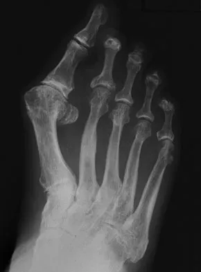
Figure 1 shows the radiograph of a 60-year-old woman who underwent a previous operation for great toe pain 20 years ago. She has had increasing pain over the past 5 years and now reports pain with any motion, swelling, and clicking. She also reports pain under the ball of foot. What is the most appropriate management to alleviate her metatarsalgia and great toe pain?


Explanation

The patient has a failed Silastic implant. Nonsurgical management will not work at this point. A Keller resection will only exacerbate her metatarsalgia. Implant removal with structural bone grafting and MTP fusion is the most appropriate choice because restoration of length is needed to alleviate the forefoot pain and bone grafting is required to fuse the MTP joint because there is an abundance of osteolysis. Total toe implants do not offer good long-term outcomes and are very difficult to fit into the large exploded-out cavity of the proximal phalanx. Hecht PJ, Gibbons MJ, Wapner KL, et al: Arthrodesis of the first metatarsophalangeal joint to salvage failed silicone implant arthroplasty. Foot Ankle Int 1997;18:383-390.

Question 2

A 47-year-old man with Charcot-Marie-Tooth (CMT) disease was treated with a fifth metatarsal head resection for a symptomatic bunionette 2 years ago. What is the most likely complication seen at this time?


Explanation

CMT is characterized by a cavovarus foot position that increases weight-bearing stresses along the lateral border. Removal of the fifth metatarsal head carries the risk of creating a transfer lesion at the fourth metatarsal head, particularly with a cavovarus foot. Claw toes are common in CMT, but the fifth toe would be flail in this situation. Ulceration is unlikely given the lack of underlying bone. Peroneal atrophy is associated with CMT but would not be a complication of this procedure. Charcot arthropathy is a neuropathic process frequently seen in individuals with diabetes mellitus. Kitaoka HB, Holiday AD Jr: Metatarsal head resection for bunionette: Long-term followup. Foot Ankle 1991;11:345-349.


Question 3

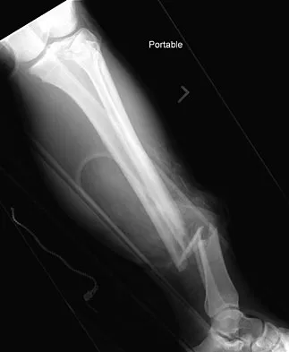
A 19-year-old man was struck by a car and is seen in the emergency department with a grade IIIC open distal tibia and fibula fracture. Examination reveals that the toes are cool and dusky with a sluggish capillary refill. Angiography reveals a lesion in the posterior tibial artery amenable to repair. There is no sensation on the plantar aspect of the foot, and he is unable to flex his toes. A clinical photograph and radiograph are shown in Figures 2a and 2b. What is the next most appropriate step in management?


Explanation

In the past, loss of plantar sensation in this grade IIIC tibial fracture would have been an indication for below-knee amputation regardless of the potential for vascular repair. However the 2002 LEAP study divided 55 patients with loss of plantar sensation into two groups, the insensate amputation group and the insensate limb salvage group, with 55% of patients in the insensate salvage group regaining normal sensation 2 years after injury. Furthermore, those in the salvage group who remained insensate after 2 years had equivalent outcomes to those in the amputation group. Because of these findings, limb salvage with vascular repair and external stabilization with delayed closure is deemed appropriate treatment. Immediate intramedullary fixation is not indicated. Because ischemia, contusion, and stretch can adversely affect the tibial nerve, the additional insult of exploration of the nerve is also not advisable given the soft-tissue compromise. Bosse MJ, McCarthy ML, Jones AL, et al: The insensate foot following severe lower extremity trauma: An indication for amputation? J Bone Joint Surg Am 2005;87:2601-2608. Lange RH, Bach AW, Hansen ST Jr, et al: Open tibial fractures with associated vascular injuries: Prognosis for limb salvage. J Trauma 1985;25:203-208. Mackenzie EJ, Bosse MJ, Kellam JF, et al: Factors influencing the decision to amputate or reconstruct after high-energy lower extremity trauma. J Trauma 2002;52:641-649.


Question 4

The pathophysiology of a claw toe deformity includes muscular imbalance caused by which of the following relatively strong structures?


Explanation

The dynamic forces acting to maintain the position of the proximal phalanx at the head of the metatarsal are a balance between the extensor digitorum longus and the weaker intrinsic muscles. With hyperextension at the metatarsophalangeal joint, the intrinsic muscles become less efficient as plantar flexors. Consequently, the hyperextension deformity progresses in the metatarsophalangeal joint as the opposition of the intrinsic muscles to the extensor tendon lessens. This is in contrast to the situation in the interphalangeal joints, where the stronger flexors overpower the weaker intrinsic muscles, which act as the extensors. This combination of events leads to hyperextension at the metatarsophalangeal joint and flexion deformities at the interphalangeal joints, resulting in claw toe. Mizel MS, Yodlowski ML: Disorders of the lesser metatarsophalangeal Joints. J Am Acad Orthop Surg 1995;3:166-173.


Question 5

A 26-year-old woman is seen in the emergency department with an intra-articular distal tibia fracture and a fibular fracture (pilon). The patient, her husband, and three small children have recently immigrated to the United States from Mexico. The husband and wife have both been in a migrant labor camp but have no immediate relatives in the States. What factor is most important when considering her recommended care and treatment?


Explanation

With documented use of a competent interpreter, informed consent should not be an issue. In Hispanic families, the husband often makes the ultimate decision regarding proceeding with surgery; however, he would not be expected to withhold recommended treatment. Hispanics may have a higher risk of comorbidities, but you do not expect this to be a significant concern with this patient. Claustrophobia and some fear of the unfamiliar may make additional imaging studies more difficult to arrange, but not impossible. The real concern is that with no extended family and three small children, the postoperative demand on the patient could significantly jeopardize her ability to comply with weight-bearing restrictions and overall ambulatory demands. Discharge planning and appropriate help may be paramount for a good outcome.


Question 6

A 57-year-old man with type II diabetes mellitus was successfully treated for a first occurrence forefoot full-thickness (Wagner II) diabetic foot ulcer underlying the third metatarsal head with associated hammertoe with a series of weight-bearing total contact casts. There was no evidence of osteomyelitis. The ulcer is now fully healed. He is insensate to the Semmes-Weinstein 5.07 (10 gm) monofilament. What is the next most appropriate step in management?


Explanation

This is the first occurrence of diabetic foot-specific morbidity. The patient has a foot deformity, a history of a diabetic foot ulcer, and is insensate to the monofilament. He is at moderate risk for the development of a recurrent ulcer. This is best avoided with therapeutic footwear. Commercially available depth-inlay shoes should be combined with a custom accommodative foot orthosis to accommodative the deformity. Pinzur MS, Slovenkai MP, Trepman E, et al: Guidelines for diabetic foot care: Recommendations endorsed by the Diabetes Committee of the American Orthopaedic Foot and Ankle Society. Foot Ankle Int 2005;26:113-119.


Question 7

A 28-year-old man has had a 2-year history of progressive lateral ankle pain. History reveals that he underwent a triple arthrodesis at age 13 for a tarsal coalition. The pain has been refractory to braces, custom inserts, and nonsteroidal anti-inflammatory drugs. Weight-bearing radiographs of the ankle and foot are shown in Figures 3a through 3d. Surgical management should include which of the following?


Explanation

The patient has a valgus-supination triple arthrodesis malunion. Weight-bearing radiographs show excessive residual valgus through the subtalar joint, producing lateral subfibular impingement, and residual forefoot abduction and midfoot supination through the talonavicular joint, lateralizing the weight-bearing forces through the foot. The deformity is best managed with a medial displacement calcaneal osteotomy and transverse tarsal derotational osteotomy. Ankle arthroscopy and lateral ligament reconstruction are indicated in the event of ligament instability. Tendon transfer, lateral column lengthening, and heel cord lengthening are used for treatment of adult flatfoot from posterior tibial tendon insufficiency. Ankle arthrodesis and ankle arthroplasty are not indicated in this patient because the lateral ankle symptoms are the result of the underlying deformity in the hindfoot, the patient is young, and the ankle joint is relatively normal. Haddad SL, Myerson MS, Pell RF IV: Clinical and radiographic outcome of revision surgery for failed triple arthrodesis. Foot Ankle Int 1997;18:489-499.


Question 8

If heel varus corrects with a Coleman block test, then the hindfoot deformity is flexible. This test proves that the varus is due to a


Explanation

The Coleman block test is used to evaluate the effect of the forefoot on the rearfoot varus. If the deformity corrects with the block, then the hindfoot deformity is flexible and the varus position is secondary to the plantar flexed first ray or valgus position of the forefoot. A rearfoot orthotic will not correct the forefoot cause of the deformity. The patient still may need a lateralizing calcaneal osteotomy to realign the hindfoot. Younger AS, Hansen ST Jr: Adult cavovarus foot. J Am Acad Orthop Surg 2005;13:302-315.


Question 9

A 27-year-old man now reports dorsiflexion and inversion weakness after an automobile collision 6 months ago in which compartment syndrome developed isolated to the anterior and deep posterior compartments. Examination reveals the development of a progressive cavovarus deformity, but the ankle and hindfoot remain flexible. In addition to Achilles tendon lengthening, which of the following procedures is most likely to improve the motor balance of his foot and ankle?


Explanation

Compartment syndrome of the anterior and deep posterior compartments results in anterior tibialis and posterior tibialis tendon weakness, respectively. Furthermore, the long flexors to the hallux and lesser toes will be weak as well. The intact peroneus longus overpowers the weak anterior tibialis tendon, resulting in plantar flexion of the first metatarsal, cavus, and hindfoot varus. Therefore, transferring the peroneus longus to the dorsolateral midfoot reduces the first metatarsal plantar flexion torque, and possibly augments ankle dorsiflexion torque. Hansen ST: Functional Reconstruction of the Foot and Ankle. Philadelphia, PA, Lippincott, Williams & Wilkins, 2000, pp 433-435.


Question 10

Figures 4a through 4c show the radiographs of a 43-year-old woman who sustained a twisting injury to her right ankle. She has ankle pain and tenderness medially and laterally. To help determine the optimal treatment, an external rotation stress radiograph of the ankle is obtained. This test is designed to evaluate the integrity of what structure?


Explanation

In the presence of a supination external rotation-type fracture of the distal fibula (Weber type B), stability of the ankle is best assessed by performing an external rotation stress AP view of the ankle. This test is used to assess the integrity of the deltoid ligament. The presence of a deltoid ligament rupture results in instability and generally is best managed surgically. The gravity stress test can also be used. Egol KA, Amirtharajah M, Tejwani NC, et al: Ankle stress test for predicting the need for surgical fixation of isolated fibular fractures. J Bone Joint Surg Am 2004;86:2393-2398. McConnell T, Creevy W, Tornetta P III: Stress examination of supination external rotation-type fibular fractures. J Bone Joint Surg Am 2004;86:2171-2178.


Question 11

A 29-year-old patient sustains a closed, displaced joint depression intra-articular calcaneus fracture. In discussing potential complications of surgical intervention through an extensile lateral approach, which of the following is considered the most common complication following surgery?


Explanation

Delayed wound healing and wound dehiscence is the most common complication of surgical management of calcaneal fractures through an extensile lateral approach, occurring in up to 25% of patients. Most wounds ultimately heal with local treatment; the deep infection rate is approximately 1% to 4% in closed fractures. Posttraumatic arthritis may develop despite open reduction and internal fixation, but the percentages remain low. Peroneal tendinitis may occur from adhesions within the tendon sheath or from prominent hardware but is relatively uncommon. Nonunion of a calcaneal fracture is rare. Sanders RW, Clare MP: Fractures of the calcaneus, in Coughlin MJ, Mann RA, Saltzman CL (eds): Surgery of the Foot and Ankle, ed 8. Philadelphia, PA, Mosby-Elsevier, 2007, vol 2, pp 2017-2073.


Question 12

Figures 5a and 5b show the radiographs of a 56-year-old man who was seen in the emergency department following a twisting injury to his left ankle. Examination in your office 3 days later reveals marked swelling and diffuse tenderness to palpation about the ankle and leg. What is the next most appropriate step in management?


Explanation

The radiographs show an isolated posterior malleolus fracture which, given the injury mechanism, is highly suspicious for a Maisonneuve injury. As with any suspected extremity injury, radiographs including the joints above and below the level of injury are acutely indicated. Although MRI may reveal a ligamentous injury to the ankle and CT may show asymmetry of the ankle mortise or syndesmosis, both studies are considerably more costly and are not indicated in the absence of a complete radiographic work-up. Technetium bone scan is nonspecific and would be of limited value in this instance, as would repeat radiographs of the ankle. Walling AK, Sanders RW: Ankle fractures, in Coughlin MJ, Mann RA, Saltzman CL (eds): Surgery of the Foot and Ankle, ed 8. Philadelphia, PA, Mosby-Elsevier, 2007, vol 2, pp 1973-2016.


Question 13

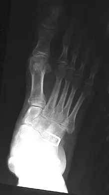
A 61-year-old man has a symptomatic bunionette that is refractory to nonsurgical management. A radiograph is shown in Figure 6. What is the optimal surgical correction?


Explanation

The patient has a bunionette with a large 4-5 intermetatarsal angle. This requires not only ostectomy of the lateral prominence but metatarsal osteotomy to decrease the intermetatarsal angle. Excising the head results in a flail joint and creates the possibility of a transfer lesion. Condylectomy can reduce plantar pressures but does not address the bunionette. The joint surface is well maintained, thus there are no indications for resection. Coughlin MJ: Treatment of bunionette deformity with longitudinal diaphyseal osteotomy with distal soft tissue repair. Foot Ankle 1991;11:195-203.


Question 14

A 25-year-old woman with a healed proximal tibiofibular fracture treated with an intramedullary nail 2 years ago is currently wearing an ankle-foot orthosis (AFO) and reports a persistent foot drop. She is unhappy with the AFO and has not seen any functional improvement despite months of physical therapy. Serial electromyograms (EMG) show no recent change over the past year. Examination and EMG findings are consistent with a tibialis anterior 1/5, extensor hallucis longus 2/5, extensor digitorum longus 2/5, posterior tibial tendon (PTT) 5/5, peroneals 3/5, flexor hallucis longus 5/5, and gastrocsoleus 5/5. No discrete nerve lesion was identified. The patient has a flexible equinovarus contracture. What is the most appropriate management?


Explanation

This pattern of injury is consistent with an unrecognized compartment syndrome of the anterior and lateral compartments. Transfer of the PTT through a long incision in the interosseous membrane corrects the foot drop deformity, and allows adequate dorsiflexion provided that the tendon to be transferred has a strength of 5/5. Muscles/tendons typically lose one grade of strength after transfer. Transfer into the tendons at the level of the ankle prevents overtensioning or pullout of a PTT tendon that is not long enough. Debridement of the scarred muscle in the anterior compartment decreases the risk of scarring down to the tendon transfer. Transfer of the peroneus longus is not preferred given its relative lack of strength and line of pull. Continued therapy and bracing are unlikely to lead to further improvement at 2 years after injury. An ankle fusion would correct the foot drop but would not address the tendon imbalances between the tibialis anterior and the peroneus longus, and the PTT and the peroneus brevis. Hansen ST Jr: Functional Reconstruction of the Foot and Ankle. Philadelphia, PA, Lippincott Williams & Wilkins, 2000, p 192. Atesalp AS, Yildiz C, Komurcu M, et al: Posterior tibial tendon transfer and tendo-Achilles lengthening for equinovarus foot deformity due to severe crush injury. Foot Ankle Int 2002;23:1103-1106. Scott AC, Scarborough N: The use of dynamic EMG in predicting the outcome of split posterior tibial tendon transfers in spastic hemiplegia. J Pediatr Orthop 2006;26:777-780.


Question 15

When using a two-incision approach for open reduction and internal fixation of a Hawkins III talar fracture-dislocation involving the talar neck and body, what anatomic structure must be preserved to optimize outcome?


Explanation

A Hawkins III fracture-dislocation generally presents with posteromedial displacement with the deltoid ligament intact. Therefore, the only remaining blood supply is the deltoid branch of the artery of the tarsal canal originating from the posterior tibial artery. Often, the medial malleolus is fractured, assisting in reduction and visualization of fracture reduction. If the medial malleolus is intact, a medial malleolus osteotomy allows visualization of the reduction without compromising the last remaining blood supply to the talus. Mulfinger GL, Trueta J: The blood supply of the talus. J Bone Joint Surg Br 1970;52:160-167.


Question 16

A 10-year-old boy who is active in soccer has had activity-related heel pain for the past 3 months. Examination reveals tenderness over the posterior heel and a tight Achilles tendon. Radiographs demonstrate a 2-cm cyst in the anterior body of the calcaneus. His physes have not closed. Based on these findings, what is the most appropriate management?


Explanation

The most likely diagnosis is Sever's disease, which is considered either an apophysitis or a para-apophyseal stress fracture. It is common in athletic children and is associated with a tight Achilles tendon. Cast immobilization may be necessary if activity reduction fails. Calcaneal cysts are quite common and do not require any further diagnostic testing or treatment unless they occupy the full width of the calcaneus or one third of the length of the calcaneus. Ogden JA, Ganey TM, Hill JD, et al: Sever's injury: A stress fracture of the immature calcaneal metaphysis. J Ped Orthop 2004;24:488-492.


Question 17

A 35-year-old woman states that she stepped on a piece of glass 6 months ago and reports numbness and shooting pain along the plantar lateral forefoot. She had previously received steroid injections in the 3 to 4 webspace. Examination reveals mild tenderness along the plantar fascia; no Tinel's sign is noted plantar medially and no Mulder's click is noted distally. An MRI scan is shown in Figure 7. What is the most likely cause of the numbness?


Explanation

The MRI scan reveals a laceration through the abductor hallucis musculature and lateral plantar nerve, producing numbness along its distribution. There is no evidence of a foreign body on the MRI scan. Baxter's nerve, or nerve to the abductor digiti quinti muscle, is the first branch off the lateral plantar nerve and impingement of this nerve typically produces a Tinel's sign along the nerve branch deep to the abductor hallucis muscle. Interdigital neuroma would be suggested by the presence of a Mulder's click. A digital nerve laceration would exhibit isolated numbness more distally. Baxter DE, Pfeffer GB: Treatment of chronic heel pain by surgical release of the first branch of the lateral plantar nerve. Clin Orthop Relat Res 1992;279:229-236.


Question 18

A 69-year-old man reports pain over his bunion while wearing shoes and pain in the joint with push-off when barefoot. Nonsurgical management has failed to provide relief. Radiographs are shown in Figures 8a and 8b. What is the surgical procedure of choice?


Explanation

Arthrodesis is indicated for severe bunion and hallux valgus deformities, but particularly with extensive degenerative disease of the first metatarsophalangeal joint. The other bunionectomy procedures have different indications, none of which include symptomatic first metatarsophalangeal degenerative disease. Richardson EG(ed): Orthopaedic Knowledge Update: Foot and Ankle 3. Rosemont, IL, American Academy of Orthopaedic Surgeons, 2004, pp 3-15.


Question 19

A 65-year-old man has chronic Achilles insertional tendinitis that is refractory to nonsurgical management. A radiograph is shown in Figure 9. Preoperative counseling should include a discussion of the realistic duration of postoperative recovery. You should inform the patient that his expected recovery will last


Explanation

An older patient with calcaneal enthesopathy may take a year or more to recover after tendon debridement and calcaneal ostectomy. Young patients, and those with purely tendon pathology, may recover more quickly. McGarvey WC, Palumbo RC, Baxter DE, et al: Insertional Achilles tendinitis: Surgical treatment through a central tendon splitting approach. Foot Ankle Int 2002;23:19-25.


Question 20

Figures 10a and 10b show the clinical photograph and MRI scan of a plantar foot lesion. If excisional biopsy is performed, what is the most likely complication?


Explanation

The MRI scan shows plantar fibromatosis. The treatment is usually nonsurgical. If surgery is indicated, wide local excision with excision of the entire plantar fascia is usually indicated. The main problem with simple excision of the lesion is the high chance of recurrence. The other listed complications are those that are a result of the wide local excision. Aluisio FV, Mair SD, Hall RL: Plantar fibromatosis: Treatment of primary and recurrent lesions and factors associated with recurrence. Foot Ankle Int 1996;17:672-678.


Question 21

A patient with rheumatoid arthritis with both ankle and subtalar involvement was treated as shown in Figures 11a and 11b. What complication is unique to this type of fixation?


Explanation

The interlocking screws at the proximal end of the rod can act as a stress riser and lead to fracture. Postoperative pain at this level should prompt inclusion of this diagnosis in the differential. Removing the screws following bone union can decrease the chances of this occurring. A short rod that avoids the diaphyseal area may also be beneficial. Rotatory deformity is controlled by the perpendicularly oriented distal transfixion screws. Talar osteonecrosis would be unusual since the dissection can be minimized with an intramedullary rod. Any type of hardware can fail if the construct does not lead to a solid arthrodesis. Nunley JA, Pfeffer GB, Sanders RW, et al (eds): Advanced Reconstruction: Foot and Ankle. Rosemont, IL, American Academy of Orthopaedic Surgeons, 2004, pp 236-237. Thordarson DB, Chang D: Stress fractures and tibial cortical hypertrophy after tibiotalocalcaneal arthrodesis with an intramedullary nail. Foot Ankle Int 1999;20:497-500.


Question 22

A 68-year-old man fell off a 20-foot mountain cliff and was seen in the emergency department the following morning. A radiograph is shown in Figure 12. He is a nonsmoker with medical comorbidities of hypertension and hypercholesterolemia that is well controlled with medicine and diet. Capillary refill and sensation are intact distally and the patient is able to move his toes with mild discomfort. Serosanguinous fracture blisters are present laterally, and the foot is swollen and red. What is the most appropriate management?


Explanation

Whereas a patient age of older than 50 years used to be a contraindication for open reduction and internal fixation of displaced intra-articular calcaneal fractures, new data suggest that the presence of associated medical comorbidities that affect wound healing such as smoking, diabetes mellitus, and peripheral vascular disease are more relevant to postoperative functional outcome. Surgical treatment of Sanders II and III displaced intra-articular calcaneal fractures with initial Bohler angles of > 15 degrees results in better outcomes as compared to nonsurgical management. Indications for primary fusion might include Sanders IV fractures in which articular congruity or Bohler angles cannot be restored. Given the condition of the soft tissues at presentation, delayed fixation is recommended. Herscovici D Jr, Widmaier J, Scaduto JM, et al: Operative treatment of calcaneal fractures in elderly patients. J Bone Joint Surg Am 2005;87:1260-1264. Buckley R, Tough S, McCormack R, et al: Operative compared with nonoperative treatment of displaced intra-articular calcaneal fractures: A prospective, randomized, controlled multicenter trial. J Bone Joint Surg Am 2002;84:1733-1744.


Question 23

A 45-year-old woman has had intense pain in her foot for the last 3 days. She also reports a mild fever and difficulty with shoe wear. Examination reveals a swollen, slightly erythematous warm foot with tenderness at the great toe metatarsophalangeal joint and pain with passive motion of the joint. An AP radiograph is shown in Figure 13. Which of the following will best aid in determining a definitive diagnosis?


Explanation

The patient has gouty arthropathy of the first metatarsophalangeal joint. This definitive diagnosis is achieved with aspiration of the joint and polarized light microscopy that shows needle-shaped negatively birefringent monosodium urate crystals. Differential diagnoses of infectious arthritis and pseudogout are also definitively made through joint aspiration. Although rheumatoid arthritis is a possibility, a serum rheumatoid factor is not always diagnostic and a patient with rheumatoid arthritis may have concomitant gouty arthritis. The radiographic findings are not typical of diabetes mellitus or of a patient with Charcot arthropathy. Wise CM, Agudelo CA: Diagnosis and management of complicated gout. Bull Rheum Dis 1998;47:2-5.


Question 24

Figures 14a and 14b show the clinical photographs of a patient who was stranded in a subzero region for several days. The photographs were taken the morning after arrival in the hospital. The patient is otherwise healthy and fit, and takes no medication. He has no clinical signs of sepsis. He reports burning pain and tingling in both feet. What is the best treatment?


Explanation

The patient has no clinical or observed signs of sepsis. The skin just proximal to the gangrenous tissue appears somewhat hyperemic and is clearly viable. These wounds should be managed much like burn wounds. Moist dressings should be used until the tissue clearly demarcates. Much of the insult may simply be superficial and only require late debridement. McAdams TR, Swenson DR, Miller RA: Frostbite: An orthopedic perspective. Am J Orthop 1999;28:21-26.


Question 25

The peroneus tertius is a commonly used landmark for arthroscopic portal placement. What is the function of this tendon?


Explanation

The peroneus tertius, although absent in 10% of the population, originates on the distal third of the extensor surface of the fibula and inserts onto the base of the fifth metatarsal, possibly extending to the fascia over the fourth interosseous space. The muscle is located in the anterior compartment of the leg and is innervated by the deep peroneal nerve. The tendon produces dorsiflexion and eversion when walking and can be used as an insertion point during tendon transfers to assist dorsiflexion. This tendon is peculiar to humans and is a proximally migrated deep extensor of the fifth toe. Joshi SD, Joshi SS, Athavale SA: Morphology of the peroneus tertius muscle. Clin Anat 2006;19:611-614. Williams PL, Bannister LH, Berry MM, et al (eds): Gray's Anatomy, ed 38. London, Churchill Livingston, 1995, p 883.


Question 26

A 32-year-old man sustains a Hawkins type II talar neck fracture. At 8 weeks postoperatively, an AP radiograph shows a subchondral radiolucent band in the talar dome. What does this finding indicate?


Explanation

The Hawkins sign is a subchondral radiolucent band seen 6 to 8 weeks after a talus fracture, indicating resorption of subchondral bone. This requires an intact vascular supply, essentially ruling out avascular necrosis of the talar body.

Question 27

A 24-year-old athlete reports midfoot pain after a twisting injury. Radiographs show a "fleck sign" in the first intermetatarsal space. Which of the following anatomical structures is primarily injured?


Explanation

The "fleck sign" represents an avulsion fracture of the Lisfranc ligament. The Lisfranc ligament is an interosseous ligament connecting the lateral aspect of the medial cuneiform to the medial base of the second metatarsal.

Question 28

A 55-year-old woman presents with flexible flatfoot, inability to perform a single-leg heel raise, and >40% uncovering of the talonavicular joint on an AP weight-bearing radiograph. What is the most appropriate surgical management for this stage of posterior tibial tendon dysfunction?


Explanation

This patient has Stage IIb adult-acquired flatfoot deformity, characterized by significant forefoot abduction (talonavicular uncovering >30-40%). Appropriate treatment includes an FDL transfer, a medializing calcaneal osteotomy (MDCO), and a lateral column lengthening to correct the abduction.

Question 29

During an extensile lateral approach for open reduction and internal fixation of a displaced intra-articular calcaneus fracture, which of the following structures is at greatest risk of iatrogenic injury at the proximal extent of the vertical incision?


Explanation

The extensile lateral approach to the calcaneus involves a full-thickness flap. The vertical limb is placed just anterior to the Achilles tendon, placing the sural nerve at high risk of injury, especially at the proximal aspect of the incision.

Question 30

A 22-year-old collegiate basketball player sustains an acute fracture at the metaphyseal-diaphyseal junction of the fifth metatarsal. Intramedullary screw fixation is planned. The high rate of nonunion in this area is primarily due to a watershed blood supply involving which vessels?


Explanation

A Jones fracture occurs at the metaphyseal-diaphyseal junction of the fifth metatarsal (Zone 2). This region is a vascular watershed area between the intraosseous nutrient artery supplying the diaphysis and the metaphyseal arteries supplying the base.

Question 31

A 40-year-old man undergoes minimally invasive (percutaneous) repair of an acute Achilles tendon rupture. Postoperatively, he complains of numbness along the lateral aspect of his foot. Which structure was most likely injured during the procedure?


Explanation

The sural nerve crosses from medial to lateral near the Achilles tendon, typically 10-12 cm proximal to the calcaneal insertion. It is at significant risk of entrapment or iatrogenic injury during percutaneous or minimally invasive Achilles tendon repairs.

Question 32

A 45-year-old woman presents with a symptomatic bunion. Weight-bearing radiographs reveal a hallux valgus angle (HVA) of 42 degrees and an intermetatarsal angle (IMA) of 18 degrees. There is clinical hypermobility at the first tarsometatarsal (TMT) joint. What is the most appropriate surgical intervention?


Explanation

Severe hallux valgus (HVA >40, IMA >15) with first TMT joint hypermobility is best treated with a first TMT joint arthrodesis (Lapidus procedure). This provides powerful correction of the intermetatarsal angle and stabilizes the incompetent medial column.

Question 33

A 14-year-old boy presents with a history of recurrent ankle sprains and rigid, painful flatfeet. Radiographs reveal an elongated anterior process of the calcaneus (anteater sign). Which radiographic view best visualizes this specific pathology?


Explanation

The clinical presentation and "anteater sign" indicate a calcaneonavicular coalition. The 45-degree internal oblique radiograph of the foot is the optimal view to visualize the calcaneonavicular interval and confirm the diagnosis.

Question 34

A professional football player sustains a hyperextension injury to his first metatarsophalangeal (MTP) joint. MRI confirms a complete tear of the plantar plate with proximal retraction of the sesamoids. If left untreated, what is the most likely long-term complication?


Explanation

A complete plantar plate tear (Grade 3 turf toe) causes severe instability and altered kinematics of the first MTP joint. Without surgical repair to restore the anatomical alignment, progressive joint degeneration inevitably leads to hallux rigidus.

Question 35

A 42-year-old man sustains a high-energy closed right tibial pilon fracture (OTA 43-C3) with severe fracture blisters and massive soft tissue swelling. What is the most appropriate initial management to minimize soft tissue complications while maintaining alignment?


Explanation

High-energy pilon fractures are associated with significant soft-tissue compromise. The standard of care is a staged approach utilizing a spanning external fixator for initial length and alignment, followed by delayed ORIF when the "wrinkle sign" appears.

Question 36

A 58-year-old man with poorly controlled diabetes presents with a warm, swollen, and erythematous left foot. He denies trauma. Radiographs show periarticular fragmentation and subluxation at the midtarsal joint. There are no open wounds. What is the most appropriate initial management?


Explanation

This patient presents with acute (Eichenholtz Stage 1) Charcot arthropathy. The mainstay of initial treatment is immobilization and offloading with a total contact cast (TCC) to prevent further deformity until the acute inflammatory phase resolves.

Question 37

A 30-year-old patient presents with a purely ligamentous Lisfranc injury with 4 mm of diastasis on weight-bearing radiographs. According to recent literature, what is the most significant advantage of primary arthrodesis over open reduction and internal fixation (ORIF) for this specific injury pattern?


Explanation

Primary arthrodesis for purely ligamentous Lisfranc injuries yields similar functional outcomes to ORIF but significantly decreases the need for subsequent hardware removal and secondary salvage arthrodesis.

Question 38

A 35-year-old man falls from a height and sustains a Hawkins Type III talar neck fracture. What is the estimated rate of avascular necrosis (AVN) of the talar body following this specific injury pattern?


Explanation

Hawkins Type III fractures involve dislocation of both the subtalar and tibiotalar joints, disrupting all three major blood supplies to the talar body. This results in a high risk of avascular necrosis, approximately 75-90%.

Question 39

A 40-year-old construction worker falls from a ladder, sustaining a closed intra-articular calcaneus fracture. Which of the following is considered an absolute contraindication to utilizing an extensile lateral approach for open reduction and internal fixation?


Explanation

Peripheral artery disease with compromised vascularity (absent pedal pulses) is an absolute contraindication to the extensile lateral approach due to the unacceptably high risk of wound necrosis and deep infection. Smoking and diabetes are considered relative contraindications.

Question 40

A 55-year-old woman presents with medial foot pain and a progressive flatfoot deformity. She has a flexible hindfoot valgus and is unable to perform a single-leg heel rise. According to the Johnson and Strom classification modified by Myerson, what stage of posterior tibial tendon dysfunction does this patient have, and what is the most appropriate surgical management if conservative treatment fails?


Explanation

Stage II PTTD is characterized by a flexible flatfoot deformity and the inability to perform a single heel rise. Surgical management typically involves an FDL tendon transfer to the navicular and a medial displacement calcaneal osteotomy (MDCO) to correct the hindfoot valgus.

Question 41

A 22-year-old man with a history of frequent ankle sprains presents with bilateral cavovarus feet. A Coleman block test demonstrates that the hindfoot varus corrects to neutral when the first metatarsal is allowed to plantarflex off the block. What does this test result indicate regarding the primary driver of this patient's deformity?


Explanation

A flexible hindfoot that corrects on the Coleman block test indicates the varus is driven by a rigidly plantarflexed first ray. In conditions like Charcot-Marie-Tooth disease, this is typically due to the overpull of the peroneus longus relative to the weak tibialis anterior.

Question 42

A 60-year-old man with poorly controlled diabetes mellitus presents with a swollen, red, warm, and painless right foot. Radiographs show periarticular fragmentation and subluxation at the tarsometatarsal joints. According to the Eichenholtz classification, what stage does this represent, and what is the most appropriate initial management?


Explanation

Stage I (Fragmentation) of Charcot arthropathy is characterized by acute inflammation, osteopenia, fragmentation, and joint subluxation. The gold standard for initial management is strict immobilization and offloading, typically with a total contact cast.

Question 43

A 45-year-old man sustains a severe, high-energy, closed tibial pilon fracture with significant soft tissue swelling and fracture blisters. What is the most appropriate staged treatment protocol to minimize soft tissue complications in this injury?


Explanation

High-energy pilon fractures with severe soft tissue compromise are best managed with a staged approach. Immediate spanning external fixation restores length and alignment, followed by definitive ORIF 10-21 days later when the soft tissue envelope has recovered.

Question 44

A 38-year-old recreational basketball player sustains an acute Achilles tendon rupture. Based on current literature comparing functional bracing with early mobilization to surgical repair, which of the following statements is true?


Explanation

Recent high-level evidence demonstrates that non-operative management utilizing functional bracing and early mobilization yields equivalent functional outcomes and similar re-rupture rates compared to surgical repair, while avoiding surgical wound complications.

Question 45

A 45-year-old woman presents with a painful bunion. Weight-bearing radiographs demonstrate a hallux valgus angle (HVA) of 45 degrees, an intermetatarsal angle (IMA) of 18 degrees, and clinical hypermobility of the first tarsometatarsal (TMT) joint. Which of the following surgical procedures is most appropriate?


Explanation

A severe hallux valgus deformity (HVA > 40°, IMA > 15°) associated with first TMT joint hypermobility is best treated with a first TMT arthrodesis (Lapidus procedure). This provides stable long-term correction and directly addresses the hypermobile segment.

Question 46

A 28-year-old soccer player sustains a twisting ankle injury. Radiographs show a widened medial clear space on the gravity stress view, consistent with a syndesmotic injury. During operative fixation, what is the most important factor in achieving a good long-term clinical outcome?


Explanation

The most critical factor determining clinical outcomes in syndesmotic injuries is the anatomic reduction of the fibula within the incisura. Malreduction is highly associated with poor functional outcomes and early post-traumatic ankle arthritis.

Question 47

A 24-year-old man presents with chronic anterolateral ankle pain. MRI demonstrates a 12 mm x 10 mm osteochondral lesion of the anterolateral talar dome with intact overlying cartilage. What is the most appropriate initial surgical management?


Explanation

For symptomatic primary osteochondral lesions of the talus smaller than 1.5 cm² (150 mm²), arthroscopic bone marrow stimulation (microfracture) is the initial surgical treatment of choice. Larger or cystic lesions often require OATS or allografting.

Question 48

A 35-year-old construction worker falls from a height and sustains a closed, displaced intra-articular calcaneus fracture. CT reveals a Sanders Type IV fracture pattern. What is the most appropriate definitive surgical management?


Explanation

Sanders Type IV calcaneus fractures are highly comminuted with four or more articular fragments. Primary subtalar arthrodesis is recommended for these injuries due to the high failure rate and poor functional outcomes associated with open reduction and internal fixation (ORIF).

Question 49

A 24-year-old collegiate football player sustains a purely ligamentous Lisfranc injury. Stress radiographs show 3 mm of widening between the base of the first and second metatarsals. What is the most appropriate management?


Explanation

Primary arthrodesis is preferred over ORIF for purely ligamentous Lisfranc injuries. It yields lower rates of hardware failure and reoperation, while providing superior functional outcomes compared to joint-preserving fixation.

Question 50

A 50-year-old woman presents with severe bunion deformity. Examination reveals a hypermobile first tarsometatarsal (TMT) joint. Radiographs show a hallux valgus angle (HVA) of 45 degrees and an intermetatarsal angle (IMA) of 18 degrees. Which of the following procedures is most appropriate?


Explanation

A Lapidus procedure (first TMT arthrodesis) is indicated for severe hallux valgus deformities (IMA >15 degrees) accompanied by first ray hypermobility. It effectively stabilizes the medial column and reliably corrects large intermetatarsal angles.

Question 51

The blood supply to the body of the talus is primarily provided by the artery of the tarsal canal. From which of the following parent vessels does this artery arise?


Explanation

The artery of the tarsal canal is a branch of the posterior tibial artery and provides the dominant blood supply to the body of the talus. It anastomoses with the artery of the sinus tarsi to form a crucial vascular sling under the talar neck.

Question 52

A 45-year-old female presents with progressive flattening of her left foot and medial ankle pain. Examination shows she is unable to perform a single-leg heel raise. Hindfoot valgus is correctable, but there is marked forefoot abduction. What is the most appropriate surgical intervention for this Stage IIB adult-acquired flatfoot deformity?


Explanation

Stage IIB posterior tibial tendon dysfunction involves a flexible hindfoot with significant forefoot abduction (>40% talonavicular uncoverage). Treatment requires FDL transfer, MDCO for hindfoot valgus, and lateral column lengthening (e.g., Evans osteotomy) to adequately correct the forefoot abduction.

Question 53

A 22-year-old man with Charcot-Marie-Tooth disease presents with a rigid cavovarus deformity. The Coleman block test demonstrates that the hindfoot varus is fully correctable when the first ray drops off the block. What is the primary anatomic driver of this patient's hindfoot deformity?


Explanation

In a cavovarus foot, a correctable Coleman block test indicates that a plantarflexed first ray is the primary driver displacing the hindfoot into varus during weight-bearing. Surgical correction must include a dorsiflexion osteotomy of the first metatarsal to address this primary pathology.

Question 54

A 28-year-old professional basketball player suffers an acute transverse fracture of the fifth metatarsal base, 1.5 cm distal to the tuberosity (Zone II). What is the recommended treatment to minimize the risk of nonunion?


Explanation

Zone II (Jones) fractures occur at the metaphyseal-diaphyseal junction, a vascular watershed area prone to nonunion. Intramedullary screw fixation is recommended for competitive athletes to ensure reliable healing and facilitate a faster return to play.

Question 55

During surgical repair of an acute Achilles tendon rupture, the surgeon dissects through the paratenon. The sural nerve is at greatest risk of injury in which location relative to the Achilles tendon insertion?


Explanation

The sural nerve crosses the lateral border of the Achilles tendon on average 9.8 cm proximal to its calcaneal insertion. Care must be taken during proximal dissection, particularly in percutaneous or minimally invasive repairs, to avoid iatrogenic injury.

Question 56

A 55-year-old diabetic male presents with an acutely swollen, red, and warm right foot without open wounds. Pulses are bounding. Radiographs show normal bone architecture with no fractures or dislocations. Laboratory markers for infection are normal. What is the most appropriate initial management?


Explanation

The presentation is classic for acute Eichenholtz Stage 0 Charcot arthropathy. Immediate total contact casting is the standard of care to offload the foot, prevent skeletal fragmentation, and allow the acute inflammatory process to subside.

Question 57

A 60-year-old male with symptomatic end-stage ankle osteoarthritis fails conservative management. He is considering an ankle arthrodesis. What is the optimal position for ankle fusion to maximize functional gait?


Explanation

The optimal position for an ankle arthrodesis is neutral dorsiflexion, approximately 5 degrees of valgus, and 5 to 10 degrees of external rotation (or matching the contralateral side). This alignment maximizes compensatory motion in the transverse tarsal joints and optimizes the patient's gait cycle.

Question 58

A 24-year-old football player sustains a hyperplantarflexion injury to his right foot. Weight-bearing radiographs show a 3 mm diastasis between the base of the first and second metatarsals. What is the primary stabilizing structure commonly injured in this scenario?


Explanation

The Lisfranc ligament is a strong interosseous ligament connecting the medial cuneiform to the base of the second metatarsal. It is the primary stabilizer of the tarsometatarsal joint complex.

Question 59

A 45-year-old weekend warrior sustains an acute Achilles tendon rupture. He elects for non-operative management with functional bracing. Compared to operative repair, which of the following is true regarding his outcomes?


Explanation

Recent literature shows that with functional rehabilitation protocols, non-operative management of acute Achilles tendon ruptures yields equivalent functional outcomes to surgery. It has a comparable or only slightly higher re-rupture rate, while avoiding surgical complications.

Question 60

A 30-year-old male is involved in a high-speed motor vehicle collision and sustains a Hawkins Type III talar neck fracture. What is the approximate risk of developing avascular necrosis (AVN) of the talar body?


Explanation

Hawkins Type III fractures involve subluxation or dislocation of both the subtalar and tibiotalar joints. The risk of AVN is historically reported between 75-100% due to disruption of the major blood supplies to the talar body.

Question 61

A 55-year-old female presents with a painful bunion. Weight-bearing radiographs show a hallux valgus angle (HVA) of 45 degrees and an intermetatarsal angle (IMA) of 18 degrees. There is hypermobility of the first tarsometatarsal (TMT) joint. What is the most appropriate surgical intervention?


Explanation

The Lapidus procedure (first TMT arthrodesis) is indicated for severe hallux valgus (IMA > 15 degrees) especially in the presence of first ray hypermobility. It provides powerful correction and stabilizes the medial column.

Question 62

During the extensile lateral approach for open reduction and internal fixation of a displaced intra-articular calcaneus fracture, the surgeon must be careful to protect a neurovascular structure located immediately deep to the peroneal tendons at the level of the calcaneocuboid joint. What is this structure?


Explanation

The sural nerve courses along the lateral aspect of the foot, typically just posterior and inferior to the lateral malleolus. It must be carefully mobilized and protected during the extensile lateral approach to the calcaneus.

Question 63

A 52-year-old patient with poorly controlled diabetes presents with a red, hot, swollen left foot for 2 weeks. There is no history of trauma. Radiographs show fragmentation, periarticular debris, and subluxation at the midfoot. What is the most appropriate initial management?


Explanation

This patient is presenting in Eichenholtz stage I (acute/fragmentation) of Charcot arthropathy. The mainstay of initial treatment is immobilization and offloading, typically with a total contact cast, until the acute inflammatory phase resolves.

Question 64

A 35-year-old construction worker falls from a height and sustains a severely displaced, closed pilon fracture (OTA/AO 43-C3). The ankle is grossly swollen with fracture blisters. What is the preferred initial management strategy?


Explanation

For severe pilon fractures with significant soft tissue compromise, a staged approach is standard. Spanning external fixation allows for soft tissue recovery before definitive open reduction and internal fixation, minimizing severe wound complications.

Question 65

A 60-year-old woman complains of progressive medial left ankle pain and a collapsing arch. On examination, she is unable to perform a single-leg heel raise on the left. Radiographs show a talonavicular uncoverage of 30% but preserved joint spaces and flexible hindfoot valgus. Which of the following is the most appropriate surgical treatment?


Explanation

The patient has Stage II posterior tibial tendon dysfunction (flexible flatfoot). The gold standard surgical treatment involves soft tissue reconstruction (FDL transfer) combined with a bony procedure (calcaneal osteotomy) to correct the deformity.

Question 66

A 22-year-old collegiate basketball player sustains a fracture at the metaphyseal-diaphyseal junction of the fifth metatarsal. He wishes to return to play as soon as possible. Intramedullary screw fixation is planned. What is a critical technical consideration to prevent failure?


Explanation

When fixing a Jones fracture with an intramedullary screw, it is critical that the screw is robust (typically 4.5 mm or larger) and that the threads pass distal to the fracture site to achieve solid purchase in the diaphyseal isthmus.

Question 67

A 15-year-old boy with Charcot-Marie-Tooth disease presents with bilateral cavovarus foot deformities. A Coleman block test is performed and the hindfoot corrects to neutral. What does this indicate about his deformity?


Explanation

The Coleman block test evaluates hindfoot flexibility. If the hindfoot varus corrects when the first ray is allowed to drop off the block, the varus is flexible and primarily driven by a rigid plantarflexed first ray.

Question 68

A 26-year-old professional football player sustains a hyperextension injury to his first MTP joint. MRI reveals a complete tear of the plantar plate with proximal retraction of the sesamoids. He is unable to push off. What is the most appropriate treatment?


Explanation

This is a Grade 3 turf toe injury with a complete tear of the plantar plate/capsule complex and sesamoid retraction. In high-level athletes, this necessitates surgical repair to restore push-off strength and prevent chronic instability.

Question 69

A 20-year-old track athlete has an insidious onset of midfoot pain. Plain radiographs are normal, but an MRI demonstrates a complete, non-displaced stress fracture in the central third of the navicular. What is the most appropriate initial management?


Explanation

Navicular stress fractures have a high risk of nonunion due to the relatively avascular central third. The initial gold standard for a non-displaced navicular stress fracture is strict non-weight bearing in a cast for 6-8 weeks.

Question 70

A 13-year-old boy presents with frequent ankle sprains and rigid flatfeet. Radiographs reveal an elongated anterior process of the calcaneus (the "anteater nose" sign). Which of the following is the most likely diagnosis?


Explanation

The "anteater nose" sign on a lateral foot radiograph is classic for a calcaneonavicular coalition. A talocalcaneal coalition may show the "C-sign" on the lateral view.

Question 71

A 28-year-old skier presents with lateral ankle pain and a snapping sensation behind the fibula following an acute dorsiflexion injury. Examination reveals apprehension and palpable subluxation of tendons over the lateral malleolus with resisted eversion. Which structure is most likely injured?


Explanation

The superior peroneal retinaculum (SPR) restrains the peroneal tendons within the retromalleolar groove. Injury to the SPR leads to peroneal tendon subluxation or dislocation, which is common in skiing injuries.

Question 72

During fixation of a pronation-external rotation ankle fracture, the surgeon performs a Cotton test which demonstrates widening of the medial clear space and the tibiofibular clear space. A syndesmotic screw is planned. Which of the following statements regarding syndesmotic screw fixation is most accurate?


Explanation

Current evidence indicates that routine removal of syndesmotic screws is not necessary unless they are symptomatic. Broken screws do not adversely affect functional outcomes and often indicate restored syndesmotic micro-motion.

Question 73

A 14-year-old boy sustains an ankle injury during a soccer match. Radiographs demonstrate a Salter-Harris III fracture of the anterolateral aspect of the distal tibia epiphysis. Which of the following ligaments exerts the primary deforming force in this injury?


Explanation

The AITFL attaches to the anterolateral distal tibia (Chaput tubercle) and causes a Salter-Harris III avulsion fracture in adolescents when the foot is externally rotated. This is known as a Tillaux fracture.

Question 74

The Lisfranc ligament complex is critical for midfoot stability. Which of the following correctly describes the anatomical attachments of the primary interosseous Lisfranc ligament?


Explanation

The Lisfranc ligament is a strong interosseous ligament crucial for midfoot stability. It originates from the medial cuneiform and inserts onto the base of the second metatarsal.

Question 75

A 21-year-old collegiate basketball player sustains a fracture of the fifth metatarsal at the metaphyseal-diaphyseal junction without distal extension. He is eager to return to play this season. Which of the following treatments provides the fastest return to sport with the lowest nonunion rate in this athletic population?


Explanation

In high-level athletes with an acute Jones fracture (Zone 2), intramedullary screw fixation is recommended. This approach yields faster return to play and lower nonunion rates compared to nonoperative management.

Question 76

A 42-year-old man sustains a complete acute rupture of the Achilles tendon. The injury occurred in the hypovascular "watershed" region. At what distance proximal to the calcaneal insertion does this hypovascular zone typically occur?


Explanation

The Achilles tendon has a hypovascular watershed region that makes it vulnerable to rupture and poor healing. This zone is typically located 2 to 6 cm proximal to its insertion on the calcaneus.

Question 77

A 28-year-old professional rugby player sustains a purely ligamentous Lisfranc injury after an axial load to a plantarflexed foot. He elects to undergo surgical intervention. According to recent literature, which of the following is the primary advantage of primary arthrodesis over open reduction and internal fixation (ORIF) for this specific injury pattern?


Explanation

Primary arthrodesis for purely ligamentous Lisfranc injuries results in comparable functional outcomes to ORIF but significantly decreases the need for secondary surgeries. ORIF often requires planned hardware removal and has a higher rate of subsequent midfoot arthritis requiring salvage arthrodesis.

Question 78

A 35-year-old man sustains a Hawkins Type II talar neck fracture and undergoes open reduction and internal fixation. At his 8-week follow-up, an anteroposterior radiograph of the ankle reveals a subchondral radiolucent band in the talar dome. What is the clinical significance of this radiographic finding?


Explanation

This finding describes the Hawkins sign, which is subchondral osteopenia in the talar dome following a talar neck fracture. It indicates that there is sufficient vascular supply to the talar body to allow for bone resorption, effectively ruling out complete avascular necrosis.

Question 79

A 55-year-old woman presents with progressive medial foot pain and a "fallen arch." Examination reveals a flexible flatfoot deformity with an inability to perform a single-leg heel rise. Weight-bearing radiographs show 45% uncovering of the talonavicular joint. Which of the following surgical combinations is most appropriate?


Explanation

This patient has Stage IIb posterior tibial tendon dysfunction (PTTD), characterized by a flexible flatfoot with significant forefoot abduction (>30% talonavicular uncovering). Management requires FDL transfer, MDCO to correct hindfoot valgus, and lateral column lengthening (e.g., Evans osteotomy) to correct the severe forefoot abduction.

Question 80

A 16-year-old boy with Charcot-Marie-Tooth (CMT) disease presents with bilateral progressive cavovarus foot deformities. A Coleman block test normalizes the hindfoot varus. Which of the following muscle imbalances is the primary initiator of this deformity?


Explanation

In CMT, the classical muscle imbalance involves a strong peroneus longus overpowering a weak tibialis anterior, causing plantarflexion of the first ray. This drives forefoot pronation and a compensatory, flexible hindfoot varus that corrects on a Coleman block test.

Question 81

A 45-year-old construction worker falls from a ladder and sustains a displaced, intra-articular calcaneus fracture (Sanders Type III). He undergoes open reduction and internal fixation via a classic extensile lateral approach. Which of the following represents the most frequent complication associated specifically with this surgical approach?


Explanation

The extensile lateral approach to the calcaneus is notorious for wound healing complications, including edge necrosis and dehiscence, occurring in up to 10-25% of cases. Careful handling of the full-thickness flap and adherence to subperiosteal dissection principles are required to minimize this risk.

Question 82

A 32-year-old recreational athlete sustains an acute mid-substance Achilles tendon rupture. He elects to undergo percutaneous surgical repair to minimize scar size. During this procedure, which of the following structures is at greatest risk of iatrogenic injury?


Explanation

The sural nerve crosses from medial to lateral posterior to the Achilles tendon and is highly vulnerable during percutaneous or minimally invasive Achilles tendon repairs. Surgeons must carefully spread the soft tissues down to the paratenon when placing percutaneous sutures laterally.

Question 83

A 52-year-old woman presents with severe bunion pain. Clinical examination demonstrates gross sagittal plane hypermobility of the first tarsometatarsal (TMT) joint. Radiographs show a hallux valgus angle of 42 degrees and an intermetatarsal angle (IMA) of 18 degrees. Which of the following procedures is most appropriate to minimize recurrence?


Explanation

A first TMT joint arthrodesis (Lapidus procedure) is indicated for severe hallux valgus (IMA > 15 degrees) in the presence of first ray hypermobility. This procedure corrects the intermetatarsal angle while simultaneously stabilizing the hypermobile first TMT joint, preventing deformity recurrence.

Question 84

A 21-year-old Division I basketball player sustains an acute foot injury during a game. Radiographs demonstrate a transverse fracture of the proximal fifth metatarsal at the metaphyseal-diaphyseal junction, extending into the fourth-fifth intermetatarsal articulation. What is the most appropriate management to ensure the fastest and most reliable return to play?


Explanation

This is a Zone 2 proximal fifth metatarsal fracture (Jones fracture). In an elite athlete, intramedullary screw fixation is recommended to reduce the risk of nonunion and expedite the return to competitive sports.

None

Clinic OS
Medically Verified Content by
Prof. Clinic OS
Consultant Orthopedic & Spine Surgeon
Chapter Index