العربية
Part of the Master Guide

AAOS & ABOS Foot & Ankle Board Review MCQs (Set 2): Ankle Fractures, Lisfranc, Diabetic Foot

Orthopedic Foot & Ankle 2026 MCQs: Board Review Questions & Answers (Part 1)

23 Apr 2026 93 min read 93 Views
Figure for Foot & Ankle 2009 MCQs - Part 1 - Question 1

Key Takeaway

Discover the latest medical recommendations for Orthopedic Foot & Ankle 2026 MCQs: Board Review Questions & Answers (Part 1). Top-rated Orthopedic Foot & Ankle 2026 MCQs bank. Practice with clinical case questions, orthopedic surgery board review, and evidence-based answers updated for 2026.

Orthopedic Foot & Ankle 2026 MCQs: Board Review Questions & Answers (Part 1)

Comprehensive 100-Question Exam


00:00

Start Quiz

Question 1

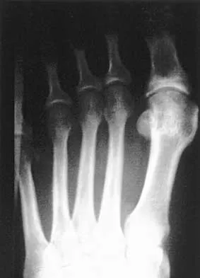
Figure 1 shows the radiograph of a 60-year-old woman who underwent a previous operation for great toe pain 20 years ago. She has had increasing pain over the past 5 years and now reports pain with any motion, swelling, and clicking. She also reports pain under the ball of foot. What is the most appropriate management to alleviate her metatarsalgia and great toe pain?


Explanation

The patient has a failed Silastic implant. Nonsurgical management will not work at this point. A Keller resection will only exacerbate her metatarsalgia. Implant removal with structural bone grafting and MTP fusion is the most appropriate choice because restoration of length is needed to alleviate the forefoot pain and bone grafting is required to fuse the MTP joint because there is an abundance of osteolysis. Total toe implants do not offer good long-term outcomes and are very difficult to fit into the large exploded-out cavity of the proximal phalanx. Hecht PJ, Gibbons MJ, Wapner KL, et al: Arthrodesis of the first metatarsophalangeal joint to salvage failed silicone implant arthroplasty. Foot Ankle Int 1997;18:383-390.

Question 2

A 47-year-old man with Charcot-Marie-Tooth (CMT) disease was treated with a fifth metatarsal head resection for a symptomatic bunionette 2 years ago. What is the most likely complication seen at this time?


Explanation

CMT is characterized by a cavovarus foot position that increases weight-bearing stresses along the lateral border. Removal of the fifth metatarsal head carries the risk of creating a transfer lesion at the fourth metatarsal head, particularly with a cavovarus foot. Claw toes are common in CMT, but the fifth toe would be flail in this situation. Ulceration is unlikely given the lack of underlying bone. Peroneal atrophy is associated with CMT but would not be a complication of this procedure. Charcot arthropathy is a neuropathic process frequently seen in individuals with diabetes mellitus. Kitaoka HB, Holiday AD Jr: Metatarsal head resection for bunionette: Long-term followup. Foot Ankle 1991;11:345-349.

Question 3

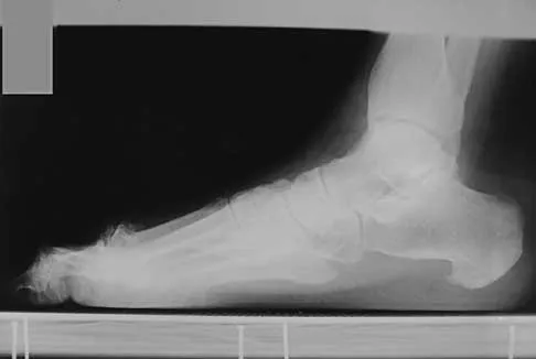
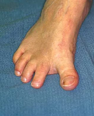
A 19-year-old man was struck by a car and is seen in the emergency department with a grade IIIC open distal tibia and fibula fracture. Examination reveals that the toes are cool and dusky with a sluggish capillary refill. Angiography reveals a lesion in the posterior tibial artery amenable to repair. There is no sensation on the plantar aspect of the foot, and he is unable to flex his toes. A clinical photograph and radiograph are shown in Figures 2a and 2b. What is the next most appropriate step in management?


Explanation

2b In the past, loss of plantar sensation in this grade IIIC tibial fracture would have been an indication for below-knee amputation regardless of the potential for vascular repair. However the 2002 LEAP study divided 55 patients with loss of plantar sensation into two groups, the insensate amputation group and the insensate limb salvage group, with 55% of patients in the insensate salvage group regaining normal sensation 2 years after injury. Furthermore, those in the salvage group who remained insensate after 2 years had equivalent outcomes to those in the amputation group. Because of these findings, limb salvage with vascular repair and external stabilization with delayed closure is deemed appropriate treatment. Immediate intramedullary fixation is not indicated. Because ischemia, contusion, and stretch can adversely affect the tibial nerve, the additional insult of exploration of the nerve is also not advisable given the soft-tissue compromise. Bosse MJ, McCarthy ML, Jones AL, et al: The insensate foot following severe lower extremity trauma: An indication for amputation? J Bone Joint Surg Am 2005;87:2601-2608. Lange RH, Bach AW, Hansen ST Jr, et al: Open tibial fractures with associated vascular injuries: Prognosis for limb salvage. J Trauma 1985;25:203-208. Mackenzie EJ, Bosse MJ, Kellam JF, et al: Factors influencing the decision to amputate or reconstruct after high-energy lower extremity trauma. J Trauma 2002;52:641-649.

Question 4

The pathophysiology of a claw toe deformity includes muscular imbalance caused by which of the following relatively strong structures?


Explanation

The dynamic forces acting to maintain the position of the proximal phalanx at the head of the metatarsal are a balance between the extensor digitorum longus and the weaker intrinsic muscles. With hyperextension at the metatarsophalangeal joint, the intrinsic muscles become less efficient as plantar flexors. Consequently, the hyperextension deformity progresses in the metatarsophalangeal joint as the opposition of the intrinsic muscles to the extensor tendon lessens. This is in contrast to the situation in the interphalangeal joints, where the stronger flexors overpower the weaker intrinsic muscles, which act as the extensors. This combination of events leads to hyperextension at the metatarsophalangeal joint and flexion deformities at the interphalangeal joints, resulting in claw toe. Mizel MS, Yodlowski ML: Disorders of the lesser metatarsophalangeal Joints. J Am Acad Orthop Surg 1995;3:166-173.

Question 5

A 26-year-old woman is seen in the emergency department with an intra-articular distal tibia fracture and a fibular fracture (pilon). The patient, her husband, and three small children have recently immigrated to the United States from Mexico. The husband and wife have both been in a migrant labor camp but have no immediate relatives in the States. What factor is most important when considering her recommended care and treatment?


Explanation

With documented use of a competent interpreter, informed consent should not be an issue. In Hispanic families, the husband often makes the ultimate decision regarding proceeding with surgery; however, he would not be expected to withhold recommended treatment. Hispanics may have a higher risk of comorbidities, but you do not expect this to be a significant concern with this patient. Claustrophobia and some fear of the unfamiliar may make additional imaging studies more difficult to arrange, but not impossible. The real concern is that with no extended family and three small children, the postoperative demand on the patient could significantly jeopardize her ability to comply with weight-bearing restrictions and overall ambulatory demands. Discharge planning and appropriate help may be paramount for a good outcome.

Question 6

A 57-year-old man with type II diabetes mellitus was successfully treated for a first occurrence forefoot full-thickness (Wagner II) diabetic foot ulcer underlying the third metatarsal head with associated hammertoe with a series of weight-bearing total contact casts. There was no evidence of osteomyelitis. The ulcer is now fully healed. He is insensate to the Semmes-Weinstein 5.07 (10 gm) monofilament. What is the next most appropriate step in management?


Explanation

This is the first occurrence of diabetic foot-specific morbidity. The patient has a foot deformity, a history of a diabetic foot ulcer, and is insensate to the monofilament. He is at moderate risk for the development of a recurrent ulcer. This is best avoided with therapeutic footwear. Commercially available depth-inlay shoes should be combined with a custom accommodative foot orthosis to accommodative the deformity. Pinzur MS, Slovenkai MP, Trepman E, et al: Guidelines for diabetic foot care: Recommendations endorsed by the Diabetes Committee of the American Orthopaedic Foot and Ankle Society. Foot Ankle Int 2005;26:113-119.

Question 7

A 28-year-old man has had a 2-year history of progressive lateral ankle pain. History reveals that he underwent a triple arthrodesis at age 13 for a tarsal coalition. The pain has been refractory to braces, custom inserts, and nonsteroidal anti-inflammatory drugs. Weight-bearing radiographs of the ankle and foot are shown in Figures 3a through 3d. Surgical management should include which of the following?


Explanation

3b 3c 3d The patient has a valgus-supination triple arthrodesis malunion. Weight-bearing radiographs show excessive residual valgus through the subtalar joint, producing lateral subfibular impingement, and residual forefoot abduction and midfoot supination through the talonavicular joint, lateralizing the weight-bearing forces through the foot. The deformity is best managed with a medial displacement calcaneal osteotomy and transverse tarsal derotational osteotomy. Ankle arthroscopy and lateral ligament reconstruction are indicated in the event of ligament instability. Tendon transfer, lateral column lengthening, and heel cord lengthening are used for treatment of adult flatfoot from posterior tibial tendon insufficiency. Ankle arthrodesis and ankle arthroplasty are not indicated in this patient because the lateral ankle symptoms are the result of the underlying deformity in the hindfoot, the patient is young, and the ankle joint is relatively normal. Haddad SL, Myerson MS, Pell RF IV: Clinical and radiographic outcome of revision surgery for failed triple arthrodesis. Foot Ankle Int 1997;18:489-499.

Question 8

If heel varus corrects with a Coleman block test, then the hindfoot deformity is flexible. This test proves that the varus is due to a


Explanation

The Coleman block test is used to evaluate the effect of the forefoot on the rearfoot varus. If the deformity corrects with the block, then the hindfoot deformity is flexible and the varus position is secondary to the plantar flexed first ray or valgus position of the forefoot. A rearfoot orthotic will not correct the forefoot cause of the deformity. The patient still may need a lateralizing calcaneal osteotomy to realign the hindfoot. Younger AS, Hansen ST Jr: Adult cavovarus foot. J Am Acad Orthop Surg 2005;13:302-315.

Question 9

A 27-year-old man now reports dorsiflexion and inversion weakness after an automobile collision 6 months ago in which compartment syndrome developed isolated to the anterior and deep posterior compartments. Examination reveals the development of a progressive cavovarus deformity, but the ankle and hindfoot remain flexible. In addition to Achilles tendon lengthening, which of the following procedures is most likely to improve the motor balance of his foot and ankle?


Explanation

Compartment syndrome of the anterior and deep posterior compartments results in anterior tibialis and posterior tibialis tendon weakness, respectively. Furthermore, the long flexors to the hallux and lesser toes will be weak as well. The intact peroneus longus overpowers the weak anterior tibialis tendon, resulting in plantar flexion of the first metatarsal, cavus, and hindfoot varus. Therefore, transferring the peroneus longus to the dorsolateral midfoot reduces the first metatarsal plantar flexion torque, and possibly augments ankle dorsiflexion torque. Hansen ST: Functional Reconstruction of the Foot and Ankle. Philadelphia, PA, Lippincott, Williams & Wilkins, 2000, pp 433-435.

Question 10

Figures 4a through 4c show the radiographs of a 43-year-old woman who sustained a twisting injury to her right ankle. She has ankle pain and tenderness medially and laterally. To help determine the optimal treatment, an external rotation stress radiograph of the ankle is obtained. This test is designed to evaluate the integrity of what structure?


Explanation

4b 4c In the presence of a supination external rotation-type fracture of the distal fibula (Weber type B), stability of the ankle is best assessed by performing an external rotation stress AP view of the ankle. This test is used to assess the integrity of the deltoid ligament. The presence of a deltoid ligament rupture results in instability and generally is best managed surgically. The gravity stress test can also be used. Egol KA, Amirtharajah M, Tejwani NC, et al: Ankle stress test for predicting the need for surgical fixation of isolated fibular fractures. J Bone Joint Surg Am 2004;86:2393-2398. McConnell T, Creevy W, Tornetta P III: Stress examination of supination external rotation-type fibular fractures. J Bone Joint Surg Am 2004;86:2171-2178.

Question 11

A 29-year-old patient sustains a closed, displaced joint depression intra-articular calcaneus fracture. In discussing potential complications of surgical intervention through an extensile lateral approach, which of the following is considered the most common complication following surgery?


Explanation

Delayed wound healing and wound dehiscence is the most common complication of surgical management of calcaneal fractures through an extensile lateral approach, occurring in up to 25% of patients. Most wounds ultimately heal with local treatment; the deep infection rate is approximately 1% to 4% in closed fractures. Posttraumatic arthritis may develop despite open reduction and internal fixation, but the percentages remain low. Peroneal tendinitis may occur from adhesions within the tendon sheath or from prominent hardware but is relatively uncommon. Nonunion of a calcaneal fracture is rare. Sanders RW, Clare MP: Fractures of the calcaneus, in Coughlin MJ, Mann RA, Saltzman CL (eds): Surgery of the Foot and Ankle, ed 8. Philadelphia, PA, Mosby-Elsevier, 2007, vol 2, pp 2017-2073.

Question 12

Figures 5a and 5b show the radiographs of a 56-year-old man who was seen in the emergency department following a twisting injury to his left ankle. Examination in your office 3 days later reveals marked swelling and diffuse tenderness to palpation about the ankle and leg. What is the next most appropriate step in management?


Explanation

5b The radiographs show an isolated posterior malleolus fracture which, given the injury mechanism, is highly suspicious for a Maisonneuve injury. As with any suspected extremity injury, radiographs including the joints above and below the level of injury are acutely indicated. Although MRI may reveal a ligamentous injury to the ankle and CT may show asymmetry of the ankle mortise or syndesmosis, both studies are considerably more costly and are not indicated in the absence of a complete radiographic work-up. Technetium bone scan is nonspecific and would be of limited value in this instance, as would repeat radiographs of the ankle. Walling AK, Sanders RW: Ankle fractures, in Coughlin MJ, Mann RA, Saltzman CL (eds): Surgery of the Foot and Ankle, ed 8. Philadelphia, PA, Mosby-Elsevier, 2007, vol 2, pp 1973-2016.

Question 13

A 61-year-old man has a symptomatic bunionette that is refractory to nonsurgical management. A radiograph is shown in Figure 6. What is the optimal surgical correction?


Explanation

The patient has a bunionette with a large 4-5 intermetatarsal angle. This requires not only ostectomy of the lateral prominence but metatarsal osteotomy to decrease the intermetatarsal angle. Excising the head results in a flail joint and creates the possibility of a transfer lesion. Condylectomy can reduce plantar pressures but does not address the bunionette. The joint surface is well maintained, thus there are no indications for resection. Coughlin MJ: Treatment of bunionette deformity with longitudinal diaphyseal osteotomy with distal soft tissue repair. Foot Ankle 1991;11:195-203.

Question 14

A 25-year-old woman with a healed proximal tibiofibular fracture treated with an intramedullary nail 2 years ago is currently wearing an ankle-foot orthosis (AFO) and reports a persistent foot drop. She is unhappy with the AFO and has not seen any functional improvement despite months of physical therapy. Serial electromyograms (EMG) show no recent change over the past year. Examination and EMG findings are consistent with a tibialis anterior 1/5, extensor hallucis longus 2/5, extensor digitorum longus 2/5, posterior tibial tendon (PTT) 5/5, peroneals 3/5, flexor hallucis longus 5/5, and gastrocsoleus 5/5. No discrete nerve lesion was identified. The patient has a flexible equinovarus contracture. What is the most appropriate management?


Explanation

This pattern of injury is consistent with an unrecognized compartment syndrome of the anterior and lateral compartments. Transfer of the PTT through a long incision in the interosseous membrane corrects the foot drop deformity, and allows adequate dorsiflexion provided that the tendon to be transferred has a strength of 5/5. Muscles/tendons typically lose one grade of strength after transfer. Transfer into the tendons at the level of the ankle prevents overtensioning or pullout of a PTT tendon that is not long enough. Debridement of the scarred muscle in the anterior compartment decreases the risk of scarring down to the tendon transfer. Transfer of the peroneus longus is not preferred given its relative lack of strength and line of pull. Continued therapy and bracing are unlikely to lead to further improvement at 2 years after injury. An ankle fusion would correct the foot drop but would not address the tendon imbalances between the tibialis anterior and the peroneus longus, and the PTT and the peroneus brevis. Hansen ST Jr: Functional Reconstruction of the Foot and Ankle. Philadelphia, PA, Lippincott Williams & Wilkins, 2000, p 192. Atesalp AS, Yildiz C, Komurcu M, et al: Posterior tibial tendon transfer and tendo-Achilles lengthening for equinovarus foot deformity due to severe crush injury. Foot Ankle Int 2002;23:1103-1106. Scott AC, Scarborough N: The use of dynamic EMG in predicting the outcome of split posterior tibial tendon transfers in spastic hemiplegia. J Pediatr Orthop 2006;26:777-780.

Question 15

When using a two-incision approach for open reduction and internal fixation of a Hawkins III talar fracture-dislocation involving the talar neck and body, what anatomic structure must be preserved to optimize outcome?


Explanation

A Hawkins III fracture-dislocation generally presents with posteromedial displacement with the deltoid ligament intact. Therefore, the only remaining blood supply is the deltoid branch of the artery of the tarsal canal originating from the posterior tibial artery. Often, the medial malleolus is fractured, assisting in reduction and visualization of fracture reduction. If the medial malleolus is intact, a medial malleolus osteotomy allows visualization of the reduction without compromising the last remaining blood supply to the talus. Mulfinger GL, Trueta J: The blood supply of the talus. J Bone Joint Surg Br 1970;52:160-167.

Question 16

A 10-year-old boy who is active in soccer has had activity-related heel pain for the past 3 months. Examination reveals tenderness over the posterior heel and a tight Achilles tendon. Radiographs demonstrate a 2-cm cyst in the anterior body of the calcaneus. His physes have not closed. Based on these findings, what is the most appropriate management?


Explanation

The most likely diagnosis is Sever's disease, which is considered either an apophysitis or a para-apophyseal stress fracture. It is common in athletic children and is associated with a tight Achilles tendon. Cast immobilization may be necessary if activity reduction fails. Calcaneal cysts are quite common and do not require any further diagnostic testing or treatment unless they occupy the full width of the calcaneus or one third of the length of the calcaneus. Ogden JA, Ganey TM, Hill JD, et al: Sever's injury: A stress fracture of the immature calcaneal metaphysis. J Ped Orthop 2004;24:488-492.

Question 17

A 35-year-old woman states that she stepped on a piece of glass 6 months ago and reports numbness and shooting pain along the plantar lateral forefoot. She had previously received steroid injections in the 3 to 4 webspace. Examination reveals mild tenderness along the plantar fascia; no Tinel's sign is noted plantar medially and no Mulder's click is noted distally. An MRI scan is shown in Figure 7. What is the most likely cause of the numbness?


Explanation

The MRI scan reveals a laceration through the abductor hallucis musculature and lateral plantar nerve, producing numbness along its distribution. There is no evidence of a foreign body on the MRI scan. Baxter's nerve, or nerve to the abductor digiti quinti muscle, is the first branch off the lateral plantar nerve and impingement of this nerve typically produces a Tinel's sign along the nerve branch deep to the abductor hallucis muscle. Interdigital neuroma would be suggested by the presence of a Mulder's click. A digital nerve laceration would exhibit isolated numbness more distally. Baxter DE, Pfeffer GB: Treatment of chronic heel pain by surgical release of the first branch of the lateral plantar nerve. Clin Orthop Relat Res 1992;279:229-236.

Question 18

A 69-year-old man reports pain over his bunion while wearing shoes and pain in the joint with push-off when barefoot. Nonsurgical management has failed to provide relief. Radiographs are shown in Figures 8a and 8b. What is the surgical procedure of choice?


Explanation

8b Arthrodesis is indicated for severe bunion and hallux valgus deformities, but particularly with extensive degenerative disease of the first metatarsophalangeal joint. The other bunionectomy procedures have different indications, none of which include symptomatic first metatarsophalangeal degenerative disease. Richardson EG(ed): Orthopaedic Knowledge Update: Foot and Ankle 3. Rosemont, IL, American Academy of Orthopaedic Surgeons, 2004, pp 3-15.

Question 19

A 65-year-old man has chronic Achilles insertional tendinitis that is refractory to nonsurgical management. A radiograph is shown in Figure 9. Preoperative counseling should include a discussion of the realistic duration of postoperative recovery. You should inform the patient that his expected recovery will last


Explanation

An older patient with calcaneal enthesopathy may take a year or more to recover after tendon debridement and calcaneal ostectomy. Young patients, and those with purely tendon pathology, may recover more quickly. McGarvey WC, Palumbo RC, Baxter DE, et al: Insertional Achilles tendinitis: Surgical treatment through a central tendon splitting approach. Foot Ankle Int 2002;23:19-25.

Question 20

Figures 10a and 10b show the clinical photograph and MRI scan of a plantar foot lesion. If excisional biopsy is performed, what is the most likely complication?


Explanation

10b The MRI scan shows plantar fibromatosis. The treatment is usually nonsurgical. If surgery is indicated, wide local excision with excision of the entire plantar fascia is usually indicated. The main problem with simple excision of the lesion is the high chance of recurrence. The other listed complications are those that are a result of the wide local excision. Aluisio FV, Mair SD, Hall RL: Plantar fibromatosis: Treatment of primary and recurrent lesions and factors associated with recurrence. Foot Ankle Int 1996;17:672-678.

Question 21

A patient with rheumatoid arthritis with both ankle and subtalar involvement was treated as shown in Figures 11a and 11b. What complication is unique to this type of fixation?


Explanation

11b The interlocking screws at the proximal end of the rod can act as a stress riser and lead to fracture. Postoperative pain at this level should prompt inclusion of this diagnosis in the differential. Removing the screws following bone union can decrease the chances of this occurring. A short rod that avoids the diaphyseal area may also be beneficial. Rotatory deformity is controlled by the perpendicularly oriented distal transfixion screws. Talar osteonecrosis would be unusual since the dissection can be minimized with an intramedullary rod. Any type of hardware can fail if the construct does not lead to a solid arthrodesis. Nunley JA, Pfeffer GB, Sanders RW, et al (eds): Advanced Reconstruction: Foot and Ankle. Rosemont, IL, American Academy of Orthopaedic Surgeons, 2004, pp 236-237. Thordarson DB, Chang D: Stress fractures and tibial cortical hypertrophy after tibiotalocalcaneal arthrodesis with an intramedullary nail. Foot Ankle Int 1999;20:497-500.

Question 22

A 68-year-old man fell off a 20-foot mountain cliff and was seen in the emergency department the following morning. A radiograph is shown in Figure 12. He is a nonsmoker with medical comorbidities of hypertension and hypercholesterolemia that is well controlled with medicine and diet. Capillary refill and sensation are intact distally and the patient is able to move his toes with mild discomfort. Serosanguinous fracture blisters are present laterally, and the foot is swollen and red. What is the most appropriate management?


Explanation

Whereas a patient age of older than 50 years used to be a contraindication for open reduction and internal fixation of displaced intra-articular calcaneal fractures, new data suggest that the presence of associated medical comorbidities that affect wound healing such as smoking, diabetes mellitus, and peripheral vascular disease are more relevant to postoperative functional outcome. Surgical treatment of Sanders II and III displaced intra-articular calcaneal fractures with initial Bohler angles of > 15 degrees results in better outcomes as compared to nonsurgical management. Indications for primary fusion might include Sanders IV fractures in which articular congruity or Bohler angles cannot be restored. Given the condition of the soft tissues at presentation, delayed fixation is recommended. Herscovici D Jr, Widmaier J, Scaduto JM, et al: Operative treatment of calcaneal fractures in elderly patients. J Bone Joint Surg Am 2005;87:1260-1264. Buckley R, Tough S, McCormack R, et al: Operative compared with nonoperative treatment of displaced intra-articular calcaneal fractures: A prospective, randomized, controlled multicenter trial. J Bone Joint Surg Am 2002;84:1733-1744.

Question 23

A 45-year-old woman has had intense pain in her foot for the last 3 days. She also reports a mild fever and difficulty with shoe wear. Examination reveals a swollen, slightly erythematous warm foot with tenderness at the great toe metatarsophalangeal joint and pain with passive motion of the joint. An AP radiograph is shown in Figure 13. Which of the following will best aid in determining a definitive diagnosis?


Explanation

The patient has gouty arthropathy of the first metatarsophalangeal joint. This definitive diagnosis is achieved with aspiration of the joint and polarized light microscopy that shows needle-shaped negatively birefringent monosodium urate crystals. Differential diagnoses of infectious arthritis and pseudogout are also definitively made through joint aspiration. Although rheumatoid arthritis is a possibility, a serum rheumatoid factor is not always diagnostic and a patient with rheumatoid arthritis may have concomitant gouty arthritis. The radiographic findings are not typical of diabetes mellitus or of a patient with Charcot arthropathy. Wise CM, Agudelo CA: Diagnosis and management of complicated gout. Bull Rheum Dis 1998;47:2-5.

Question 24

Figures 14a and 14b show the clinical photographs of a patient who was stranded in a subzero region for several days. The photographs were taken the morning after arrival in the hospital. The patient is otherwise healthy and fit, and takes no medication. He has no clinical signs of sepsis. He reports burning pain and tingling in both feet. What is the best treatment?


Explanation

14b The patient has no clinical or observed signs of sepsis. The skin just proximal to the gangrenous tissue appears somewhat hyperemic and is clearly viable. These wounds should be managed much like burn wounds. Moist dressings should be used until the tissue clearly demarcates. Much of the insult may simply be superficial and only require late debridement. McAdams TR, Swenson DR, Miller RA: Frostbite: An orthopedic perspective. Am J Orthop 1999;28:21-26.

Question 25

The peroneus tertius is a commonly used landmark for arthroscopic portal placement. What is the function of this tendon?


Explanation

The peroneus tertius, although absent in 10% of the population, originates on the distal third of the extensor surface of the fibula and inserts onto the base of the fifth metatarsal, possibly extending to the fascia over the fourth interosseous space. The muscle is located in the anterior compartment of the leg and is innervated by the deep peroneal nerve. The tendon produces dorsiflexion and eversion when walking and can be used as an insertion point during tendon transfers to assist dorsiflexion. This tendon is peculiar to humans and is a proximally migrated deep extensor of the fifth toe. Joshi SD, Joshi SS, Athavale SA: Morphology of the peroneus tertius muscle. Clin Anat 2006;19:611-614. Williams PL, Bannister LH, Berry MM, et al (eds): Gray's Anatomy, ed 38. London, Churchill Livingston, 1995, p 883.

Question 26

A 28-year-old professional soccer player sustains an external rotation ankle injury. Radiographs show a widening of the medial clear space and tibiofibular clear space. Intraoperative stress testing confirms syndesmotic instability. Which of the following statements regarding syndesmotic fixation is most accurate?


Explanation

Suture button fixation (flexible fixation) for syndesmotic injuries allows for physiologic motion at the distal tibiofibular joint. Multiple studies and meta-analyses have shown that flexible fixation results in comparable, if not slightly improved, clinical outcomes and lower rates of malreduction compared to static screw fixation. Routine removal of syndesmotic screws is no longer recommended unless they become symptomatic, and screw breakage does not typically correlate with worse clinical outcomes.

Question 27

A 34-year-old male sustains a purely ligamentous Lisfranc injury of the left foot after falling off a horse with his foot caught in the stirrup. Weight-bearing radiographs demonstrate 3 mm of widening between the base of the first and second metatarsals. He is healthy and highly active. What is the most appropriate definitive management?


Explanation

For purely ligamentous Lisfranc injuries, primary arthrodesis of the medial column (first, second, and third tarsometatarsal joints) has been shown in prospective randomized trials (e.g., Ly and Coetzee) to yield better functional outcomes and a lower rate of revision surgery compared to open reduction and internal fixation (ORIF). ORIF is generally preferred for bony Lisfranc fracture-dislocations.

Question 28

A 42-year-old recreational basketball player experiences a sudden 'pop' in his posterior heel. Clinical examination demonstrates a positive Thompson test. He opts for non-operative management with a functional rehabilitation protocol. Compared to acute surgical repair, which of the following is true regarding his chosen management?


Explanation

Recent high-quality level 1 evidence demonstrates that non-operative management of acute Achilles tendon ruptures using an early functional rehabilitation protocol (incorporating early weight-bearing and mobilization) yields re-rupture rates that are comparable to those of surgical repair. Surgical repair carries a higher risk of complications such as wound healing issues, deep infection, and sural nerve injury, while non-operative management avoids these surgical risks without significantly compromising functional strength.

Question 29

A 55-year-old woman presents with progressive flattening of her right foot and medial ankle pain. Examination reveals a flexible pes planovalgus deformity, inability to perform a single-leg heel rise, and tenderness along the course of the posterior tibial tendon. The hindfoot valgus corrects to neutral when standing on her toes. Which surgical intervention is most appropriate if non-operative management fails?


Explanation

The patient presents with Stage II adult acquired flatfoot deformity (posterior tibial tendon dysfunction), characterized by a flexible deformity and an inability to perform a single-leg heel rise. The standard operative management for a flexible Stage II deformity involves joint-sparing procedures, typically a flexor digitorum longus (FDL) tendon transfer to replace the dysfunctional posterior tibial tendon, combined with a medializing calcaneal osteotomy (MCO) to correct the hindfoot valgus and restore the biomechanical axis. Arthrodesis is reserved for fixed deformities or joint degeneration (Stage III).

Question 30

A 24-year-old male with Charcot-Marie-Tooth disease presents with bilateral cavovarus foot deformities. A Coleman block test is performed. When the patient's heel and lateral border of the foot are placed on a 1-inch block while the first metatarsal hangs freely off the block, the hindfoot varus corrects to a neutral position. What does this physical examination finding indicate?


Explanation

The Coleman block test is used to evaluate a cavovarus foot to determine if the hindfoot varus is flexible and driven by a forefoot deformity (specifically a rigid, plantarflexed first metatarsal) or if it is a rigid hindfoot deformity. If the hindfoot varus corrects to neutral when the first metatarsal is allowed to drop off the block, the hindfoot deformity is flexible and forefoot-driven. Surgical management would therefore include a dorsiflexion osteotomy of the first metatarsal rather than an initial primary hindfoot arthrodesis.

Question 31

A 30-year-old man sustains a Hawkins type III talar neck fracture in a motor vehicle collision. He undergoes open reduction and internal fixation 24 hours after the injury. At his 8-week postoperative visit, a subchondral radiolucent band is observed in the talar dome on the anteroposterior radiograph of the ankle. What is the clinical significance of this radiographic finding?


Explanation

The radiographic finding described is Hawkins' sign, which is a subchondral radiolucent band seen in the talar dome, typically visible on the mortise or AP ankle view at 6 to 8 weeks post-injury. It represents subchondral osteopenia secondary to disuse and active hyperemia. The presence of Hawkins' sign is a reliable indicator that the talar body has sufficient blood supply and is not undergoing avascular necrosis.

Question 32

A 45-year-old woman presents with severe bunion pain. Weight-bearing radiographs reveal a hallux valgus angle (HVA) of 45 degrees and an intermetatarsal angle (IMA) of 20 degrees. Clinical examination reveals profound hypermobility at the first tarsometatarsal (TMT) joint. Which of the following procedures is most appropriate?


Explanation

The patient has a severe hallux valgus deformity (HVA > 40 degrees, IMA > 13-15 degrees) accompanied by first tarsometatarsal (TMT) joint hypermobility. A first TMT arthrodesis (Lapidus procedure) is the surgical treatment of choice in this scenario. It provides powerful correction of the intermetatarsal angle and addresses the underlying hypermobility at the TMT joint, reducing the risk of recurrence. Distal osteotomies are insufficient for severe deformities with joint hypermobility.

Question 33

A 28-year-old male runner presents with chronic, deep ankle pain following an inversion injury 1 year ago. MRI reveals an osteochondral lesion of the medial talar dome measuring 1.8 square centimeters. Non-operative management has failed. Which of the following is the most appropriate surgical intervention for this lesion?


Explanation

The management of osteochondral lesions of the talus (OLT) depends on the size of the lesion. Arthroscopic bone marrow stimulation (microfracture) is generally indicated for primary, smaller lesions (typically < 1.5 cm^2). For larger lesions (> 1.5 cm^2), structural restoration of the subchondral bone and cartilage is required to achieve a durable clinical outcome. Therefore, osteochondral autograft transfer (OATS) or allograft transplantation is the most appropriate treatment for an OLT measuring 1.8 cm^2.

Question 34

A 58-year-old male with poorly controlled type 2 diabetes presents with a red, hot, swollen right foot. He denies recent trauma. Radiographs reveal fragmentation, periarticular debris, and subluxation of the midfoot joints. Laboratory results show a normal WBC count and mildly elevated CRP. What is the most appropriate initial management for this condition?


Explanation

The clinical presentation and radiographic findings (fragmentation, debris, subluxation) are classic for acute Charcot neuroarthropathy (Eichenholtz stage I - development/fragmentation). The initial management for acute Charcot foot is strict immobilization and offloading, most effectively achieved with a total contact cast (TCC), to prevent further progression of the deformity until the acute inflammatory phase subsides. Surgery in the acute phase is generally contraindicated due to unacceptably high complication rates, and the normal WBC mitigates the likelihood of acute deep space infection requiring immediate drainage.

Question 35

A 40-year-old roofer falls from a ladder and sustains a displaced intra-articular calcaneus fracture (Sanders type II). He is a current smoker (1 pack per day). Open reduction and internal fixation via an extensile lateral approach is planned. Which of the following is the most critical factor regarding the timing and approach to minimize soft tissue complications in this patient?


Explanation

Wound healing complications are a major concern with the extensile lateral approach for calcaneus fractures, especially in high-risk patients such as smokers. To minimize these risks, surgery must be delayed until the soft tissue swelling has subsided, indicated by the return of skin wrinkles (the 'wrinkle sign'). This typically takes 10 to 14 days, and sometimes longer. Operating through severely swollen soft tissues significantly increases the risk of wound dehiscence, flap necrosis, and deep infection.

Question 36

A 55-year-old woman presents with a progressive flatfoot deformity. She reports pain localized medially along the posterior tibial tendon and laterally within the sinus tarsi. On examination, she is completely unable to perform a single-leg heel rise on the affected side. Weight-bearing radiographs demonstrate 45% uncovering of the talar head on the AP view and severe talonavicular sag on the lateral view. What is the most appropriate surgical management for this patient?


Explanation

This patient presents with a Stage IIb adult-acquired flatfoot deformity (posterior tibial tendon dysfunction). Stage II denotes a flexible deformity where the patient is unable to perform a single-leg heel rise. Stage IIb specifically involves significant forefoot abduction, radiographically indicated by >40% uncovering of the talar head on the AP view. Treatment for Stage IIb requires addressing both the medial column failure and the lateral column shortening. The standard surgical treatment includes an FDL transfer, a medial displacement calcaneal osteotomy (MDCO) to correct hindfoot valgus, and a lateral column lengthening (e.g., Evans osteotomy or calcaneocuboid distraction arthrodesis) to correct the severe forefoot abduction.

Question 37

A 24-year-old professional football player sustains an axial load injury to a plantarflexed foot. Weight-bearing radiographs show 3 mm of widening between the base of the first and second metatarsals without any obvious fracture fragments, consistent with a purely ligamentous Lisfranc injury. Based on recent outcome studies, which of the following treatments provides the lowest rate of hardware removal and the highest functional outcome score at 2 years for this specific injury pattern?


Explanation

Purely ligamentous Lisfranc injuries have poor healing potential compared to bony avulsion fractures. Level I evidence (e.g., Ly and Coetzee, JBJS Am) has shown that primary arthrodesis of the medial 2 or 3 tarsometatarsal joints for purely ligamentous Lisfranc injuries results in superior functional outcomes, lower rates of subsequent surgeries (hardware removal), and fewer persistent symptoms compared to open reduction and internal fixation (ORIF).

Question 38

A 42-year-old recreational basketball player presents with an acute, midsubstance Achilles tendon rupture. The surgeon and patient are discussing operative versus non-operative management, planning to utilize an early functional rehabilitation protocol. Based on Level 1 evidence, the patient should be counseled that non-operative management is associated with which of the following when compared to operative management?


Explanation

High-quality Level 1 evidence (such as the landmark Willits et al. trial) demonstrates that when an early functional rehabilitation protocol (early weight-bearing and range of motion) is utilized, the re-rupture rates between operative and non-operative management of acute Achilles tendon ruptures are statistically similar. However, operative management is associated with a significantly higher risk of soft-tissue and wound complications, including infection and sural nerve injury.

Question 39

A 62-year-old man presents with severe, end-stage post-traumatic ankle osteoarthritis and is inquiring about total ankle arthroplasty (TAA). Which of the following conditions is considered an absolute contraindication to performing a total ankle arthroplasty?


Explanation

Absolute contraindications to total ankle arthroplasty (TAA) include active or recent ankle infection, severe peripheral vascular disease, inadequate soft tissue coverage, absent lower extremity sensation, Charcot neuroarthropathy, and extensive avascular necrosis of the talus (>50%). Age, moderate BMI, mild to moderate coronal deformities (which can be corrected concurrently), and previous ligamentous surgeries are relative contraindications or factors requiring careful planning, but are not absolute contraindications.

Question 40

A 31-year-old man falls from a height of 15 feet and sustains a Hawkins type III talar neck fracture. Which of the following correctly describes the joint dislocations associated with this specific injury grade, and what is the primary blood supply to the talar body that is at greatest risk of disruption?


Explanation

The Hawkins classification of talar neck fractures is predictive of the risk of avascular necrosis (AVN). Type I is non-displaced. Type II involves subtalar subluxation/dislocation. Type III involves both subtalar and tibiotalar (ankle) dislocation. Type IV (added by Canale) includes talonavicular dislocation. The talar body's most significant blood supply is the artery of the tarsal canal (a branch of the posterior tibial artery), which supplies the majority of the talar body. In a type III fracture, the artery of the tarsal canal, the artery of the sinus tarsi, and the dorsal network are typically all disrupted, leading to a very high risk of AVN.

Question 41

A 58-year-old man with a 15-year history of poorly controlled type 2 diabetes presents with a swollen, warm, and erythematous right foot. He denies any recent trauma, systemic illness, or fever. Radiographs show fragmentation, periarticular debris, and early subluxation of the midfoot joints, but no signs of ulceration, gas, or focal osteomyelitis. Laboratory tests show a normal white blood cell count and a slightly elevated CRP.

What is the most appropriate initial management?


Explanation

This patient is presenting with acute Charcot neuroarthropathy (Eichenholtz stage I: development/fragmentation stage). The clinical picture often mimics infection, but the lack of an open ulcer, systemic signs of infection, and normal WBC point to an acute Charcot event. The gold standard for initial treatment is offloading the extremity to arrest the progression of the deformity and allow the inflammatory process to resolve. This is most effectively accomplished with a total contact cast (TCC), which is changed frequently as the edema subsides. Surgical intervention during the acute, hyperemic stage is generally contraindicated due to poor bone quality and high risk of failure.

Question 42

A 45-year-old woman complains of progressive pain and deformity of her left great toe that limits her ability to wear closed-toe shoes. Weight-bearing radiographs reveal a hallux valgus angle (HVA) of 45 degrees and an intermetatarsal angle (IMA) of 18 degrees. Clinical examination demonstrates significant hypermobility of the first tarsometatarsal (TMT) joint with dorsal elevation of the first ray. There is no evidence of metatarsophalangeal (MTP) joint arthritis. Which of the following surgical procedures is most appropriate to provide a durable correction?


Explanation

This patient has a severe hallux valgus deformity (HVA > 40 degrees, IMA > 15 degrees) combined with clinical hypermobility of the first tarsometatarsal (TMT) joint. A Lapidus procedure (first TMT arthrodesis) is the procedure of choice in this scenario. It provides powerful correction of large intermetatarsal angles and definitively addresses the underlying instability at the TMT joint, preventing recurrence. Distal osteotomies are insufficient for this degree of deformity, and proximal osteotomies without fusion do not address the TMT hypermobility. MTP arthrodesis is typically reserved for severe deformity with concomitant MTP arthritis.

Question 43

During the operative treatment of a displaced, intra-articular calcaneus fracture via an extensile lateral approach, the surgeon must reduce the lateral tuberosity and posterior facet fragments to the 'constant' fragment. Which anatomical structure maintains the position of this 'constant' fragment relative to the talus?


Explanation

The 'constant' fragment in a calcaneus fracture refers to the anteromedial fragment, which includes the sustentaculum tali. Despite severe comminution of the rest of the calcaneus, this fragment consistently remains anatomically aligned with the talus. This stability is maintained by the strong medial talocalcaneal ligament, the interosseous talocalcaneal ligament, and the superficial fibers of the deltoid ligament. During open reduction, this fragment serves as the foundational cornerstone to which the rest of the calcaneus is reduced.

Question 44

A 52-year-old woman presents with burning pain in her forefoot that is exacerbated by wearing tight, high-heeled shoes. She describes a sensation of 'walking on a bunched-up sock.' Examination reveals a palpable click and radiating pain when compressing the medial and lateral aspects of the forefoot while simultaneously applying pressure to the plantar aspect of the third web space (Mulder's sign). If surgical excision of the lesion is eventually performed, what is the expected primary histologic finding of the excised tissue?


Explanation

The patient's presentation is classic for a Morton's neuroma, most commonly occurring in the third intermetatarsal space. Despite the name, a Morton's neuroma is not a true neoplasm or a true neuroma (which would show Schwann cell proliferation). Instead, it is a compressive/entrapment neuropathy resulting from mechanical irritation of the common digital nerve under the transverse metatarsal ligament. Histologically, it is characterized by extensive perineural fibrosis, local vascular proliferation, edema, and subsequent axonal degeneration.

Question 45

A 22-year-old collegiate soccer player sustains an acute fracture at the metaphyseal-diaphyseal junction of the fifth metatarsal (Zone 2) during a match. He desires to return to competitive play as quickly and safely as possible. What is the most recommended treatment for this athlete, and what is the primary reason this specific anatomical region is prone to nonunion?


Explanation

A fracture at the metaphyseal-diaphyseal junction of the fifth metatarsal is an acute Jones fracture (Zone 2). This area is a vascular watershed zone between the metaphyseal arteries (which supply the tuberosity) and the intramedullary nutrient artery (which supplies the diaphysis). Because of this tenuous blood supply, these fractures have a high rate of delayed union and nonunion. In elite or competitive athletes, early intramedullary screw fixation is highly recommended as it significantly decreases the time to union and allows for a faster, safer return to play compared to non-operative management.

Question 46

A 35-year-old recreational basketball player sustains an acute, closed Achilles tendon rupture. He is active but prefers to avoid surgery if possible. After discussing treatment options, he elects to undergo non-operative management with an early functional rehabilitation protocol. Compared to traditional open operative repair, what is the most statistically expected outcome of his chosen management?


Explanation

Recent high-quality level I evidence and meta-analyses have demonstrated that non-operative management of acute Achilles tendon ruptures utilizing an early functional rehabilitation protocol (involving early weight-bearing in an orthosis) results in re-rupture rates that are statistically similar to operative repair. Non-operative management also avoids surgical complications such as deep infection, wound breakdown, and iatrogenic sural nerve injury, leading to a lower overall complication rate.

Question 47

A 62-year-old woman with a BMI of 28 presents with severe, end-stage post-traumatic osteoarthritis of the right ankle. Radiographs demonstrate bone-on-bone tibiotalar joint space narrowing with a 5-degree varus deformity. She is evaluating the options of total ankle arthroplasty (TAA) versus ankle arthrodesis. When counseling the patient, which of the following is considered a well-documented advantage of TAA compared to arthrodesis?


Explanation

Total Ankle Arthroplasty (TAA) preserves tibiotalar motion, which alters the biomechanical stresses on adjacent joints. Studies have shown that TAA is associated with a decreased risk, or delayed progression, of adjacent joint osteoarthritis (such as the subtalar and talonavicular joints) compared to ankle arthrodesis. However, TAA generally has a higher reoperation rate and is not recommended for patients desiring a return to high-impact sports, or those with severe, uncorrectable coronal plane deformities.

Question 48

A 55-year-old female presents with medial foot pain, difficulty standing on her toes, and a progressively flattening arch over the past year. Examination reveals a positive 'too-many-toes' sign and a flexible hindfoot that corrects to neutral on heel rise. Standing AP radiograph shows greater than 40% uncoverage of the talonavicular joint.

Based on the Johnson and Strom classification (modified by Myerson), what is the most appropriate surgical management?


Explanation

The patient has Stage IIb posterior tibial tendon dysfunction (adult-acquired flatfoot deformity), characterized by a flexible hindfoot but significant forefoot abduction (defined as >40% talonavicular uncoverage). Appropriate surgical management includes FDL transfer to replace the diseased posterior tibial tendon, medial calcaneal displacement osteotomy (to correct hindfoot valgus), and lateral column lengthening (Evans osteotomy) to correct the severe forefoot abduction. Stage IIa (<40% uncoverage) can often be treated without the lateral column lengthening. Stage III (rigid deformity) requires arthrodesis.

Question 49

A 22-year-old collegiate football player sustains a midfoot injury after an axial load was applied to a plantarflexed foot. Weight-bearing radiographs reveal a 3 mm diastasis between the base of the first and second metatarsals, with no associated fractures. What is the gold standard surgical treatment to limit the development of midfoot arthritis and maximize functional outcome in this strictly ligamentous injury?


Explanation

While Open Reduction and Internal Fixation (ORIF) has historically been the standard for Lisfranc injuries, level I evidence (such as the landmark study by Ly and Coetzee) has demonstrated that primary arthrodesis of the medial column (1st, 2nd, and 3rd tarsometatarsal joints) yields superior functional outcomes and a lower reoperation rate compared to ORIF specifically for strictly ligamentous Lisfranc injuries. ORIF remains the standard for purely bony or predominantly bony Lisfranc fracture-dislocations.

Question 50

A 28-year-old male sustains an acute high ankle sprain. Examination reveals a positive external rotation stress test and positive squeeze test. An MRI confirms an isolated full-thickness tear of the anterior inferior tibiofibular ligament (AITFL) and the interosseous membrane up to 5 cm proximal to the joint line. Intraoperatively, the syndesmosis is unstable to the hook test. If dynamic suture-button fixation is chosen over static syndesmotic screw fixation, what is an established clinical advantage?


Explanation

Suture-button fixation for syndesmotic instability provides dynamic stabilization. Unlike static syndesmotic screws, the suture-button allows for normal, physiologic micro-motion (rotation and proximal-distal translation) of the fibula during the gait cycle. It also does not require routine removal and facilitates an earlier return to weight-bearing and functional activities. Current literature indicates equivalent or lower rates of malreduction compared to traditional screw fixation.

Question 51

A 58-year-old male with poorly controlled type 2 diabetes and profound peripheral neuropathy presents with a red, hot, swollen right foot for 2 weeks. There are no skin breaks or ulcerations. He denies fever or chills. His WBC count is normal, and ESR is mildly elevated at 35 mm/hr. Radiographs demonstrate soft tissue swelling but no bony destruction, fragmentation, or subluxation.

What is the initial treatment of choice?


Explanation

The clinical presentation is classic for Eichenholtz stage 0 Charcot neuroarthropathy, characterized by a red, hot, swollen foot with normal or osteopenic radiographs but no distinct fragmentation or subluxation. The mainstay of treatment in the acute (Stage 0 or 1) phase of Charcot arthropathy is total contact casting (TCC) and strict non-weight bearing to arrest the progression of joint destruction until the acute inflammatory phase resolves (skin temperature equalizes with the contralateral limb).

Question 52

A 50-year-old male presents with chronic dorsal pain in his first metatarsophalangeal (MTP) joint, exacerbated by walking. He has failed conservative management including a stiff-soled shoe with a Morton extension. X-rays reveal dorsal osteophytes and mild to moderate joint space narrowing primarily in the dorsal aspect of the joint, while the plantar joint space is preserved (Coughlin and Shurnas Grade 2). He desires to maintain motion. What is the most appropriate surgical intervention?


Explanation

Coughlin and Shurnas Grade 2 hallux rigidus presents with moderate osteophyte formation, dorsal joint space narrowing, and preserved plantar joint cartilage. Cheilectomy (removal of the dorsal osteophyte and the dorsal third of the metatarsal head) is the treatment of choice for Grade 1 and 2 hallux rigidus to relieve dorsal impingement and preserve motion. Arthrodesis is typically reserved for Grade 3 or 4 hallux rigidus (diffuse joint space loss). Keller arthroplasty is largely historical or reserved for elderly, low-demand patients due to the risk of transfer metatarsalgia and cock-up deformity.

Question 53

A 31-year-old male falls from a ladder and sustains a Hawkins Type III talar neck fracture (fracture of the talar neck with subtalar and tibiotalar dislocation).

He is at high risk for avascular necrosis (AVN) of the talar body. Which of the following anatomical structures provides the majority of the blood supply to the talar body that is compromised in this injury?


Explanation

The talar body receives its blood supply from a retrograde extraosseous arterial ring. The largest and most significant contributor to the talar body's blood supply is the artery of the tarsal canal, which is a branch of the posterior tibial artery. The artery of the tarsal sinus (formed by branches of the dorsalis pedis and perforating peroneal) supplies the lateral aspect. The deltoid branches supply the medial aspect. A Hawkins III fracture disrupts all three major sources, leading to a near 100% risk of AVN.

Question 54

A 21-year-old collegiate basketball player sustains an acute foot injury. Radiographs show a transverse fracture at the metaphyseal-diaphyseal junction of the fifth metatarsal (Zone 2) without displacement. He is eager to return to play as quickly and safely as possible. What is the recommended treatment to minimize the risk of nonunion and expedite his return to sports?


Explanation

A fracture at the metaphyseal-diaphyseal junction of the fifth metatarsal is a true Jones fracture (Zone 2). Because this area is a vascular watershed zone, it has a high rate of delayed union or nonunion with conservative management. In high-level or elite athletes desiring an expedited return to play, intramedullary screw fixation is the gold standard. It significantly decreases the time to union and return to sports compared to non-operative treatment.

Question 55

A 24-year-old competitive runner presents with severe, bilateral anterolateral leg pain that begins 15 minutes into her runs. The pain is accompanied by numbness in the first dorsal web space of both feet and a transient inability to actively dorsiflex her ankles. Symptoms completely resolve after 30 minutes of rest. Intracompartmental pressure testing reveals an elevated anterior compartment pressure of 45 mmHg immediately post-exercise. Compression of which nerve is directly responsible for her sensory and motor symptoms?


Explanation

The patient's presentation is classic for chronic exertional compartment syndrome (CECS) of the anterior compartment. The anterior compartment contains the deep peroneal nerve, which provides motor innervation to the ankle dorsiflexors (tibialis anterior, EHL, EDL) and sensory innervation to the first dorsal web space. Increased pressure in the anterior compartment compresses the deep peroneal nerve, leading to transient foot drop and first web space numbness. The superficial peroneal nerve courses through the lateral compartment.

Question 56

A 52-year-old woman presents with progressive medial ankle pain and a new-onset flatfoot deformity. On examination, she has a flexible hindfoot, a 'too many toes' sign, and is unable to perform a single-leg heel rise. Weight-bearing radiographs reveal a flexible pes planovalgus deformity with >40% talonavicular uncoverage on the AP view, indicative of significant forefoot abduction. What is the most appropriate surgical management for this stage of posterior tibial tendon dysfunction (Stage IIb)?


Explanation

The patient has Stage IIb posterior tibial tendon dysfunction (PTTD), characterized by a flexible hindfoot deformity with significant forefoot abduction (>40% talonavicular uncoverage). While Stage IIa (minimal forefoot abduction) is effectively treated with an FDL transfer and medializing calcaneal osteotomy, Stage IIb requires the addition of a lateral column lengthening (such as an Evans osteotomy or calcaneocuboid distraction arthrodesis) to correct the forefoot abduction and restore the talonavicular joint alignment.

Question 57

A 35-year-old male recreational athlete sustains an acute Achilles tendon rupture. He is discussing operative repair versus non-operative management utilizing a strict early functional rehabilitation protocol. Based on current randomized controlled trials (e.g., Willits et al.), what is the expected difference in outcomes between these two treatment strategies?


Explanation

Current high-level evidence, including the landmark study by Willits et al., demonstrates that when an early functional rehabilitation protocol (early weight-bearing and range of motion) is employed, there is no statistically significant difference in rerupture rates, functional outcomes, or plantarflexion strength between operative and non-operative management of acute Achilles tendon ruptures. However, operative management carries a significantly higher risk of complications, primarily related to wound healing and infection.

Question 58

A 24-year-old male athlete sustains a purely ligamentous Lisfranc injury. Weight-bearing radiographs demonstrate 4 mm of diastasis between the bases of the first and second metatarsals without associated fractures. Which of the following treatments has been shown to provide the best long-term functional outcome and lowest reoperation rate for this specific injury pattern?


Explanation

For purely ligamentous Lisfranc injuries, multiple studies (such as those by Ly and Coetzee) have demonstrated that primary arthrodesis of the medial column (1st, 2nd, and 3rd tarsometatarsal joints) yields superior functional outcomes, higher return to pre-injury activity levels, and lower rates of subsequent surgeries for hardware removal or salvage arthrodesis compared to open reduction and internal fixation (ORIF).

Question 59

A 32-year-old man undergoes open reduction and internal fixation for a Hawkins type III talar neck fracture following a high-energy motor vehicle collision. At his 8-week postoperative visit, an AP radiograph of the ankle demonstrates a subchondral radiolucent band in the dome of the talus. What does this radiographic finding indicate?


Explanation

The subchondral radiolucent band described is known as Hawkins sign. It represents subchondral osteopenia secondary to disuse and hyperemia. The presence of this sign indicates that there is intact vascularity to the talar body, effectively ruling out avascular necrosis (AVN). Its absence, however, does not definitively confirm AVN, though it raises the clinical suspicion.

Question 60

A 55-year-old man with a 15-year history of poorly controlled type 2 diabetes and profound peripheral neuropathy presents with a red, hot, swollen right foot. He denies trauma. He is afebrile with normal white blood cell count and inflammatory markers. Radiographs reveal fragmentation, periarticular debris, and subluxation of the midfoot joints. There are no skin ulcerations. What is the most appropriate initial management?


Explanation

The patient's presentation is classic for an acute Charcot neuroarthropathy (Eichenholtz Stage 1 - Fragmentation). In the absence of an open ulcer or systemic signs of infection, the initial treatment is non-operative and consists of offloading to prevent further progressive deformity. A total contact cast (TCC) and strict non-weight bearing are the gold standards for managing acute Charcot arthropathy.

Question 61

A 45-year-old woman presents with persistent forefoot pain and a prominent bunion. Weight-bearing radiographs demonstrate a hallux valgus angle (HVA) of 42 degrees, an intermetatarsal angle (IMA) of 18 degrees, and notable hypermobility at the first tarsometatarsal (TMT) joint. Which of the following surgical procedures is most appropriate to address her pathology?


Explanation

The patient has a severe hallux valgus deformity (IMA > 15 degrees, HVA > 40 degrees) accompanied by first TMT joint hypermobility. A first TMT arthrodesis (Lapidus procedure) is the procedure of choice in this scenario, as it allows for large corrections of the IMA while simultaneously addressing the apex of the deformity and stabilizing the hypermobile medial column. Distal osteotomies are insufficient for IMA correction of this magnitude.

Question 62

A 22-year-old collegiate basketball player sustains a fracture at the metaphyseal-diaphyseal junction of the fifth metatarsal (Zone 2) during a game. Radiographs show a non-displaced fracture without evidence of intramedullary sclerosis. He wishes to return to athletic competition as soon as safely possible. What is the most appropriate management?


Explanation

Zone 2 fractures of the fifth metatarsal (Jones fractures) involve a vascular watershed area and have a high rate of delayed union or nonunion. In elite or high-level athletes who desire an expedited return to play, intramedullary screw fixation is the standard of care. It has been shown to significantly reduce the time to union and return to sports compared to conservative management.

Question 63

A 28-year-old woman presents with persistent anterolateral ankle pain 1 year after a severe inversion injury. MRI demonstrates a 1.8 cm^2 osteochondral lesion of the anterolateral talar dome with deep subchondral cystic changes. She has failed a 6-month trial of conservative management. What is the most appropriate surgical treatment?


Explanation

Osteochondral lesions of the talus (OLT) that are large (> 1.5 cm^2) or associated with significant subchondral cystic changes have a high failure rate with marrow stimulation techniques like microfracture. For large or cystic lesions, structural bone grafting and cartilage restoration via an osteochondral autograft transfer system (OATS) or fresh osteochondral allograft is indicated to restore the articular contour and provide structural support.

Question 64

A 40-year-old construction worker falls from a ladder and sustains a displaced, intra-articular calcaneus fracture. He undergoes open reduction and internal fixation utilizing an extensile lateral approach. Which of the following is the most common complication associated with this specific surgical approach?


Explanation

The extensile lateral approach to the calcaneus is notorious for soft tissue complications. Wound edge necrosis, dehiscence, and subsequent infection are the most common complications, occurring in approximately 10% to 25% of cases. The risk is minimized by creating a full-thickness 'no-touch' flap, respecting the vascular supply from the lateral calcaneal artery, and delaying surgery until the 'wrinkle sign' appears.

Question 65

A 30-year-old man undergoes open reduction and internal fixation for a Weber C ankle fracture with syndesmotic instability. The syndesmosis is stabilized with two 3.5-mm metallic screws crossing four cortices. Postoperatively, he recovers well and begins weight-bearing at 6 weeks. According to current orthopedic literature, what is the recommendation regarding the routine removal of metallic syndesmotic screws in asymptomatic patients?


Explanation

Current literature supports that routine removal of metallic syndesmotic screws is not necessary in asymptomatic patients. Studies have shown that functional outcomes do not significantly differ between patients who have their screws removed and those who retain them, even if the screws break or loosen. Retained hardware only requires removal if it causes localized pain or irritation.

Question 66

A 24-year-old professional football player sustains a hyperplantarflexion injury to his midfoot. Weight-bearing radiographs show a 2.5 mm diastasis between the base of the first and second metatarsals. MRI confirms a purely ligamentous disruption of the Lisfranc complex without associated fractures. What is the most appropriate definitive management to minimize reoperation rates and maximize functional outcome in this athlete?


Explanation

For purely ligamentous Lisfranc injuries, multiple randomized controlled trials (such as Ly and Coetzee) have demonstrated that primary arthrodesis of the medial column (1st, 2nd, and 3rd TMT joints) results in superior short- and medium-term functional outcomes and significantly lower reoperation rates compared to ORIF. ORIF is typically preferred for bony Lisfranc fracture-dislocations to preserve joint motion, but purely ligamentous injuries have a high rate of hardware failure and post-traumatic arthritis when treated with ORIF.

Question 67

A 34-year-old woman presents with bilateral foot pain, lateral column overload, and frequent ankle sprains. Examination shows a bilateral cavovarus foot type. To evaluate the flexibility of the hindfoot, you perform a Coleman block test. When the patient stands with her heel and lateral border of the foot on the block and the first metatarsal suspended freely off the block, her hindfoot varus corrects entirely to neutral. What does this physical examination finding indicate?


Explanation

The Coleman block test evaluates the flexibility of the hindfoot in a cavovarus deformity. If suspending the first ray (dropping it off the block) allows the hindfoot to correct to neutral, the hindfoot is flexible. This indicates that the hindfoot varus is a compensatory, non-fixed deformity driven by a rigidly plantarflexed first ray. Treatment should therefore focus on correcting the forefoot (e.g., first metatarsal dorsiflexion osteotomy) rather than fusing the hindfoot.

Question 68

A 52-year-old man presents with chronic weakness in his posterior ankle 4 months after feeling a 'pop' while playing tennis. He has a palpable gap 6 cm proximal to the calcaneal insertion of the Achilles tendon. MRI confirms a chronic Achilles tendon rupture with a 5.5 cm gap with the foot in resting equinus. Which of the following is the most appropriate surgical treatment?


Explanation

Chronic Achilles ruptures with a gap of greater than 5 cm typically cannot be repaired end-to-end, even with V-Y fascial advancement alone. An FHL transfer is the procedure of choice for large defects (> 3-5 cm). The FHL provides well-vascularized tissue to bridge the gap, has an in-phase firing pattern with the Achilles, and supplies supplemental plantarflexion power.

Question 69

A 62-year-old man with end-stage post-traumatic ankle osteoarthritis presents for surgical consultation. He has a BMI of 28, is a non-smoker, and has well-controlled hypertension. Examination reveals severe tibiotalar arthritis but an intact and perfectly aligned hindfoot. His ankle range of motion is 5 degrees of dorsiflexion to 20 degrees of plantarflexion. Which of the following would be considered an absolute contraindication to a total ankle arthroplasty (TAA) in this patient?


Explanation

Significant avascular necrosis (typically >50%) of the talar body is an absolute contraindication to total ankle arthroplasty (TAA) due to inadequate bone stock, which leads to a very high risk of talar component subsidence and failure. Other absolute contraindications include active infection, severe peripheral neuropathy (e.g., Charcot arthropathy), and absent or non-functioning leg musculature. Age <65 and mild coronal deformities are relative, not absolute, contraindications.

Question 70

A 28-year-old woman presents with persistent deep ankle pain following an inversion ankle sprain 6 months ago. MRI reveals an osteochondral lesion on the posteromedial aspect of the talar dome measuring 8 mm x 8 mm (64 mm^2), with intact overlying cartilage and no significant subchondral cystic changes. Conservative management has failed. What is the most appropriate next step in management?


Explanation

For symptomatic osteochondral lesions of the talus (OLT) that are less than 1.5 cm^2 (150 mm^2) and lack extensive subchondral cysts, arthroscopic bone marrow stimulation (microfracture) is the gold standard first-line surgical treatment. OATS or structural allografts are typically reserved for larger lesions (>1.5 cm^2), lesions with large cysts, or those that have failed prior microfracture.

Question 71

A 55-year-old woman presents with progressively worsening right foot pain. On examination, she has a flexible flatfoot, a positive 'too-many-toes' sign, and an inability to perform a single-limb heel raise. Radiographs reveal uncovering of the talonavicular joint of 45% indicating severe forefoot abduction. What is the most appropriate surgical treatment algorithm for this stage of adult acquired flatfoot deformity?


Explanation

This patient presents with Stage IIb adult acquired flatfoot deformity (posterior tibial tendon dysfunction). Stage IIb is characterized by a flexible hindfoot (Stage II) but with significant forefoot abduction (typically >30-40% talonavicular uncoverage). Treatment requires an FDL transfer, a medial displacement calcaneal osteotomy (MDCO) to restore the heel axis, AND a lateral column lengthening (e.g., Evans osteotomy or calcaneocuboid fusion) to specifically correct the severe forefoot abduction. Triple arthrodesis is reserved for rigid (Stage III) deformities.

Question 72

A 58-year-old man with poorly controlled type 2 diabetes presents with a red, hot, swollen left foot. He reports no trauma. Radiographs show soft tissue swelling but no acute fractures, dislocations, or bony destruction. Inflammatory markers are mildly elevated. He is diagnosed with acute Eichenholtz stage 0 Charcot neuroarthropathy. What is the most appropriate initial management?


Explanation

Acute (Eichenholtz Stage 0 or I) Charcot neuroarthropathy presents with a red, hot, swollen extremity that mimics infection. Radiographs in Stage 0 may be normal or show only soft tissue swelling. The mainstay of treatment in the acute phase is strict offloading to prevent progressive deformity and devastating bone destruction. This is most effectively achieved with a total contact cast (TCC). Surgery is generally contraindicated in the acute inflammatory phase unless there is an unstable deformity causing impending soft tissue compromise.

Question 73

A 42-year-old woman presents with a painful bunion. Weight-bearing radiographs demonstrate a hallux valgus angle (HVA) of 45 degrees and an intermetatarsal angle (IMA) of 18 degrees. Clinical examination reveals profound hypermobility of the first tarsometatarsal (TMT) joint in the sagittal plane. What is the most appropriate surgical procedure?


Explanation

This patient has severe hallux valgus (HVA >40 degrees, IMA >15 degrees) combined with first TMT hypermobility. A distal osteotomy (Chevron) cannot adequately correct a large IMA of 18 degrees. The Lapidus procedure (first TMT arthrodesis) is highly effective for large IMA corrections and uniquely addresses the TMT hypermobility. A proximal osteotomy is also an option for severe deformities without hypermobility, but Lapidus is the classic choice when hypermobility is present.

Question 74

A 22-year-old elite college basketball player sustains an inversion injury to his foot. Radiographs demonstrate an acute, non-displaced fracture of the fifth metatarsal at the metaphyseal-diaphyseal junction extending into the fourth-fifth intermetatarsal articulation.

To minimize the risk of nonunion and expedite his return to play, what is the best treatment option?


Explanation

The clinical scenario and imaging describe a Jones fracture (Zone 2 fracture of the 5th metatarsal base). Because of the watershed blood supply in this region, these fractures are highly prone to delayed union and nonunion. In elite athletes, early intramedullary screw fixation is recommended to significantly reduce the time to union, lower the nonunion rate, and allow for a faster return to competitive sports compared to conservative cast immobilization.

Question 75

A 40-year-old male sustains a displaced intra-articular calcaneus fracture. He undergoes open reduction and internal fixation via an extensile lateral approach. Which of the following technical factors or patient characteristics most significantly increases his risk of postoperative wound necrosis and deep infection?


Explanation

Smoking is the single most significant modifiable patient risk factor for wound healing complications following the extensile lateral approach for calcaneus fractures, increasing the risk of wound necrosis and infection by up to 3 to 4 times. Waiting for the wrinkle sign (decreased swelling), using a full-thickness 'no-touch' subperiosteal flap, and proper incision placement are all standard techniques explicitly used to minimize the risk of wound complications.

Question 76

A 35-year-old male weekend warrior sustained an acute Achilles tendon rupture 2 days ago. He prefers nonoperative management but asks about the risks compared to surgery. According to recent high-level evidence, what is the most significant difference between operative and nonoperative management when an early functional rehabilitation protocol is employed?


Explanation

According to multiple randomized controlled trials (e.g., Willits et al.), when early functional rehabilitation protocols are utilized, the re-rupture rates between operative and nonoperative management of acute Achilles tendon ruptures are statistically similar. However, operative management is associated with a significantly higher risk of soft-tissue complications, including infection and wound breakdown. Functional outcomes and return to sports rates are generally equivalent when early motion is instituted.

Question 77

A 25-year-old female sustains a severe midfoot sprain after a fall from a horse. Weight-bearing radiographs show a 4 mm diastasis between the base of the first and second metatarsals. MRI confirms a purely ligamentous Lisfranc injury with complete disruption of the Lisfranc ligament. Compared to open reduction and internal fixation (ORIF), primary arthrodesis for this specific injury pattern is associated with which of the following?


Explanation

Purely ligamentous Lisfranc injuries are notoriously unstable and have a high rate of post-traumatic arthritis and hardware failure when treated with ORIF. Studies (such as Coetzee et al.) have shown that primary arthrodesis of the medial columns (first, second, and third tarsometatarsal joints) for purely ligamentous injuries results in lower rates of planned secondary surgeries (such as hardware removal) and comparable or slightly better functional outcomes compared to ORIF.

Question 78

A 28-year-old male sustains a Hawkins Type II talar neck fracture following a motor vehicle collision. The primary blood supply to the talar body is at significant risk for disruption. Which of the following vessels provides the majority of the blood supply to the talar body?


Explanation

The major blood supply to the talar body is the artery of the tarsal canal, which is a branch of the posterior tibial artery. The artery of the sinus tarsi, derived from the perforating peroneal and dorsalis pedis arteries, provides a secondary supply. The deltoid branches supply the medial aspect of the body. In a Hawkins II fracture (talar neck fracture with subtalar dislocation), the artery of the tarsal canal is frequently disrupted, placing the talar body at risk for avascular necrosis.

Question 79

A 55-year-old woman complains of medial ankle pain and progressive flattening of her left foot. Examination reveals an inability to perform a single-leg heel raise and a flexible planovalgus deformity (Stage II Adult Acquired Flatfoot Deformity). She undergoes a flexor digitorum longus (FDL) transfer and a medializing calcaneal osteotomy. Which of the following best describes the primary biomechanical rationale of the calcaneal osteotomy in this setting?


Explanation

Adult acquired flatfoot deformity (AAFD) Stage II is characterized by a flexible planovalgus foot. By translating the posterior calcaneal tuberosity medially, the medializing calcaneal osteotomy (MCO) shifts the mechanical axis of the hindfoot. This converts the pull of the Achilles tendon from an evertor (which exacerbates the valgus deformity) into an invertor, thereby decreasing the stress on the medial soft tissue reconstructions, such as the FDL transfer.

Question 80

A 65-year-old man presents with end-stage post-traumatic ankle osteoarthritis and is evaluating surgical options between ankle arthrodesis and total ankle arthroplasty (TAA). Which of the following is considered a primary indication favoring TAA over ankle arthrodesis?


Explanation

Total ankle arthroplasty (TAA) is indicated for end-stage ankle arthritis. Because ankle arthrodesis alters foot biomechanics and significantly increases stress on adjacent joints, pre-existing advanced arthritis of the ipsilateral subtalar or talonavicular joints is a primary indication for TAA over arthrodesis. Absolute contraindications for TAA include active or prior deep infection, severe peripheral neuropathy (Charcot), significant talar body avascular necrosis, and severe, uncorrectable malalignment.

Question 81

A 45-year-old female presents with a painful bunion. Weight-bearing radiographs show a hallux valgus angle of 35 degrees and an intermetatarsal angle (IMA) of 17 degrees. Clinical examination of the first tarsometatarsal (TMT) joint demonstrates significant hypermobility in the sagittal plane. What is the most appropriate surgical intervention?


Explanation

Hallux valgus associated with first tarsometatarsal (TMT) joint hypermobility and a significant intermetatarsal angle (IMA > 15 degrees) is best treated with a first TMT arthrodesis, also known as the Lapidus procedure. This provides powerful correction of the IMA and stabilizes the medial column, addressing the primary deforming force and reducing the risk of recurrence. Distal osteotomies are indicated for mild deformities without hypermobility.

Question 82

A 40-year-old roofer falls 15 feet, sustaining a closed, displaced, intra-articular calcaneus fracture (Sanders Type III). Open reduction and internal fixation via an extensile lateral approach is planned. During the approach, which of the following structures is at greatest risk of iatrogenic injury if the full-thickness flap is not appropriately mobilized and protected?


Explanation

The extensile lateral approach is the standard workhorse approach for open reduction and internal fixation of displaced intra-articular calcaneus fractures. The subperiosteal flap must be elevated as a single full-thickness unit (including the periosteum, peroneal tendons, and sural nerve) to preserve its blood supply. The sural nerve crosses the lateral aspect of the hindfoot and is at highest risk of iatrogenic injury or neuroma formation at both the proximal (vertical) and distal (horizontal) limbs of the L-shaped incision.

Question 83

A 21-year-old collegiate basketball player experiences acute lateral foot pain during practice. Radiographs reveal a transverse fracture of the fifth metatarsal at the metaphyseal-diaphyseal junction (Zone 2). To minimize the risk of nonunion and allow the fastest safe return to play, what is the most appropriate management?


Explanation

A fracture at the metaphyseal-diaphyseal junction of the fifth metatarsal (Zone 2) is a true Jones fracture. This area represents a vascular watershed zone, predisposing these fractures to delayed union and nonunion. In high-level or elite athletes, intramedullary screw fixation is considered the gold standard to significantly decrease the risk of nonunion, allow for an accelerated rehabilitation protocol, and provide a faster, more predictable return to play compared to nonoperative cast immobilization.

Question 84

A 58-year-old male with poorly controlled diabetes mellitus presents with a swollen, erythematous, warm, and painless right foot. Radiographs demonstrate periarticular fragmentation, debris, and subluxation of the tarsometatarsal joints. According to the modified Eichenholtz classification, what is the appropriate stage of this disease process, and what is the most appropriate initial management?


Explanation

Charcot neuroarthropathy in the acute phase presents with a warm, erythematous, and swollen foot. According to the modified Eichenholtz classification, Stage I (Development/Fragmentation) is characterized by joint subluxation, periarticular fragmentation, and debris. The gold standard for initial management of Stage I is strict offloading and immobilization, most effectively achieved with a total contact cast (TCC), to prevent progressive deformity while the bones coalesce (Stage II) and consolidate (Stage III).

Question 85

A 26-year-old professional football player presents after a severe hyperextension injury to his great toe. He has significant plantar ecchymosis, swelling, and gross instability with resisted plantarflexion of the first MTP joint. MRI confirms a complete tear of the plantar plate with proximal retraction of the sesamoids. Which of the following is the most appropriate management?


Explanation

Turf toe is a sprain of the first MTP joint plantar plate, typically resulting from a severe hyperextension injury. Grade 3 injuries involve a complete disruption of the plantar plate complex, manifesting with gross instability, weakness in push-off, and proximal retraction of the sesamoids on imaging. In competitive athletes, surgical repair of the plantar plate is indicated to restore function, push-off strength, and prevent chronic MTP joint instability, progressive deformity, and early osteoarthritis.

Question 86

A 54-year-old female presents with medial ankle pain and progressive flattening of her left foot arch. On examination, she has a flexible hindfoot valgus, flexible forefoot varus, and is unable to perform a single-leg heel raise. Weight-bearing radiographs reveal a talonavicular coverage angle of 45 degrees. A trial of custom orthotics and physical therapy has failed to provide relief. What is the most appropriate surgical intervention?


Explanation

The patient presents with Stage IIb adult acquired flatfoot deformity (posterior tibial tendon dysfunction). Stage IIb is characterized by a flexible hindfoot valgus and significant forefoot abduction (typically indicated by a talonavicular coverage angle > 30-40 degrees). In addition to a flexor digitorum longus (FDL) transfer and a medializing calcaneal osteotomy to correct the hindfoot valgus, a lateral column lengthening (e.g., Evans osteotomy or calcaneocuboid distraction arthrodesis) is required to correct the forefoot abduction. A triple arthrodesis is reserved for Stage III (rigid deformity).

Question 87

A 28-year-old male runner presents with chronic, deep anterolateral ankle pain following a severe inversion injury 18 months ago. Non-operative management, including immobilization and physical therapy, has failed. MRI reveals a 1.8 square centimeter osteochondral lesion on the lateral talar dome with underlying subchondral cysts measuring 5 mm in depth. Which of the following is the most appropriate surgical treatment?


Explanation

The management of osteochondral lesions of the talus (OLT) depends on the size of the lesion and the presence of subchondral cysts. While arthroscopic bone marrow stimulation (microfracture) is the first-line surgical treatment for lesions smaller than 1.5 square centimeters without significant cystic changes, larger lesions (> 1.5 square centimeters) and those with deep subchondral cysts are better treated with structural bone grafting to restore the subchondral architecture. Osteochondral autograft transfer (OATS) provides a viable hyaline cartilage surface and addresses the subchondral bony defect simultaneously.

Question 88

A 65-year-old female presents with a painful bunion. Weight-bearing radiographs reveal a hallux valgus angle (HVA) of 42 degrees and an intermetatarsal angle (IMA) of 16 degrees. There is evidence of hypermobility at the first tarsometatarsal (TMT) joint, but no degenerative changes are noted at the first metatarsophalangeal (MTP) joint. Which of the following surgical procedures is most appropriate?


Explanation

This patient presents with a severe hallux valgus deformity (HVA > 40 degrees, IMA > 13 degrees) combined with first tarsometatarsal (TMT) joint hypermobility. A first TMT joint arthrodesis (Lapidus procedure) provides correction of the severe deformity and stabilizes the hypermobile medial column, preventing recurrence. Distal osteotomies (like a chevron osteotomy) are inadequate for severe deformities and do not address TMT hypermobility. First MTP arthrodesis is typically reserved for severe hallux valgus associated with MTP joint degenerative changes.

Question 89

A 35-year-old roofer falls from a height and sustains a closed, displaced intra-articular calcaneus fracture. He undergoes open reduction and internal fixation via an extensile lateral approach. Which of the following structures is at greatest risk of injury when reflecting the full-thickness fasciocutaneous flap?


Explanation

The extensile lateral approach to the calcaneus involves creating a full-thickness fasciocutaneous flap to preserve the precarious vascular supply. The sural nerve travels behind the lateral malleolus and along the lateral aspect of the foot. It crosses both the vertical and horizontal limbs of the standard extensile lateral incision and is at significant risk of injury (either direct transection or traction neuritis) during flap elevation and retraction. Strict adherence to subperiosteal dissection and a 'no-touch' technique for the apex of the flap minimize this risk.

Question 90

Which of the following patient presentations represents an absolute contraindication to a primary total ankle arthroplasty (TAA) for end-stage ankle osteoarthritis?


Explanation

Active or a history of significant Charcot neuroarthropathy (loss of protective sensation and severe dysvascular or neuropathic states) is generally considered an absolute contraindication to total ankle arthroplasty due to an unacceptably high risk of implant loosening, subsidence, and catastrophic failure. Relative contraindications include significant malalignment (>15 degrees), severe obesity, younger age with high physical demand, and avascular necrosis of the talus. Prior trauma and adjacent joint arthritis (which may actually favor TAA over fusion to preserve motion) are not contraindications.

Question 91

A 42-year-old recreational basketball player sustains an acute closed Achilles tendon rupture. He opts for percutaneous surgical repair. During the procedure, the surgeon places sutures blindly through the proximal stump of the Achilles tendon. Which of the following anatomical structures is most susceptible to iatrogenic injury during this specific step?


Explanation

During percutaneous or minimally invasive repair of the Achilles tendon, blind or semi-blind suture passage through the proximal tendon stump places the sural nerve at significant risk. The sural nerve crosses from medial to lateral, crossing the lateral border of the Achilles tendon approximately 10 cm proximal to the calcaneal insertion, and travels distally. Passing sutures from lateral to medial in the proximal stump must be done with extreme caution, often requiring small stab incisions to spread down to the tendon, to avoid entrapping or transecting the nerve.

Question 92

A 24-year-old collegiate football lineman presents with midfoot pain after his foot was axially loaded while plantarflexed. Weight-bearing radiographs reveal a 3 mm diastasis between the base of the first and second metatarsals. An MRI confirms a complete tear of the Lisfranc ligament. He undergoes open reduction and internal fixation. Which of the following fixation constructs is considered the biomechanical gold standard to restore the primary stabilizing function of the Lisfranc complex?


Explanation

The Lisfranc ligament connects the lateral aspect of the medial cuneiform to the medial base of the second metatarsal, representing the primary structural tie between the medial and middle columns of the foot. The biomechanical 'home run' screw mimics this anatomical trajectory, crossing from the medial cuneiform to the base of the second metatarsal to effectively reduce and stabilize the diastasis, acting as a surrogate for the torn interosseous ligament.

Question 93

A 58-year-old man with poorly controlled diabetes mellitus presents with a unilateral swollen, warm, and erythematous left foot. He denies trauma, fever, or chills, and there are no cutaneous ulcers. Radiographs reveal soft tissue swelling without obvious bony fragmentation. You suspect acute Charcot neuroarthropathy but wish to rule out an infectious etiology. Which of the following clinical bedside tests is most helpful in differentiating early acute Charcot neuroarthropathy from infection?


Explanation

The clinical presentation of acute Charcot neuroarthropathy mimics infection (cellulitis or early osteomyelitis) with a red, hot, swollen foot. A classic bedside test to differentiate the two is elevating the affected leg for 5 to 10 minutes. In a patient with acute Charcot neuroarthropathy, the erythema is primarily due to autonomic neuropathy and resultant arteriovenous shunting (hyperemia), which typically resolves or significantly diminishes with elevation. In contrast, erythema secondary to an infectious process will persist despite limb elevation.

Question 94

A 14-year-old boy presents with frequent ankle sprains and a rigid, painful flatfoot. On examination, he has decreased subtalar motion and peroneal muscle spasm. Radiographs reveal a 'C-sign' on the lateral view. A CT scan confirms a middle facet talocalcaneal coalition involving approximately 60% of the joint surface. There are secondary degenerative changes noted in the posterior facet. What is the most appropriate definitive surgical management?


Explanation

The patient has a symptomatic talocalcaneal coalition, supported by the radiographic 'C-sign'. The standard surgical treatment for symptomatic talocalcaneal coalitions that fail conservative management is resection, provided the coalition involves less than 50% of the joint surface area and there are no significant degenerative changes. In this scenario, the coalition involves 60% of the middle facet joint surface and there are secondary degenerative changes in the posterior facet. Therefore, resection is contraindicated due to a high rate of failure and persistent pain, making a subtalar (or triple) arthrodesis the most appropriate definitive management to relieve pain and stabilize the hindfoot.

Question 95

A 26-year-old male sustains a pronation-external rotation ankle injury. Radiographs show a high fibular fracture (Maisonneuve). During surgical fixation, a syndesmotic injury is confirmed. The surgeon elects to use a flexible, suture-button construct rather than static syndesmotic screws. According to recent literature, what is the primary biomechanical and clinical advantage of using a suture-button construct for syndesmotic fixation?


Explanation

Flexible suture-button constructs have become popular for syndesmotic fixation. Their primary advantages include allowing for physiologic micromotion at the syndesmosis (dynamic stabilization), which more closely replicates native kinematics and may lead to earlier functional recovery. Additionally, it avoids the need for routine hardware removal, which is a common requirement or secondary procedure when using traditional rigid metal screws, as rigid screws can loosen, back out, or break upon weight-bearing.

Question 96

A 28-year-old male sustains an axial load injury to his plantarflexed foot while playing football. Non-weight-bearing radiographs are unremarkable. However, weight-bearing radiographs demonstrate a 3 mm diastasis between the base of the first and second metatarsals. MRI confirms a complete rupture of the primary stabilizing ligament of this joint without associated fractures. Which of the following statements regarding the definitive surgical management of this injury is most strongly supported by current literature?


Explanation

Purely ligamentous Lisfranc injuries are prone to poor outcomes with ORIF due to the lack of primary bone healing, leading to high rates of hardware failure, hardware removal, and post-traumatic arthritis. Multiple randomized controlled trials and long-term follow-up studies have demonstrated that primary arthrodesis for purely ligamentous Lisfranc injuries results in superior functional outcomes, fewer unplanned reoperations (such as isolated hardware removal), and a faster return to pre-injury activity levels compared to ORIF.

Question 97

A 35-year-old woman presents with deep, aching, posteromedial ankle pain. She denies any specific traumatic event. MRI demonstrates an osteochondral lesion of the posteromedial talar dome measuring 1.8 square centimeters with underlying cystic changes. She has failed 6 months of non-operative management including immobilization and physical therapy. What is the most appropriate surgical intervention?


Explanation

The management of osteochondral lesions of the talus (OLT) depends significantly on the size and characteristics of the lesion. Arthroscopic bone marrow stimulation (microfracture) is generally indicated as first-line surgical treatment for lesions smaller than 1.5 square centimeters. For larger lesions (> 1.5 square centimeters) or those with significant subchondral cystic changes, structural restoration is required because microfracture has a high failure rate in this scenario. Osteochondral autograft transfer (OATS) or structural allograft provides viable hyaline cartilage and structural bone to fill the defect. Retrograde drilling is reserved for intact cartilage with underlying cysts.

Question 98

A 52-year-old woman presents with progressive medial ankle pain and flattening of her left foot arch over the past year. On examination, she is unable to perform a single-leg heel rise on the left. Weight-bearing radiographs reveal a talonavicular uncoverage of 45%, a Meary's angle of 15 degrees apex plantar, and no subtalar or talonavicular arthrosis. Which of the following surgical combinations is most appropriate for her condition?


Explanation

The patient has Stage IIb adult-acquired flatfoot deformity (posterior tibial tendon dysfunction). Stage II is characterized by a flexible deformity. Stage IIb specifically involves significant forefoot abduction (defined as > 40% talonavicular uncoverage on an AP radiograph). The appropriate surgical management for Stage IIb requires addressing both the medial column weakness and the lateral column shortening. An FDL transfer restores the dynamic medial longitudinal arch stabilizer, while a medial displacement calcaneal osteotomy (MDCO) realigns the hindfoot valgus. Due to the significant forefoot abduction, a lateral column lengthening is also mandatory to correct the deformity. Joint-sparing procedures are preferred over arthrodesis in flexible, non-arthritic deformities.

Question 99

A 68-year-old man with severe post-traumatic osteoarthritis of the right ankle is being evaluated for surgical intervention. He reports significant pain with weight-bearing activities. His past medical history is significant for well-controlled type 2 diabetes mellitus, hypertension, active Charcot neuroarthropathy of the midfoot, and a remote history of a deep vein thrombosis. Radiographs show bone-on-bone tibiotalar arthritis with 5 degrees of coronal plane varus deformity. Which of the following is an ABSOLUTE contraindication to performing a total ankle arthroplasty (TAA) in this patient?


Explanation

Total ankle arthroplasty (TAA) is a viable option for end-stage ankle arthritis, but strict patient selection is crucial for success. Absolute contraindications for TAA include active infection, severe peripheral neuropathy, absent plantar sensation, active or prior Charcot neuroarthropathy, avascular necrosis involving more than 50% of the talar body, and severe uncorrectable malalignment. Age over 65 is generally considered a good indication due to lower functional demands compared to younger patients. Mild coronal malalignment (up to 10-15 degrees) can usually be corrected with concurrent soft tissue balancing or osteotomies. Well-controlled diabetes is a relative risk factor but not an absolute contraindication.

Question 100

A 21-year-old collegiate basketball player presents with acute lateral foot pain after a sudden pivoting maneuver during practice. Radiographs and an MRI confirm an acute, non-displaced transverse fracture of the fifth metatarsal at the metaphyseal-diaphyseal junction (Zone 2). What is the recommended treatment to minimize his risk of nonunion and expedite his return to competitive play?


Explanation

Fractures of the fifth metatarsal metaphyseal-diaphyseal junction (Zone 2, Jones fractures) occur in a vascular watershed area and have a high propensity for delayed union or nonunion if treated non-operatively, reaching up to 15-30% in some series. In elite or highly competitive athletes, the standard of care to minimize the risk of nonunion, reduce the time to clinical union, and expedite the return to play is prompt operative fixation, most commonly utilizing a solid, solid-core intramedullary screw. Non-operative management with non-weight bearing casting carries an unacceptably high risk of prolonged absence from sports for elite athletes.

None

Clinic OS
Medically Verified Content by
Prof. Clinic OS
Consultant Orthopedic & Spine Surgeon
Chapter Index