العربية
Part of the Master Guide

AAOS & ABOS Foot & Ankle Board Review MCQs (Set 2): Ankle Fractures, Lisfranc, Diabetic Foot

Orthopedic Foot & Ankle 2026 MCQs: Board Review Questions & Answers (Part 2)

23 Apr 2026 83 min read 66 Views
Figure for Foot & Ankle 2006 MCQs - Part 2 - Question 26

Key Takeaway

This article provides essential research regarding Orthopedic Foot & Ankle 2026 MCQs: Board Review Questions & Answers (Part 2). Top-rated Orthopedic Foot & Ankle 2026 MCQs bank. Practice with clinical case questions, orthopedic surgery board review, and evidence-based answers updated for 2026.

Orthopedic Foot & Ankle 2026 MCQs: Board Review Questions & Answers (Part 2)

Comprehensive 100-Question Exam


00:00

Start Quiz

Question 1

What nerve is at the highest risk for injury with a percutaneous repair of an Achilles tendon injury?


Explanation

Cadaver and clinical studies have shown that the sural nerve is at the highest risk for injury with a percutaneous repair of the Achilles tendon.

Question 2

Which of the following tendons is the primary antagonist of the posterior tibialis tendon?


Explanation

The primary action of the posterior tibialis tendon is inversion of the foot; secondarily, it plantar flexes the ankle. The anterior tibialis tendon also inverts the foot and only partially antagonizes the posterior tibialis tendon. The primary action of the peroneus longus is plantar flexion of the first ray. It secondarily everts the posterior tibialis tendon. The action of the flexor digitorum longus tendon is synergistic with the posterior tibialis tendon. The primary action of the peroneus brevis tendon is eversion; therefore, it is the primary antagonist of the posterior tibialis tendon. Sarrafian SK: Anatomy of the Foot and Ankle: Descriptive, Topographic, Functional, ed 2. Philadelphia, PA, JB Lippincott, 1993, pp 550-551.

Question 3

Which of the following is considered the most common infectious organism causing osteochondritis in pediatric puncture wounds of the foot?


Explanation

Pseudomonas aeruginosa is the most common infectious organism causing osteochondritis in pediatric puncture wounds of the foot. Eikenella corrodens is found in human bites, and Pasteurella multocida is characteristically seen with animal bites. Serratia marcescens and Proteus mirabilis have been reported but are much less likely. Jacobs RF, Adelman L, Sack CM, et al: Management of pseudomonas osteochondritis complicating puncture wounds of the foot. Pediatrics 1982;69:432-435.

Question 4

An 18-year-old man sustains an injury to his lateral ankle after being kicked while playing soccer. He reports persistent pain on the lateral ankle as well as a popping sensation with attempted ankle dorsiflexion and eversion. Which of the following structures anatomically restrains the retracted structure shown in Figure 12?


Explanation

The peroneus brevis and peroneus longus muscles are the main evertors of the hindfoot. As they descend along the posterior fibula, they pass through the retromalleolar sulcus, formed by the concavity of the retromalleolar fibula. This sulcus is deepened by a fibrocartilaginous rim. The superior peroneal retinaculum covers the fibular groove and stabilizes the peroneal tendons within the retromalleolar sulcus. It originates from the posterolateral ridge of the fibula and inserts onto the lateral calcaneus. Richardson EG (ed): Orthopaedic Knowledge Update: Foot and Ankle 3. Rosemont, IL, American Academy of Orthopaedic Surgeons, 2003, pp 81-89.

Question 5

A 22-year-old man who sustained a Gustilo-Anderson grade IIIC open fracture of the right tibia and fibula was treated with an immediate open transtibial amputation. After two serial debridements, he underwent wound closure with a posterior myocutaneous soft-tissue flap. What is the preferred method of early rehabilitation?


Explanation

There is no evidence that early weight bearing enhances ultimate rehabilitation. At the other extreme, weight bearing should not be delayed for a prolonged period of time. In a young, healthy individual, the rigid plaster dressing appears to be the safest method of protecting the wound during the early postoperative period. If the wound appears to be secure, early partial weight bearing can be safely initiated. Burgess EM, Romano RL, Zettl JH: The Management of Lower Extremity Amputations. Washington, DC, US Government Printing Office, 1969, also at: www.prs-research.org.

Question 6

Figure 13 shows the clinical photograph of a 66-year-old man who has had an increasingly painful right foot deformity for the past 3 years. Examination reveals that the subtalar joint is fixed in 15 degrees of valgus, and forefoot supination can be corrected to 10 degrees from neutral. Nonsurgical management has failed to provide relief. Treatment should now consist of


Explanation

The most important determining factor for correction of an adult flatfoot without an arthrodesis is the flexibility of the subtalar and transverse tarsal joints. Rigid deformities cannot be corrected with a medial sliding calcaneal osteotomy with FDL transfer or a subtalar arthroereisis. Isolated subtalar or talonavicular arthrodesis does not correct the deformities entirely. If the patient has forefoot supination that can be corrected to less than 7 degrees, an isolated subtalar fusion is a possible alternative.

Question 7

When evaluating a patient with hallux rigidus, what is the most important clinical factor indicating the need for an arthrodesis as opposed to a cheilectomy?


Explanation

Cheilectomy has been shown to provide satisfactory pain relief and improved function in long-term studies. It is important to select patients appropriately when choosing a cheilectomy versus an arthrodesis. Pain at the midrange of motion and loss of more than 50% of the metatarsal head cartilage are predictors of a poor outcome following cheilectomy, and these patients should receive an arthrodesis. Coughlin MJ, Shurnas PS: Hallux rigidus: Grading and long-term results of operative treatment. J Bone Joint Surg Am 2003;85:2072-2088.

Question 8

A patient who has recalcitrant medial plantar heel pain and pain directly over the medial side of the heel undergoes open release of the plantar fascia. After releasing a portion of the plantar fascia, the deep fascia of the abductor hallucis muscle is released to relieve pressure on which of the following structures?


Explanation

The deep fascia of the abductor hallucis muscle is released to relieve pressure on the first branch of the lateral plantar nerve. The tibial nerve lies more proximal to this area. The medial plantar nerve has already passed dorsally and medially, while the sural nerve lies on the lateral side of the foot. The flexor hallucis brevis muscle lies deep to the plantar fascia, not the abductor fascia. Baxter DE, Pfeffer GB: Treatment of chronic heel pain by surgical release of the first branch of the lateral plantar nerve. Clin Orthop 1992;279:229-236.

Question 9

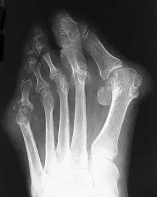
A 47-year-old woman has a painful bunion of the right foot, and shoe wear modifications have failed to provide relief. Examination reveals a severe hallux valgus with dorsal subluxation of the second toe. Radiographs are shown in Figures 14a and 14b. The most appropriate management should include


Explanation

14b The radiographs do not show significant arthrosis of the hallux metatarsophalangeal joint; therefore, arthrodesis is unnecessary. Orthotics will not correct the deformity. A distally based osteotomy will not achieve sufficient correction of the incongruity of deformity, and a Keller resection is not indicated in the younger population. The treatment of choice is a proximal metatarsal osteotomy with second toe correction.

Question 10

What is the most appropriate orthosis for hallux rigidus?


Explanation

A Morton's extension limits excursion of the first metatarsophalangeal joint. It also functions as a ground reaction stabilizer during the toe-off phase of gait and thus reduces torque and joint reaction force at the first metatarsophalangeal joint. The metatarsal arch pad and full-length semi-rigid longitudinal arch support may help by dorsiflexing the first metatarsal relative to the phalanx and thus decompress the first metatarsophalangeal joint. However, they are not as biomechanically effective as the Morton's extension. Both medial hindfoot and lateral forefoot posting are contraindicated because they increase ground reaction at the first metatarsophalangeal joint. Coughlin MJ: Arthritides, in Coughlin MJ, Mann RA (eds): Surgery of the Foot and Ankle, ed 7. St Louis, MO, Mosby, 1999, p 611.

Question 11

While experts disagree whether the postpolio syndrome is caused by a reactivation of the dormant virus or by an attritional aging phenomena of muscles that have been overworked over a period of time, both groups recommend which of the following guidelines for optimizing function in this population?


Explanation

Most leaders in orthopaedic surgery support Jacqueline Perry's theory that the postpolio syndrome is an attritional degenerative process that is the result of overuse of muscles and joints that are unable to adequately tolerate overload, and have little functional reserve. For that reason, aerobic conditioning and exercise are important. Overload and exhaustion of involved muscles should be avoided.

Question 12

Figures 15a through 15c show the radiographs of a 23-year-old football player who was injured when another player fell on his flexed and planted foot. He reports severe pain in the midfoot with a feeling of numbness on the dorsum of the foot, and he is unable to bear weight on the limb. Examination reveals mild swelling. Management should consist of


Explanation

15b 15c Myerson and associates studied the outcomes of 19 patients with tarsometatarsal joint injuries during athletic activity. Injuries were classified as first- or second-degree sprains of the tarsometatarsal joint or a third-degree sprain with diastasis between the metatarsals or cuneiforms. Poor functional results were seen in those with a delay in diagnosis and with inadequate treatment. For patients with third-degree sprains, poor results were obtained with nonsurgical management. These patients required open reduction and internal fixation for optimal return to function. The anatomic reduction is critical to the outcome; therefore, open reduction is preferred. Baxter DE: The Foot and Ankle in Sport, ed 1. St Louis, MO, Mosby, 1995, pp 107-123. Curtis MJ, Myerson M, Szura B: Tarsometatarsal joint injuries in the athlete. Am J Sports Med 1993;21:497-502. Kuo RS, Tejwani NC, DiGiovanni CW, et al: Outcome after open reduction and internal fixation of Lisfranc joint injuries. J Bone Joint Surg Am 2000;82:1609-1618.

Question 13

Which of the following methods best aids in diagnosis of an interdigital neuroma?


Explanation

History and physical examination are still the gold standard for diagnosis of an interdigital neuroma. Ultrasound and MRI may be helpful adjuncts but are dependent on equipment and operator expertise. Web space injection may be helpful for diagnostic and therapeutic purposes. Electromyography and nerve conduction velocity studies are of little benefit for distal lesions. Richardson EG (ed): Orthopaedic Knowledge Update: Foot and Ankle 3. Rosemont, IL, American Academy of Orthopaedic Surgeons, 2004, pp 145-147.

Question 14

A 58-year-old man has had a 3-year history of recurrent ulcerations of the left ankle and instability despite multiple attempts at custom bracing, contact casting, and surgical debridement. He has an ankle-brachial index of 0.76. A clinical photograph and radiographs are shown in Figures 16a through 16c. Treatment should now consist of


Explanation

16b 16c Nonsurgical management has failed to provide relief; therefore, the treatment of choice is arthrodesis with an intramedullary nail. Amputation may be indicated if the arthrodesis fails. The patient does have adequate circulation for an attempt at salvage. Total ankle arthroplasty is not indicated in a neuropathic patient. Pinzur MS, Kelikian A: Charcot ankle fusion with a retrograde locked intramedullary nail. Foot Ankle Int 1997;18:699-704.

Question 15

Figures 17a and 17b show the radiographs of a 32-year-old professional athlete who sustained an injury to the first metatarsal. A view of the opposite noninjured side is shown in Figure 17c. Management of the fracture should consist of


Explanation

17b 17c Parameters for first metatarsal fracture management are different than for shaft fractures of the central second, third, and fourth metatarsals. The first metatarsal carries a greater load and if malunited, can create transfer lesions by virtue of uneven weight distribution; therefore, nonsurgical management is not indicated for this patient. Percutaneous pinning is not as likely to result in an anatomic reduction as open reduction and internal fixation. As his livelihood depends on an expeditious return to function, the choice of open reduction and internal fixation allows for earlier motion and rehabilitation. Richardson EG (ed): Orthopaedic Knowledge Update: Foot and Ankle 3. Rosemont, IL, American Academy of Orthopaedic Surgeons, 2004, pp 64-65.

Question 16

Which of the following are considered appropriate nonsurgical bracing/orthotic options for a supple adult-acquired flatfoot deformity with forefoot abduction, secondary to posterior tibial tendon insufficiency?


Explanation

The initial stages of posterior tibial tendon insufficiency, where the deformity remains supple, may be treated with bracing or an orthotic for pain relief. The Arizona brace was introduced in 1988, and assists in pain relief and deformity correction by minimizing hindfoot valgus alignment, lateral calcaneal displacement, and medial ankle collapse. It is particularly helpful in those patients with advanced disease that cannot tolerate an ankle-foot orthosis. All other choices are incorrect because of the addition of lateral posting, which is not advantageous in valgus deformities. The addition of medial posting to any of the above choices would render them correct alternatives. A heel lift is applicable in Achilles tendon disorders, not posterior tibial tendon disorders. Chao W, Wapner KL, Lee TH, et al: Nonoperative management of posterior tibial tendon dysfunction. Foot Ankle Int 1996;17:736-741.

Question 17

A 28-year-old man underwent open reduction and internal fixation of a closed, displaced, intra-articular calcaneal fracture 8 weeks ago. Examination now reveals that the lateral wound is red and draining purulent material. Cultures obtained from the wound grow out Staphylococcus aureus. Radiographs show early healing of the fracture. What is the next most appropriate step in management?


Explanation

Intravenous antibiotics alone will not adequately treat this infection. At 8 weeks after surgery, the hardware must be removed because Staphylococcus aureus is a virulent microbe. VAC therapy alone is not adequate without debridement and hardware removal, but it may play a role in postoperative wound care. Calcanectomy is a salvage procedure for calcaneal osteomyelitis or recalcitrant heel ulceration. Benirschke SK, Kramer PA: Wound healing complications in closed and open calcaneal fractures. J Orthop Trauma 2004;18:1-6. Lim EV, Leung JP: Complications of intra-articular calcaneal fractures. Clin Orthop 2001;391:7-16.

Question 18

A 37-year-old man with a history of congenital flatfoot reports worsening pain on the medial aspect of his ankle for the past year. The pain is worse with weight bearing and is better with rest and the use of an ankle brace. What findings are shown on the MRI scans shown in Figures 18a through 18c?


Explanation

18b 18c The MRI scans reveal an enlarged posterior tibial tendon, with degenerative signal within the tendon and an excessive amount of fluid in its sheath. This is a type II tear, as noted by Conti and associates, which is the most commonly seen tear. Slovenkai MP: Clinical and radiographic evaluation (Adult flatfoot: Posterior tibial tendon dysfunction). Foot Ankle Clin 1997;2:241-260.

Question 19

A 60-year-old man reports increasing pain in his right foot with limited ankle dorsiflexion and anterior ankle pain after sustaining a fracture of the calcaneus in a fall several years ago. Bracing, nonsteroidal anti-inflammatory drugs, and cortisone injections have failed to provide significant relief. Radiographs are shown in Figures 19a and 19b. What is the next most appropriate step in management?


Explanation

19b Following a calcaneal fracture, the patient has severe subtalar arthritis with loss of talar declination and shortening of the heel; therefore, the treatment of choice is subtalar distraction arthrodesis. Orthotics will not provide significant relief as bracing has failed. Ankle arthrodesis will not be beneficial because the arthritis is in the subtalar joint. Subtalar arthroscopy would only be helpful for a small area of arthrosis, and calcaneal osteotomy would not be beneficial given the extent of the arthritis of the subtalar joint.

Question 20

A 58-year-old woman sustained a ruptured Achilles tendon 1 year ago, and management consisted of an ankle-foot orthosis. She now reports increasing difficulty with ambulation and increasing pain. An MRI scan shows a 6-cm defect in the right Achilles tendon. Management should now consist of


Explanation

With a gap of less than 4 cm, a V-Y repair would be appropriate without a tendon transfer. For gaps greater than 5 cm, a lengthening with augmentation is the most appropriate treatment. Therefore, the treatment of choice is an Achilles tendon turndown with flexor hallucis longus tendon transfer. The plantaris tendon is not a strong enough repair, and direct repair is not possible given the large defect in the Achilles tendon. Continued use of the ankle-foot orthosis will not provide adequate relief for this patient.

Question 21

A 29-year-old woman reports dysesthesias and burning after undergoing bunion surgery that consisted of a proximal crescentic first metatarsal osteotomy 6 months ago. Examination reveals a positive Tinel's sign at the proximal aspect of the healed incision. What injured nerve is responsible for her continued symptoms?


Explanation

Painful incisional neuromas after bunion surgery frequently involve the dorsomedial cutaneous branch of the superficial peroneal nerve. This is the medial branch of the superficial peroneal nerve that terminates as the dorsomedial cutaneous nerve to the hallux. Branches of the deep peroneal nerve to this area are rare, and no branches to this area exist from the sural nerve. The saphenous nerve branches are generally more proximal, and the medial plantar nerve lies plantarly. Kenzora JE: Sensory nerve neuromas: Leading to failed foot surgery. Foot Ankle 1986;7:110-117.

Question 22

Figure 20 shows the clinical photograph of a man who has had diabetes mellitus controlled with oral medication for the past 10 years. He wears soft-soled shoes and only uses leather-soled shoes for important business meetings. Examination reveals palpable dorsalis pedis and posterior tibial pulses, although they are somewhat diminished. He is insensate to pressure with the Semmes-Weinstein 5.07 monofilament. The ulcer heals after treatment with a full contact cast. What is the best course of action at this time?


Explanation

The patient has not undergone a trial of foot-specific patient education and accommodative/therapeutic shoe wear. He must use therapeutic shoe wear at all times, as even the occasional use of pressure-concentrating shoe wear has a high likelihood of leading to the development of a diabetic foot ulcer. Pinzur MS, Kernan-Schroeder D, Emmanuele NV, et al: Development of a nurse-provided health system strategy for diabetic foot care. Foot Ank Int 2001;22:744-746. Pinzur MS, Shields N, Goelitz B, et al: American Orthopaedic Foot & Ankle Society shoe survey of diabetic patients. Foot & Ankle Int 1999;20:703-707.

Question 23

Figures 21a and 21b show the clinical photograph and radiograph of a 15-year-old girl who has a deformity of her feet. Her parents are concerned because there is a family history of Charcot-Marie-Tooth disease. The patient reports some mild instability of the ankle and has noticed mild early callosities; however, she is not having any significant pain. Coleman block testing reveals a forefoot valgus and supple hindfoot. She has weakness to eversion and dorsiflexion. Initial management should consist of


Explanation

21b Initial management of a young patient with a cavovarus deformity of the foot and a family history of Charcot-Marie-Tooth disease should focus on mobilization and strengthening of the weakening muscular units and an accommodative insert. Surgical intervention should be delayed until progression of the deformity begins to cause symptoms and/or weakness of the muscular units, resulting in contractures of the antagonistic muscle units. Pinzur MS: Charcot's foot. Foot Ankle Clin 2000;5:897-912. Holmes JR, Hansen ST Jr: Foot and ankle manifestations of Charcot-Marie-Tooth disease. Foot Ankle 1993;14:476-486.

Question 24

A 50-year-old woman reports a burning sensation on the plantar aspect of her left forefoot, distal to the metatarsal heads between her third and fourth digits. Palpation of the third web space recreates her symptoms. Which of the following will most accurately aid in confirming a diagnosis?


Explanation

The diagnosis of an interdigital neuroma is best made by a thorough history and careful physical examination. Radiographs are helpful in excluding other pathologic processes such as a metatarsal stress fracture. MRI and ultrasound have both been reported to aid in the diagnosis of an interdigital neuroma. Richardson EG (ed): Orthopaedic Knowledge Update: Foot and Ankle 3. Rosemont, IL, American Academy of Orthopaedic Surgeons, 2003, pp 145-151.

Question 25

A 21-year-old collegiate track athlete increased her training 4 months ago in anticipation of starting the season. Two months into her training program, she reported pain followed by a 1-month history of diffuse pain in the first metatarsophalangeal joint that was aggravated by weight bearing. A removable walker boot partially relieved the pain, and she was able to complete the season. Her pain has now returned; however, she denies any history of injury. Examination reveals tenderness over the medial sesamoid but no deformities. A radiograph and bone scan are shown in Figures 22a and 22b. What is the best treatment option at this time?


Explanation

22b The radiograph reveals either a fractured or bipartite sesamoid. The bone scan shows asymmetrically increased uptake over the medial sesamoid. Given the history and physical examination, a stress fracture is the most likely diagnosis. Medial sesamoidectomy reliably improves pain, and athletes return to sports on an average of 7 weeks after excision. Immobilization typically requires more than 4 to 8 weeks and is not always successful; however, it would be appropriate management for a patient who is not an elite athlete. Sanders R: Fractures of the midfoot and forefoot, in Coughlin MJ, Mann RA (eds): Surgery of the Foot and Ankle, ed 7. St Louis, MO, Mosby, 1999, pp 1601-1603.

Question 26

A 24-year-old male presents with bilateral cavovarus foot deformity and reports frequent ankle sprains. Examination shows a plantarflected first ray and weakness in certain muscle groups. In the pathogenesis of a cavovarus foot deformity in Charcot-Marie-Tooth disease, which muscle typically retains its strength and drives the initial plantar flexion of the first ray?


Explanation

In Charcot-Marie-Tooth (CMT) disease, there is a characteristic pattern of muscle weakness. The tibialis anterior and peroneus brevis typically weaken early. The peroneus longus and tibialis posterior maintain their strength longer. The relatively strong peroneus longus unopposed by the weak tibialis anterior leads to plantarflexion of the first ray, causing a forefoot-driven cavovarus deformity.

Question 27

A 25-year-old athlete sustains a hyperplantarflexion injury to his midfoot. Weight-bearing radiographs show a 3 mm diastasis between the base of the first and second metatarsals. He is diagnosed with a purely ligamentous Lisfranc injury. Which of the following surgical interventions has been shown to result in better functional outcomes and lower rates of hardware removal for a purely ligamentous Lisfranc injury?


Explanation

Studies (such as those by Ly and Coetzee) have shown that for purely ligamentous Lisfranc injuries, primary arthrodesis of the medial column (1st, 2nd, and 3rd TMT joints) results in superior functional outcomes, less need for subsequent hardware removal, and a lower rate of secondary procedures compared to ORIF.

Question 28

A 65-year-old patient with end-stage post-traumatic ankle osteoarthritis undergoes a total ankle arthroplasty using the standard anterior approach. During the superficial dissection and placement of retractors, which of the following nerves is at greatest risk of iatrogenic injury?


Explanation

The standard anterior approach to the ankle utilizes an internervous plane between the superficial peroneal nerve (SPN) and the deep peroneal nerve. The SPN, particularly its medial dorsal cutaneous branch, crosses the operative field from lateral to medial and is at high risk of injury during superficial dissection and anterior retraction.

Question 29

A 21-year-old collegiate track athlete presents with insidious onset of vague midfoot pain. Plain radiographs are normal, but an MRI confirms a navicular stress fracture extending through the dorsal cortical margin without complete displacement or fragmentation. What is the most appropriate initial management for this patient?


Explanation

For uncomplicated, non-displaced navicular stress fractures, the gold standard for initial conservative management is strict non-weight-bearing in a short leg cast for 6 to 8 weeks. Weight-bearing protocols have an unacceptably high rate of delayed union or nonunion due to the precarious blood supply to the central third of the navicular.

Question 30

A 28-year-old professional soccer player sustains an acute, non-displaced fracture at the metaphyseal-diaphyseal junction of the fifth metatarsal during a game. He wishes to return to play as quickly and safely as possible. What is the most appropriate definitive management for this patient to minimize the risk of nonunion and allow early return to sport?


Explanation

Acute fractures of the fifth metatarsal metaphyseal-diaphyseal junction (Jones fractures) in high-level or professional athletes are typically treated with intramedullary screw fixation. This provides a significantly faster time to union and return to sport, and lowers the risk of nonunion compared to conservative management.

Question 31

A 55-year-old female presents with severe pain and stiffness in her first metatarsophalangeal (MTP) joint. Radiographs demonstrate marked dorsal osteophytes, total loss of joint space, and subchondral sclerosis consistent with Coughlin and Shurnas Grade 3 hallux rigidus. Conservative measures have failed. Which of the following surgical procedures is considered the gold standard for long-term pain relief and functional improvement?


Explanation

For advanced hallux rigidus (Coughlin and Shurnas Grade 3 or 4), arthrodesis of the first MTP joint remains the gold standard. It provides excellent long-term pain relief, functional stability, and high patient satisfaction rates. Cheilectomy is generally reserved for Grades 1 and 2 where the joint space is relatively preserved.

Question 32

A 45-year-old avid runner presents with posterior heel pain. MRI shows insertional Achilles tendinosis with retrocalcaneal bursitis and a Haglund's deformity. She has failed 6 months of nonoperative management. If surgical intervention is planned, what percentage of the Achilles tendon insertion can typically be detached without requiring augmentation?


Explanation

During surgical debridement of insertional Achilles tendinopathy and excision of a Haglund's deformity, it is often necessary to detach a portion of the Achilles tendon. Biomechanical studies have shown that up to 50% of the tendon's insertion can be detached without a significant risk of avulsion or need for primary augmentation. Detachment greater than 50% generally warrants suture anchor repair or FHL transfer.

Question 33

A 23-year-old football player presents with an acute rotational ankle injury. A syndesmotic sprain is suspected. On a standard AP radiograph of the ankle, what is the normal threshold for the tibiofibular clear space, measured 1 cm proximal to the tibial plafond?


Explanation

The tibiofibular clear space is the distance between the lateral border of the posterior tibial malleolus and the medial border of the fibula, measured 1 cm proximal to the joint line. A normal value is less than 6 mm on both AP and mortise views. Values greater than 6 mm are highly suggestive of syndesmotic injury.

Question 34

A 30-year-old male is involved in a high-speed motor vehicle collision and sustains a talar neck fracture with subluxation of the subtalar joint and complete dislocation of the tibiotalar joint. The talonavicular joint remains anatomically reduced. According to the Hawkins classification, what type of fracture is this, and what is the approximate risk of avascular necrosis (AVN) of the talar body?


Explanation

The Hawkins classification describes talar neck fractures: Type I is non-displaced (0-10% AVN risk). Type II is a talar neck fracture with subtalar dislocation or subluxation (20-50% AVN risk). Type III involves subtalar and tibiotalar dislocation (>75% AVN risk, sometimes quoted as 75-90% in modern series). Type IV involves subtalar, tibiotalar, and talonavicular dislocation.

Question 35

A 42-year-old construction worker falls from a ladder, sustaining a joint-depressed, intra-articular calcaneus fracture. During an extensile lateral approach for open reduction and internal fixation (ORIF), the surgeon must carefully plan the incision to avoid a nerve that crosses the lateral hindfoot. Which of the following structures is most at risk during flap elevation?


Explanation

The extensile lateral approach for calcaneus ORIF involves creating a full-thickness subperiosteal flap. The sural nerve crosses the lateral aspect of the hindfoot and is at significant risk of transection or stretch injury during the incision and retraction. The vertical limb is placed just anterior to the Achilles tendon and the horizontal limb in the transition zone between the plantar and lateral skin to safely mobilize the sural nerve within the flap.

Question 36

A 24-year-old male presents with a long-standing history of frequent ankle sprains and progressive foot deformity. Clinical examination reveals a cavovarus foot posture with a positive "peek-a-boo" heel sign. During the Coleman block test, the hindfoot varus corrects completely to neutral. Based on this physical examination finding, which of the following surgical interventions is most appropriate?


Explanation

The Coleman block test is utilized to evaluate hindfoot flexibility in a cavovarus foot deformity. If the hindfoot varus corrects when the first ray is dropped off a block (neutralizing the plantarflexed first ray), the deformity is forefoot-driven, and the hindfoot is flexible. Therefore, joint-sparing, forefoot-correcting procedures (e.g., 1st metatarsal dorsiflexion osteotomy) and tendon transfers (peroneus longus to peroneus brevis to decrease plantarflexion force on the 1st ray) are appropriate. If the hindfoot does not correct, the deformity is rigid, necessitating a calcaneal osteotomy or arthrodesis.

Question 37

A 45-year-old female undergoes a modified Lapidus procedure (first tarsometatarsal arthrodesis) for a severe hallux valgus deformity. Three months postoperatively, she returns complaining of a new, severe plantar foot pain directly beneath the second metatarsal head. Which of the following technical errors during the index procedure is the most likely cause of her new symptom?


Explanation

Transfer metatarsalgia following a Lapidus procedure is most commonly caused by dorsal elevation (malunion) or excessive shortening of the first metatarsal. Dorsal elevation unloads the first ray during the stance phase of gait, transferring disproportionate weight-bearing stress to the lesser metatarsals, typically the second metatarsal head. Excessive plantarflexion would instead lead to primary sesamoiditis or an intractable plantar keratosis under the first metatarsal head.

Question 38

A 32-year-old male sustains a closed talar neck fracture following a motor vehicle collision. Radiographs demonstrate a displaced fracture of the talar neck with posterior displacement of the talar body, which is extruded from both the subtalar and tibiotalar joints. The talonavicular joint remains reduced. Which of the following vessels provides the primary blood supply to the talar body and is at highest risk of catastrophic disruption in this specific fracture pattern?


Explanation

This patient has a Hawkins Type III talar neck fracture (subtalar and tibiotalar dislocation). The primary blood supply to the talar body is the artery of the tarsal canal, which is a branch of the posterior tibial artery. While the artery of the tarsal sinus supplies the head and neck, and deltoid branches supply the medial body, the artery of the tarsal canal is the most critical source for the body. A Hawkins III fracture disrupts nearly all vascular sources, most importantly the artery of the tarsal canal, leading to an avascular necrosis (AVN) rate traditionally reported to approach 75-100%.

Question 39

A 55-year-old female presents with medial ankle pain and a progressive flatfoot deformity. Examination demonstrates a flexible hindfoot valgus and inability to perform a single-leg heel raise. Weight-bearing radiographs reveal greater than 40% talonavicular uncoverage and significant forefoot abduction. In addition to a flexor digitorum longus (FDL) to navicular transfer and medializing calcaneal osteotomy, which of the following procedures is indicated to adequately correct her deformity?


Explanation

The patient presents with Stage IIb adult-acquired flatfoot deformity (flexible deformity with significant forefoot abduction characterized by >40% talonavicular uncoverage). Surgical reconstruction for Stage IIb requires restoring the medial column (FDL transfer) and correcting the hindfoot/midfoot deformity. A medializing calcaneal osteotomy corrects hindfoot valgus, but a lateral column lengthening (Evans calcaneal osteotomy or calcaneocuboid distraction arthrodesis) is specifically indicated to correct the substantial forefoot abduction. A triple arthrodesis is a joint-sacrificing procedure generally reserved for Stage III (rigid) deformities.

Question 40

A 22-year-old collegiate football player sustains an axial loading injury to his plantarflexed foot. Weight-bearing radiographs demonstrate subtle widening of the interval between the first and second metatarsal bases, and a "fleck sign" is noted in this space. The ligament represented by this bony avulsion normally originates from which of the following bony structures?


Explanation

The "fleck sign" is highly specific for a Lisfranc injury and represents a bony avulsion of the Lisfranc ligament. The Lisfranc ligament is an oblique, stout ligament that originates from the lateral aspect of the medial cuneiform and inserts onto the medial base of the second metatarsal. It acts as the primary stabilizer between the first and second rays, given the absence of a direct transverse intermetatarsal ligament between the bases of the first and second metatarsals.

Question 41

A 60-year-old male with long-standing, poorly controlled type 2 diabetes presents with a red, hot, swollen right foot. He denies any recent trauma, fever, or open ulcerations. Laboratory studies reveal a normal white blood cell count and a mildly elevated CRP. Radiographs show extensive periarticular fragmentation, subluxation of the tarsometatarsal joints, and bony debris, without definitive signs of osteomyelitis. What is the most appropriate initial management for this condition?


Explanation

The patient is presenting with acute Charcot neuroarthropathy, specifically Eichenholtz Stage I (Developmental/Fragmentation phase), which is characterized by a red, hot, swollen foot and radiographically by bone fragmentation, joint subluxation, and debris. In the absence of an open ulcer, this is an inflammatory rather than an infectious process. The gold standard for initial treatment is immobilization with a total contact cast (TCC) and strict non-weight-bearing to arrest the acute inflammatory phase, prevent further progressive deformity, and allow progression to the coalescence phase.

Question 42

A 40-year-old roofer falls from a ladder and sustains a displaced intra-articular Sanders Type IIB calcaneus fracture. He is indicated for open reduction and internal fixation via an extensile lateral approach. Which of the following neurovascular structures is at the highest risk of iatrogenic injury during the creation of the full-thickness soft tissue flap, particularly at the superior and anterior aspect of the vertical limb?


Explanation

The extensile lateral approach to the calcaneus requires elevating a full-thickness subperiosteal "no-touch" flap to minimize the risk of wound necrosis. The sural nerve courses posterior and inferior to the lateral malleolus and provides sensation to the lateral border of the foot. It is highly susceptible to injury or retraction neuropraxia during the vertical limb and corner creation of the extensile lateral incision. The primary vascular supply to this flap is the lateral calcaneal artery.

Question 43

A 38-year-old recreational athlete sustains an acute, closed mid-substance Achilles tendon rupture. After discussing treatment options with his orthopedic surgeon, he elects for non-operative management utilizing an early functional rehabilitation protocol. Compared to open surgical repair, which of the following clinical outcomes is most strongly supported by current Level I evidence for this patient?


Explanation

Multiple Level I studies, including the landmark randomized controlled trial by Willits et al., have demonstrated that when an early functional rehabilitation protocol (weight-bearing and early ROM in a controlled brace) is strictly utilized for acute Achilles tendon ruptures, the rerupture rates between non-operative and operative management are not statistically different. However, non-operative management avoids the inherent surgical risks, thereby demonstrating a significantly decreased rate of soft-tissue complications, infections, and sural nerve injuries.

Question 44

A 26-year-old female presents with an external rotation injury to her right ankle. Weight-bearing radiographs show no fracture, but there is an isolated widening of the medial clear space to 6 mm. An MRI confirms an acute syndesmotic rupture. On a standard radiographic ankle series, which of the following parameters is considered the most reliable indicator of a normal distal tibiofibular syndesmosis?


Explanation

The tibiofibular clear space is the most reliable radiographic parameter for evaluating the syndesmosis because it is the least affected by the rotational position of the foot during the radiograph. It is measured 1 cm proximal to the tibial plafond and should be less than 6 mm on both the AP and mortise views. Tibiofibular overlap is highly dependent on rotation and thus less reliable. While medial clear space widening indicates deltoid insufficiency and lateral talar shift (often seen in syndesmotic injuries), the tibiofibular clear space is the direct measure of syndesmotic integrity.

Question 45

A 19-year-old elite collegiate basketball player presents with acute lateral foot pain after landing awkwardly. Radiographs reveal a transverse fracture of the proximal fifth metatarsal at the metaphyseal-diaphyseal junction, extending into the fourth-fifth intermetatarsal articulation. Which of the following is the most appropriate management for this patient to minimize the risk of nonunion and expedite his return to competitive play?


Explanation

The patient has sustained a Zone II fracture of the proximal fifth metatarsal, commonly referred to as a Jones fracture. This region represents a vascular watershed area, placing these fractures at a substantially higher risk for delayed union and nonunion compared to Zone I (tuberosity avulsion) fractures. In high-demand or elite athletes, early surgical intervention with percutaneous intramedullary screw fixation is the standard of care to decrease nonunion rates and allow for a more reliable, accelerated return to sport.

Question 46

A 32-year-old male sustains a high-energy motor vehicle accident resulting in a displaced talar neck fracture. Radiographs demonstrate a talar neck fracture with both subtalar and tibiotalar joint dislocation. Based on the Hawkins classification, what is his injury grade and the approximate associated risk of avascular necrosis (AVN) of the talar body?


Explanation

This is a Hawkins Type III fracture, which is characterized by a displaced talar neck fracture with dislocation of both the subtalar and tibiotalar joints. The blood supply to the talar body is disrupted from the artery of the tarsal canal, artery of the sinus tarsi, and the superior neck vessels. The risk of AVN for a Type III fracture is historically 80-100%, though some modern series show slightly lower rates. Hawkins II involves subtalar subluxation/dislocation only (20-50% AVN risk).

Question 47

A 55-year-old female presents with medial foot pain and a progressive flatfoot deformity. Examination shows a flexible hindfoot valgus and inability to perform a single-leg heel raise. Weight-bearing radiographs demonstrate >40% uncovering of the talonavicular joint. If conservative management fails, which of the following surgical interventions is most appropriate?


Explanation

The patient has Stage IIb adult acquired flatfoot deformity (posterior tibial tendon dysfunction), characterized by a flexible hindfoot valgus and forefoot abduction (>40% talonavicular uncoverage). Surgical correction requires addressing both the medial column weakness and the biomechanical deformity. An FDL transfer addresses the tendon deficiency, an MDCO corrects the hindfoot valgus, and a lateral column lengthening corrects the severe forefoot abduction.

Question 48

A 45-year-old female presents with a painful bunion. Weight-bearing radiographs reveal a Hallux Valgus Angle (HVA) of 45 degrees, an Intermetatarsal Angle (IMA) of 18 degrees, and clinical hypermobility of the first tarsometatarsal (TMT) joint. Which of the following surgical procedures is most biomechanically appropriate to correct her deformity?


Explanation

A severe hallux valgus deformity (IMA >15 degrees, HVA >40 degrees) in the presence of first TMT joint hypermobility is classically treated with a Lapidus procedure (first TMT arthrodesis). This provides powerful correction of the intermetatarsal angle and stabilizes the medial column, preventing recurrence that is common if hypermobility is ignored.

Question 49

A 40-year-old roofer falls from a ladder, sustaining a Sanders type III calcaneus fracture. An open reduction and internal fixation via an extensile lateral approach is planned. To minimize the risk of the most common postoperative wound complication associated with this approach, how should the surgical flap be managed?


Explanation

The extensile lateral approach to the calcaneus carries a significant risk of wound edge necrosis and infection. To minimize this, a full-thickness subperiosteal flap must be developed. Retractors (such as K-wires inserted into the talus) should be used to hold the flap (the 'no-touch' technique), preserving the delicate vascular supply from the lateral calcaneal artery.

Question 50

A 25-year-old professional athlete sustains a purely ligamentous Lisfranc injury with 3 mm of displacement between the medial cuneiform and second metatarsal base. Based on recent prospective studies evaluating primarily ligamentous Lisfranc injuries, primary arthrodesis compared to open reduction and internal fixation (ORIF) is associated with which of the following?


Explanation

Prospective studies (such as Coetzee and Ly) comparing ORIF to primary arthrodesis for purely ligamentous Lisfranc injuries have demonstrated that primary arthrodesis leads to comparable or superior functional outcomes in the short term, but significantly less functional deterioration over time, and lower rates of secondary surgeries (due to lack of hardware irritation/removal or post-traumatic arthritis). The 4th and 5th TMT joints should almost never be primarily fused, as they are essential for mobile foot adaptation.

Question 51

A 58-year-old male with poorly controlled type II diabetes presents with a swollen, erythematous, and warm right foot. He denies any trauma. Radiographs reveal periarticular fragmentation, subluxation of the tarsometatarsal joints, and osseous debris. According to the Eichenholtz classification, what stage does this represent, and what is the standard initial treatment?


Explanation

This clinical and radiographic picture characterizes Eichenholtz Stage 1 (Developmental/Fragmentation) Charcot arthropathy. It is marked by joint edema, warmth, periarticular fragmentation, debris, and subluxation/dislocation. The standard of care in the acute active phase is immobilization (Total Contact Cast) and strict non-weight bearing to prevent further deformity until the active phase transitions to coalescence (Stage 2).

Question 52

A 22-year-old running back sustains an acute hyperextension injury to his great toe. MRI demonstrates a complete tear of the plantar plate with proximal retraction of the sesamoids. Which of the following is an absolute indication for operative repair in this type of injury?


Explanation

Turf toe is a sprain/tear of the first MTP plantar plate complex. Operative indications for turf toe include: a large intra-articular sesamoid fracture, retraction of the sesamoids >3 mm, traumatic bunion deformity, a purely unstable joint (gross instability), or failure of conservative management. Proximal migration of the sesamoids with gross first MTP instability indicates a complete (Grade 3) rupture requiring surgical repair.

Question 53

A 20-year-old collegiate basketball player complains of lateral foot pain for 3 months. Radiographs demonstrate a radiolucent line with cortical hypertrophy distal to the fourth-fifth intermetatarsal articulation in the fifth metatarsal. What is the most appropriate management to ensure the fastest return to play with the lowest risk of nonunion?


Explanation

The clinical scenario and radiographic findings describe a Zone 3 proximal diaphyseal stress fracture of the fifth metatarsal. Because these fractures occur in a vascular watershed area and have a high risk of delayed union or nonunion (especially with cortical hypertrophy indicating chronicity), the gold standard treatment in an elite athlete to allow for rapid return to play is intramedullary screw fixation.

Question 54

During an open reduction and internal fixation of a bimalleolar equivalent ankle fracture, the surgeon performs an intraoperative Cotton test to evaluate the syndesmosis. Which fluoroscopic measurement objectively indicates syndesmotic instability requiring operative fixation?


Explanation

The Cotton test involves applying a lateral force to the fibula using a bone hook. Widening of the medial clear space > 4 mm (or asymmetry compared to the superior clear space) on a mortise view indicates deep deltoid and syndesmotic disruption. Normal tibiofibular clear space is < 6 mm on both AP and mortise views, and normal overlap is > 1 mm on the mortise view.

Question 55

A 42-year-old recreational runner presents with 6 months of posterior ankle pain. Physical exam reveals a palpable, tender nodule 4 cm proximal to the calcaneal insertion of the Achilles tendon. MRI shows fusiform thickening and mucoid degeneration involving >50% of the tendon substance. Conservative treatment has failed. During surgical debridement of the tendinosis, if more than 50% of the tendon is debrided, what is the most appropriate adjunctive procedure to preserve plantarflexion strength?


Explanation

For severe non-insertional Achilles tendinopathy where more than 50% of the tendon is compromised and requires debridement, primary repair alone is biomechanically insufficient. Augmentation is indicated. The flexor hallucis longus (FHL) tendon transfer is the procedure of choice due to its strong plantarflexion force, line of pull (in phase with the Achilles), and anatomic proximity.

Question 56

A 30-year-old male sustains a high-energy motor vehicle collision resulting in a closed injury to his foot and ankle. Radiographs reveal a displaced fracture of the talar neck with subluxation of the subtalar joint, while the tibiotalar joint remains completely congruent. According to the Hawkins classification, what is the injury type and the approximate associated risk of avascular necrosis (AVN) of the talar body?


Explanation

This is a Hawkins Type II fracture, defined as a vertical fracture of the talar neck with subluxation or dislocation of the subtalar joint, but with an intact ankle (tibiotalar) joint. The risk of avascular necrosis (AVN) for a Type II injury is historically cited as 20% to 50%. Hawkins Type I is non-displaced (0-10% AVN risk), Type III involves both subtalar and tibiotalar dislocation (high AVN risk, >80%), and Type IV adds talonavicular subluxation/dislocation.

Question 57

A 55-year-old female presents with progressive, painful flatfoot deformity. Examination reveals a flexible hindfoot valgus and an inability to perform a single-limb heel rise. When viewing the foot from behind, 'too many toes' are visible. Radiographs demonstrate an uncoverage of the talar head of 45%. Which of the following surgical strategies is most appropriate for addressing this specific stage of adult acquired flatfoot deformity?


Explanation

This patient has Stage IIb adult-acquired flatfoot deformity (posterior tibial tendon dysfunction). Stage IIb is differentiated from Stage IIa by the presence of significant forefoot abduction (typically >30% talonavicular uncoverage). While Stage IIa can be treated with a medial displacement calcaneal osteotomy (MDCO) and FDL transfer, Stage IIb requires the addition of a lateral column lengthening (e.g., Evans osteotomy) to adequately correct the transverse plane deformity (forefoot abduction).

Question 58

A 24-year-old male with a history of Charcot-Marie-Tooth disease presents with a bilateral symptomatic cavovarus foot deformity. During the Coleman block test, the patient's lateral foot and heel are placed on a 1-inch wooden block while the first metatarsal is allowed to hang freely in plantarflexion. During this test, the hindfoot varus completely corrects to neutral. What does this clinical finding indicate regarding the primary driver of the deformity?


Explanation

The Coleman block test is essential in evaluating a cavovarus foot. If allowing the first ray to plantarflex off the block corrects the hindfoot varus, it demonstrates that the hindfoot varus is flexible and primarily driven by the rigid, plantarflexed first ray hitting the ground early and forcing the hindfoot into varus. Surgical correction in this case must address the forefoot (e.g., dorsiflexion osteotomy of the 1st metatarsal) alongside any necessary soft tissue balancing.

Question 59

A 22-year-old collegiate football player sustains a hyperplantarflexion injury to his midfoot. Weight-bearing radiographs show a 3 mm diastasis between the base of the first and second metatarsals. CT scan confirms a pure ligamentous Lisfranc injury with no associated osseous fractures. To optimize functional outcomes and minimize the risk of hardware failure or post-traumatic arthritis, which of the following is the most evidence-based surgical treatment?


Explanation

Multiple studies, including the landmark prospective randomized trial by Ly and Coetzee, have demonstrated that primary arthrodesis of the medial column (first, second, and third tarsometatarsal joints) provides superior functional outcomes and lower reoperation rates compared to ORIF in cases of purely ligamentous Lisfranc injuries. ORIF has a higher rate of hardware failure, loss of reduction, and subsequent post-traumatic arthritis in purely ligamentous variants.

Question 60

A 45-year-old female presents with a painful bunion. Clinical examination demonstrates significant hypermobility of the first tarsometatarsal (TMT) joint in the sagittal plane. Weight-bearing radiographs reveal a hallux valgus angle (HVA) of 42 degrees and an intermetatarsal angle (IMA) of 16 degrees. Which of the following surgical options is most appropriate?


Explanation

The Lapidus procedure (first TMT arthrodesis) is indicated for patients with moderate to severe hallux valgus (IMA > 13 degrees) who have concurrent hypermobility of the first ray. It provides excellent correction of the intermetatarsal angle and stabilizes the medial column, preventing recurrence that is often seen if only a distal or proximal osteotomy is utilized in the presence of TMT hypermobility.

Question 61

A 21-year-old elite collegiate basketball player sustains an acute, non-displaced fracture at the metaphyseal-diaphyseal junction of the fifth metatarsal (Zone 2). To facilitate the fastest safe return to play and minimize the risk of delayed union or nonunion, what is the treatment of choice?


Explanation

Zone 2 fractures (Jones fractures) involve the metaphyseal-diaphyseal junction and are prone to nonunion due to a vascular watershed area. In elite or high-level athletes, early operative intervention with intramedullary screw fixation is recommended. It significantly decreases the time to clinical and radiographic union and lowers the rate of nonunion compared to non-operative management, allowing for an accelerated return to sport.

Question 62

A 58-year-old male with poorly controlled type 2 diabetes and peripheral neuropathy presents with a red, hot, and swollen left foot. He denies any trauma. The skin is intact with no ulceration. Radiographs show osteopenia and early fragmentation of the navicular. To differentiate between acute Charcot neuroarthropathy and osteomyelitis, which of the following nuclear medicine imaging studies is considered the most specific?


Explanation

The combination of an Indium-111 WBC scan and a Technetium-99m sulfur colloid bone marrow scan is highly specific for differentiating osteomyelitis from Charcot neuroarthropathy. An isolated WBC scan can have false positives in Charcot due to normal marrow remodeling taking up WBCs. The sulfur colloid targets bone marrow; therefore, discordant uptake (WBC uptake without corresponding marrow uptake) confirms infection (osteomyelitis), whereas concordant uptake suggests sterile Charcot remodeling.

Question 63

During open reduction and internal fixation of a pronation-external rotation (PER) ankle fracture, a syndesmotic diastasis is confirmed using the intraoperative hook test. When placing a trans-syndesmotic positional screw, which of the following represents the most accurate anatomic and biomechanical principle?


Explanation

Because the fibula is positioned posterior to the tibia at the level of the syndesmosis, the optimal trajectory for a trans-syndesmotic screw is 20 to 30 degrees from posterolateral to anteromedial to anatomically capture the tibia. Research (e.g., Tornetta et al.) has shown that the position of the ankle (dorsiflexion vs. plantarflexion) during screw placement does not significantly affect the final mortise width or postoperative range of motion. Typically, screws are placed 2-3 cm above the joint line.

Question 64

A 35-year-old male presents with an isolated, complete, and irreversible common peroneal nerve palsy following a traumatic knee dislocation 2 years ago. He has a flexible hindfoot and a passively correctable equinus contracture. Which tendon transfer is considered the gold standard to restore active dorsiflexion and prevent foot drop in this patient?


Explanation

The tibialis posterior tendon is the classic and most reliable transfer for an isolated common peroneal nerve palsy (foot drop). It is usually detached from its insertion, routed through the interosseous membrane, and secured to the dorsum of the foot (often the lateral or middle cuneiform) to restore active dorsiflexion. A concomitant Achilles lengthening is often required if an equinus contracture is present, but the primary motor substitution is the posterior tibial tendon.

Question 65

A 62-year-old male with end-stage post-traumatic ankle osteoarthritis is being evaluated in the clinic to discuss surgical options, including total ankle arthroplasty (TAA) versus ankle arthrodesis. Which of the following is widely considered an absolute contraindication to Total Ankle Arthroplasty (TAA)?


Explanation

Severe avascular necrosis (AVN) of the talar body (typically defined as >50% involvement) is an absolute contraindication for Total Ankle Arthroplasty due to the lack of viable bone stock needed to support the talar component, leading to early subsidence and catastrophic failure. Advanced age is not a contraindication (TAA is often preferred in older, lower-demand patients). Subtalar arthritis is actually an indication for TAA over fusion to preserve remaining hindfoot kinematics. Mild coronal deformity and a BMI of 32 are relative considerations, not absolute contraindications.

Question 66

A 55-year-old female presents with stage IIB posterior tibial tendon dysfunction. During her surgical reconstruction, a lateral column lengthening is performed in addition to a medializing calcaneal osteotomy and flexor digitorum longus (FDL) transfer. What is the primary biomechanical purpose of the lateral column lengthening in this setting?


Explanation

Lateral column lengthening (such as an Evans osteotomy) is primarily used in Stage IIB adult-acquired flatfoot deformity to correct forefoot abduction, which occurs due to uncoupling of the transverse tarsal joint (talonavicular joint uncoverage). While it indirectly restores the medial arch, its primary and direct effect is correcting the abduction deformity.

Question 67

A 32-year-old male undergoes anterior ankle arthroscopy for an osteochondral lesion of the talus. During the establishment of the anterolateral portal, a nerve is inadvertently injured. Which of the following functional deficits is most likely to result from this specific injury?


Explanation

The anterolateral portal places the superficial peroneal nerve at risk. Injury to this nerve leads to sensory loss or paresthesias over the dorsum of the foot, sparing the first web space (which is innervated by the deep peroneal nerve). The anteromedial portal places the saphenous nerve at risk.

Question 68

A 58-year-old patient with poorly controlled type 2 diabetes presents with a swollen, erythematous, and warm right foot without any open ulcers. Radiographs show periarticular osteopenia and early subluxation of the tarsometatarsal joints. Which of the following MRI findings best differentiates acute Charcot arthropathy from osteomyelitis in this patient?


Explanation

In acute Charcot arthropathy, MRI typically reveals bone marrow edema that is periarticular and subchondral, involving multiple adjacent bones around a joint (e.g., the midfoot). Conversely, osteomyelitis usually arises contiguous to a soft tissue ulcer, presenting with diffuse marrow edema that is not strictly periarticular, often associated with cortical destruction, sinus tracts, or abscesses.

Question 69

A 40-year-old male undergoes open reduction and internal fixation of a displaced intra-articular calcaneus fracture via an extensile lateral approach. At his 6-week follow-up, he is noted to have significant clawing of the lesser toes. What is the most likely cause of this complication?


Explanation

Clawing of the lesser toes after a calcaneus fracture is the hallmark late sequela of an unrecognized foot compartment syndrome. Ischemia and subsequent fibrotic contracture of the intrinsic muscles of the foot (specifically the interossei and lumbricals, as well as quadratus plantae) lead to an imbalance, causing the claw toe deformity.

Question 70

A 28-year-old motorcyclist sustains a Hawkins type III talar neck fracture. Which of the following arteries provides the majority of the blood supply to the talar body, placing it at the highest risk for avascular necrosis (AVN) in this injury pattern?


Explanation

The artery of the tarsal canal, a branch of the posterior tibial artery, provides the majority of the blood supply to the talar body. In a Hawkins type III fracture (displaced talar neck fracture with subluxation/dislocation of both the subtalar and ankle joints), the blood supply from the artery of the tarsal canal, artery of the tarsal sinus, and the capsular vessels is severely disrupted, leading to an AVN risk nearing 100%.

Question 71

A 45-year-old female presents with symptomatic hallux valgus that has failed nonoperative management. Weight-bearing radiographs demonstrate an intermetatarsal angle (IMA) of 18 degrees and a hallux valgus angle (HVA) of 42 degrees. There is no evidence of first tarsometatarsal hypermobility or osteoarthritis. Which of the following surgical options is most appropriate?


Explanation

A severe hallux valgus deformity (IMA > 13-15 degrees, HVA > 40 degrees) requires a proximal metatarsal osteotomy (e.g., proximal crescentic or Ludloff) or a Lapidus procedure, combined with a distal soft tissue release to achieve adequate correction. A distal chevron osteotomy cannot provide sufficient translation for large IMA corrections. First MTP arthrodesis is typically reserved for severe deformity with concomitant arthritis or rheumatoid arthritis.

Question 72

A 22-year-old American football player sustains a severe hyperextension injury to his first metatarsophalangeal (MTP) joint. Clinical examination reveals marked ecchymosis, swelling, and plantar tenderness over the joint. MRI confirms a complete rupture of the plantar plate complex with 5 mm proximal retraction of the sesamoids. What is the most appropriate management?


Explanation

The patient has a Grade 3 turf toe injury (complete rupture of the plantar plate complex). Indications for surgical repair include a complete tear with retraction of the sesamoids (loss of normal sesamoid tracking), large intra-articular loose bodies, traumatic hallux valgus, or failure of conservative treatment. Nonoperative treatment is appropriate for Grade 1 and 2 injuries, but a displaced Grade 3 tear in a high-level athlete requires surgery to restore push-off strength.

Question 73

A 38-year-old recreational athlete sustains an acute Achilles tendon rupture and elects for nonoperative management. Based on recent high-level evidence, which of the following nonoperative protocols is most strongly recommended to minimize the risk of re-rupture while optimizing functional outcome?


Explanation

Recent high-quality randomized controlled trials and meta-analyses have demonstrated that nonoperative management of acute Achilles tendon ruptures using an early functional rehabilitation protocol (early weight-bearing in a functional brace/boot with heel wedges and early active ROM) yields re-rupture rates comparable to operative management. Traditional prolonged non-weight-bearing cast immobilization is associated with higher re-rupture rates and greater calf atrophy.

Question 74

A 24-year-old skier presents with lateral ankle pain and a palpable snapping sensation behind the lateral malleolus after a forced dorsiflexion and eversion injury. On examination, the peroneal tendons subluxate anterior to the posterior cortex of the fibula with resisted ankle eversion. Injury to which of the following anatomic structures is the primary cause of this condition?


Explanation

Peroneal tendon subluxation or dislocation is caused by an injury to the superior peroneal retinaculum (SPR). The mechanism usually involves forced dorsiflexion and eversion, which causes a violent contraction of the peroneal muscles that strips, avulses, or tears the SPR from its attachment on the posterolateral fibula.

Question 75

A 27-year-old soccer player sustains an external rotation injury to his ankle. Radiographs show a proximal fibular fracture (Maisonneuve fracture). Intraoperatively, after placing a syndesmotic screw, fluoroscopic evaluation is performed. Which of the following radiographic parameters is considered the most reliable indicator of syndesmotic reduction on a standard AP and Mortise view?


Explanation

The tibiofibular clear space (measured 1 cm proximal to the tibial plafond) is the most reliable radiographic parameter for assessing syndesmotic integrity, as it is relatively unaffected by foot rotation. It should be less than 5 mm on both the AP and Mortise views. Tibiofibular overlap is highly dependent on the internal rotation of the foot, making it less reliable. The medial clear space should normally be less than 4 mm.

Question 76

A 55-year-old poorly controlled diabetic male presents with a red, hot, swollen right foot. Radiographs reveal fragmentation of the navicular and cuneiforms with a collapse of the medial longitudinal arch. Laboratory markers show a normal white blood cell count and a mildly elevated ESR. He is diagnosed with acute Eichenholtz stage I Charcot arthropathy. What is the most appropriate initial management?


Explanation

Acute (Eichenholtz stage I) Charcot arthropathy presents with erythema, edema, and warmth, often mimicking an infection. Treatment in the acute phase is strict immobilization with total contact casting and non-weight-bearing to prevent further deformity until the active inflammatory phase resolves. Surgery is generally contraindicated in the acute phase due to severe osteopenia and the high risk of hardware failure, unless there is severe instability threatening the soft tissue envelope or an associated deep infection.

Question 77

A 45-year-old female presents with progressive foot pain and a bunion deformity. Weight-bearing radiographs show a hallux valgus angle of 45 degrees, an intermetatarsal angle (IMA) of 18 degrees, and evidence of hypermobility at the first tarsometatarsal (TMT) joint. Which of the following surgical interventions is most appropriate to minimize the risk of recurrence?


Explanation

The Lapidus procedure (first TMT arthrodesis) is indicated for severe hallux valgus (IMA > 15-20 degrees), especially when associated with first ray hypermobility. Distal osteotomies are suitable for mild to moderate deformities. Proximal osteotomies can correct larger deformities but do not address TMT hypermobility, leading to a high recurrence rate in these specific patients. A first MTP arthrodesis is an excellent option for severe deformity, but is typically reserved for cases with associated severe osteoarthritis.

Question 78

A 32-year-old male sustains a severe hyperdorsiflexion injury to his right ankle in a motor vehicle collision. Radiographs reveal a displaced talar neck fracture with subluxation of the subtalar joint, but the ankle mortise and talonavicular joints remain congruent. According to the Hawkins classification, what is the approximate risk of developing avascular necrosis (AVN) of the talar body?


Explanation

The patient has a Hawkins Type II fracture, defined as a talar neck fracture with subtalar subluxation or dislocation. The risk of AVN for Type I (nondisplaced) is 0-15%. For Type II, it increases to 20-50%. For Type III (subtalar and tibiotalar dislocation), it is 70-100%. Type IV (addition of talonavicular dislocation) also carries a near 100% risk of AVN.

Question 79

A 28-year-old professional soccer player sustains a twisting injury to his ankle. Examination reveals tenderness over the anterior inferior tibiofibular ligament (AITFL) and a positive external rotation stress test. Weight-bearing radiographs show widening of the medial clear space and decreased tibiofibular overlap. What is the most reliable intraoperative method to confirm accurate syndesmotic reduction?


Explanation

Malreduction of the syndesmosis is a major cause of poor functional outcomes in ankle fractures. Standard intraoperative fluoroscopy (AP and Mortise views) is notoriously unreliable for detecting subtle syndesmotic malreductions. Postoperative or intraoperative CT scan is the most sensitive and reliable method to definitively assess the accuracy of syndesmotic reduction.

Question 80

A 22-year-old football player presents with midfoot pain and an inability to bear weight after a competitor fell on his plantarflexed foot. Radiographs show a 2 mm widening between the base of the 1st and 2nd metatarsals. An MRI confirms a complete rupture of the Lisfranc ligament with no associated fractures (purely ligamentous injury). What is the recommended operative treatment to maximize his long-term functional outcome?


Explanation

Purely ligamentous Lisfranc injuries have a poorer prognosis with ORIF compared to bony fracture-dislocations due to poor ligament healing capacity and late arch collapse or post-traumatic arthritis. Multiple landmark studies have shown that primary arthrodesis for purely ligamentous Lisfranc injuries provides better functional outcomes and fewer return trips to the operating room for hardware removal or salvage procedures compared to ORIF.

Question 81

A 40-year-old male sustains an acute, closed midsubstance Achilles tendon rupture. He is treated with functional bracing and an early mobilization rehabilitation protocol. Compared to surgical repair, which of the following statements is true regarding his non-operative management?


Explanation

Current literature, including high-quality randomized controlled trials, indicates that non-operative treatment of Achilles tendon ruptures utilizing early functional rehabilitation results in similar functional outcomes and plantarflexion strength compared to surgical repair. However, non-operative management is associated with a slightly higher risk of re-rupture (historically, though modern functional rehab minimizes this difference), whereas surgical repair carries surgical risks such as wound necrosis and sural nerve injury.

Question 82

A 45-year-old construction worker falls from a roof and sustains a closed, displaced intra-articular calcaneus fracture (Sanders Type III). He undergoes open reduction and internal fixation via an extensile lateral approach. Which of the following patient factors is the most significant independent predictor for postoperative wound necrosis and deep infection?


Explanation

The extensile lateral approach for calcaneus fractures is notorious for a high rate of wound complications (up to 25%). Current tobacco smoking is consistently identified as the most significant independent risk factor for wound edge necrosis, deep infection, and soft tissue flap failure. Delaying surgery until the 'wrinkle sign' appears (often taking 10-14 days) is actually protective against wound complications.

Question 83

A 14-year-old boy presents with frequent ankle sprains and a rigid, painful flatfoot. Clinical examination demonstrates markedly decreased subtalar motion and peroneal spasticity. Lateral radiographs demonstrate an elongation of the anterior process of the calcaneus, known as the 'anteater sign'. Which condition is most likely present?


Explanation

The 'anteater sign' on a lateral radiograph refers to the elongation of the anterior process of the calcaneus, which is pathognomonic for a calcaneonavicular coalition. This is best visualized on a 45-degree internal oblique radiograph. Talocalcaneal coalitions (often involving the middle facet) present with a 'C-sign' and a 'talar beak' on lateral radiographs.

Question 84

A 60-year-old female presents with medial ankle pain and a progressive flatfoot deformity. She is unable to perform a single-limb heel rise. Weight-bearing radiographs show an uncovered talonavicular joint and plantarflexion of the talus. She is diagnosed with Stage II posterior tibial tendon dysfunction (PTTD). What is the primary functional role of the spring ligament, which is often attenuated in this condition?


Explanation

The spring ligament complex (plantar calcaneonavicular ligament) forms a hammock under the talar head, connecting the sustentaculum tali of the calcaneus to the navicular. It provides critical static support to the medial longitudinal arch by resisting plantarflexion and medial deviation of the talar head. Its attenuation or rupture is a key pathophysiological feature of adult acquired flatfoot deformity (PTTD).

Question 85

A 24-year-old collegiate athlete presents with severe pain at the base of his great toe after being tackled while his foot was planted and dorsiflexed. Examination reveals exquisite tenderness over the plantar aspect of the first MTP joint, with gross instability on Lachman testing. Fluoroscopy demonstrates proximal migration of the sesamoids compared to the contralateral side. What is the most appropriate management?


Explanation

The patient has sustained a severe Turf Toe injury, corresponding to a Grade 3 injury (complete disruption of the plantar plate complex). Proximal migration of the sesamoids is pathognomonic for a complete tear. Given his high athletic demands, gross instability, and proximal sesamoid migration, surgical repair of the plantar plate is indicated to restore joint stability and push-off strength.

Question 86

A 45-year-old female presents with pain and medial deviation of her great toe 6 months after a chevron osteotomy and distal soft tissue release for hallux valgus. On physical examination, she has a flexible hallux varus deformity. Radiographs demonstrate a negative intermetatarsal angle and medial subluxation of the first metatarsophalangeal (MTP) joint. Which of the following intraoperative technical errors is most likely responsible for this complication?


Explanation

Hallux varus is a known complication following hallux valgus corrective surgery, characterized by medial deviation of the great toe. Iatrogenic causes include excessive release of the lateral structures (lateral capsule, adductor hallucis tendon), over-resection of the medial eminence ('staking' the metatarsal head), over-tightening of the medial capsule, and excessive lateral translation of the capital fragment causing a negative intermetatarsal angle. Under-resection or inadequate release typically leads to recurrence of hallux valgus, not varus.

Question 87

A 35-year-old male is involved in a high-speed motor vehicle collision and sustains a displaced talar neck fracture. Radiographs show a fracture of the talar neck with subluxation of the subtalar joint, while the tibiotalar and talonavicular joints remain concentrically reduced. According to the Hawkins classification, what is the grade of this injury and the approximate historically reported risk of developing avascular necrosis (AVN) of the talar body?


Explanation

The Hawkins classification is used for talar neck fractures. Type I is non-displaced (0-10% AVN risk). Type II involves subtalar subluxation or dislocation with a reduced ankle joint (historically 20-50% AVN risk). Type III involves dislocation of both the subtalar and tibiotalar joints (historically >80% AVN risk). Type IV (added by Canale and Kelly) involves subtalar, tibiotalar, and talonavicular dislocation (nearly 100% AVN risk). The scenario describes a Type II fracture.

Question 88

A 55-year-old female presents with progressive flattening of her left foot, medial arch pain, and an inability to perform a single-leg heel raise. Examination reveals a flexible flatfoot deformity with notable forefoot abduction. Weight-bearing radiographs reveal greater than 40% uncovering of the talonavicular joint on the AP view. Non-operative management has failed. Which of the following surgical strategies is most appropriate?


Explanation

The patient has Stage IIb adult acquired flatfoot deformity (AAFD) / posterior tibial tendon dysfunction. Stage II indicates a flexible deformity. Stage IIb is distinguished from IIa by the presence of significant forefoot abduction (typically >30-40% talonavicular uncoverage on AP radiograph). Surgical management for Stage IIb requires addressing the transverse plane deformity (forefoot abduction) through a lateral column lengthening (e.g., Evans osteotomy) in addition to an FDL transfer and MDCO. Triple arthrodesis is reserved for Stage III (rigid) deformity.

Question 89

A 62-year-old male with poorly controlled type 2 diabetes and profound peripheral neuropathy presents with a swollen, erythematous, and warm right foot. He denies any prior trauma or systemic symptoms. Inflammatory markers are mildly elevated. Radiographs demonstrate periarticular debris, fragmentation of the tarsometatarsal joints, and subluxation, but no soft tissue gas or focal osteomyelitis. Which of the following is the most appropriate initial management?


Explanation

This patient presents with acute (Eichenholtz Stage I) Charcot neuroarthropathy. The presentation of a warm, swollen, red foot in a diabetic patient must be differentiated from infection; however, the classic radiographic findings of fragmentation and subluxation without systemic signs or ulceration strongly point to acute Charcot. The mainstay of treatment in the acute fragmentation phase is strict immobilization (total contact casting) and offloading to prevent further deformity. Surgical reconstruction is generally contraindicated in the acute inflammatory phase unless there is severe instability or impending ulceration that cannot be managed non-operatively. Custom footwear is appropriate for Eichenholtz Stage III (consolidation) once the foot is stable.

Question 90

A 68-year-old male with end-stage post-traumatic ankle osteoarthritis is being evaluated for surgical management. Which of the following conditions is considered an ABSOLUTE contraindication to performing a total ankle arthroplasty (TAA)?


Explanation

Absolute contraindications to total ankle arthroplasty (TAA) include active infection, active Charcot neuroarthropathy (due to complete lack of protective sensation and progressive bone destruction), avascular necrosis involving a significant portion (>50%) of the talar body, and severe uncorrectable malalignment. Concomitant subtalar arthritis is an indication for combined procedures (TAA + subtalar fusion) but not a contraindication. Mild coronal plane deformities (up to 15-20 degrees) can often be corrected during the procedure. Age > 65 is actually a preferred demographic for TAA due to lower functional demands compared to young patients.

Question 91

A 22-year-old collegiate basketball player sustains a fracture at the metaphyseal-diaphyseal junction of the fifth metatarsal (Zone 2). He is counseled on the high rate of nonunion in this region, which is primarily attributed to a vascular watershed area. This watershed area exists between the metaphyseal blood supply and which of the following arterial structures?


Explanation

A Jones fracture occurs at the metaphyseal-diaphyseal junction of the fifth metatarsal (Zone 2). This area is notorious for delayed union and nonunion because it represents a vascular watershed zone. The blood supply to the fifth metatarsal comes from the metaphyseal vessels proximally and the intramedullary nutrient artery distally, which enters the medial cortex in the proximal third of the diaphysis. The watershed area between these two blood supplies leaves Zone 2 relatively avascular.

Question 92

A 45-year-old distance runner presents with chronic, recalcitrant heel pain. The pain is maximal at the medial aspect of the heel and radiates into the plantar-lateral foot. Physical exam reveals point tenderness over the medial calcaneal tuberosity and pain exacerbation with eversion and dorsiflexion of the ankle. Electromyography (EMG) reveals isolated denervation of the abductor digiti minimi muscle. Entrapment of which of the following nerves is the most likely diagnosis?


Explanation

The patient is presenting with Baxter's nerve entrapment. The first branch of the lateral plantar nerve (Baxter's nerve) can become entrapped between the deep fascia of the abductor hallucis and the medial head of the quadratus plantae. It provides motor innervation to the abductor digiti minimi and sensory innervation to the calcaneal periosteum. Denervation of the abductor digiti minimi on EMG or fatty atrophy on MRI is pathognomonic for this entrapment syndrome.

Question 93

A 21-year-old football player sustains a severe hyperextension injury to his first metatarsophalangeal (MTP) joint. Examination demonstrates gross instability of the joint with dorsal subluxation of the proximal phalanx. Weight-bearing radiographs reveal significant proximal migration of the sesamoid apparatus compared to the uninjured foot. What is the most appropriate management for this injury?


Explanation

This is a Grade III turf toe injury, which involves a complete tear of the plantar plate and flexor hallucis brevis (FHB) complex, leading to gross instability and proximal retraction of the sesamoids. While Grade I and II injuries are managed non-operatively (stiff shoe, taping, rest), surgical repair of the plantar plate and FHB complex is indicated for Grade III injuries with significant instability, widely separated intra-articular fractures, or significant proximal migration of the sesamoids to restore push-off strength and prevent chronic deformity.

Question 94

A 35-year-old construction worker falls from a height and sustains a displaced, joint-depression type intra-articular calcaneus fracture. Surgical fixation is planned via an extensile lateral approach. During the development of the full-thickness flap, which of the following neurologic structures is at greatest risk of iatrogenic injury?


Explanation

The extensile lateral approach to the calcaneus involves creating a full-thickness subperiosteal flap. The sural nerve courses along the lateral aspect of the hindfoot, posterior to the lateral malleolus, and is at significant risk during the vertical and horizontal limbs of the incision. Retracting the full-thickness flap protects the nerve if it is correctly elevated within the flap, but it remains the most commonly injured neurologic structure during this specific surgical approach.

Question 95

A 40-year-old recreational athlete sustains an acute, complete mid-substance rupture of the Achilles tendon. He is discussing operative versus non-operative management with his surgeon. Based on recent high-level randomized controlled trials and meta-analyses, which of the following statements is true regarding non-operative management utilizing an early functional rehabilitation protocol compared to surgical repair?


Explanation

Historically, non-operative management of Achilles tendon ruptures was associated with a higher re-rupture rate. However, modern high-level evidence (such as the study by Willits et al. and subsequent meta-analyses like Soroceanu et al.) has demonstrated that when early functional rehabilitation (early weight-bearing in a functional brace and early range of motion) is employed, the re-rupture rates between operative and non-operative management are statistically similar. Operative management is associated with a higher risk of complications such as infection and sural nerve injury. Long-term functional outcomes and strength are generally similar between the two groups.

Question 96

A 55-year-old patient with long-standing, poorly controlled diabetes mellitus presents with a unilaterally swollen, warm, and erythematous right foot. The patient denies any trauma and has no open wounds or ulcers. Pedal pulses are bounding. Plain radiographs demonstrate periarticular fragmentation, debris, and subluxation at the tarsometatarsal joints. Laboratory markers including CRP and ESR are mildly elevated. What is the most appropriate initial management for this condition?


Explanation

The clinical presentation of a red, hot, swollen foot in a diabetic patient with intact pulses and no ulceration strongly suggests acute Charcot neuroarthropathy (Eichenholtz stage I). The radiographic findings of fragmentation, debris, and subluxation confirm this diagnosis. The mainstay of initial treatment for acute, active Charcot arthropathy is strict offloading and immobilization, most effectively achieved with a total contact cast. Surgery in the acute phase is typically contraindicated due to severe osteopenia, active inflammation, and a high risk of failure.

Question 97

A 24-year-old rugby player sustains an axial load injury to a plantarflexed foot. Weight-bearing radiographs reveal 2.5 mm of diastasis between the bases of the first and second metatarsals. What is the precise anatomic origin and insertion of the primary ligamentous structure disrupted in this injury?


Explanation

The Lisfranc ligament is the largest and most important of the interosseous ligaments stabilizing the tarsometatarsal joint complex. It originates from the lateral aspect of the medial cuneiform and inserts onto the medial aspect of the base of the second metatarsal. This ligament provides crucial stability to the midfoot arch, and its disruption leads to diastasis between the first and second rays, hallmark findings of a Lisfranc injury.

Question 98

A 28-year-old male sustains a Hawkins Type II fracture of the talar neck and is treated with open reduction and internal fixation. He returns to the clinic for an 8-week postoperative follow-up. Which of the following radiographic findings reliably indicates that the talar body has a sufficient vascular supply and is unlikely to develop avascular necrosis?


Explanation

Hawkins sign is characterized by a subchondral radiolucency (disuse osteopenia) observed in the talar dome on an AP or mortise radiograph of the ankle, typically appearing 6 to 8 weeks following a talar neck fracture. This localized osteopenia signifies that the bone retains an intact vascular supply, allowing for the active resorption of bone. The presence of a Hawkins sign effectively rules out avascular necrosis (AVN) of the talar body, whereas diffuse relative sclerosis of the talar body indicates AVN.

Question 99

During a reconstructive procedure for a flexible stage II adult acquired flatfoot deformity (posterior tibial tendon dysfunction), the surgeon evaluates the primary static stabilizer of the talonavicular joint. Attenuation of this structure is a major contributor to the classic 'too many toes' sign. What is the most critical anatomical component of this stabilizing complex?


Explanation

The spring ligament complex, specifically the superomedial calcaneonavicular ligament, is the primary static stabilizer of the talonavicular joint and provides crucial support to the medial longitudinal arch. In adult acquired flatfoot deformity, failure of the posterior tibial tendon places increased stress on the spring ligament complex, leading to its attenuation and eventual failure. This results in talonavicular subluxation, plantarflexion of the talus, and the abductoplanovalgus deformity clinically recognized by the 'too many toes' sign.

Question 100

A 22-year-old professional running back sustains a hyperextension injury to his first metatarsophalangeal (MTP) joint during a game. Clinical examination reveals marked ecchymosis, swelling, a palpable gap, and localized plantar tenderness. MRI confirms a complete tear of the plantar plate with proximal retraction of the sesamoids. What is the most appropriate definitive management for this injury in an elite athlete?


Explanation

A Grade III turf toe injury involves a complete tear of the plantar capsuloligamentous complex (plantar plate) of the first MTP joint, often resulting in proximal migration of the sesamoids and gross instability. While Grade I and II injuries are managed nonoperatively, Grade III injuries in elite professional athletes generally require primary surgical repair. Surgery aims to restore the anatomical alignment of the sesamoids, rebuild the push-off strength of the great toe, and prevent long-term complications such as chronic instability or progressive hallux rigidus.

None

Clinic OS
Medically Verified Content by
Prof. Clinic OS
Consultant Orthopedic & Spine Surgeon
Chapter Index