العربية
Part of the Master Guide

AAOS & ABOS Foot & Ankle Board Review MCQs (Set 2): Ankle Fractures, Lisfranc, Diabetic Foot

Orthopedic Foot & Ankle 2026 MCQs: Board Review Questions & Answers (Part 2)

23 Apr 2026 53 min read 80 Views
Figure for Foot & Ankle 2000 MCQs - Part 2 - Question 26

Key Takeaway

For anyone wondering about Orthopedic Foot & Ankle 2026 MCQs: Board Review Questions & Answers (Part 2), Top-rated Orthopedic Foot & Ankle 2026 MCQs bank. Practice with clinical case questions, orthopedic surgery board review, and evidence-based answers updated for 2026.

Orthopedic Foot & Ankle 2026 MCQs: Board Review Questions & Answers (Part 2)

Comprehensive 100-Question Exam


00:00

Start Quiz

Question 1

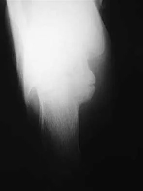
Figures 15a and 15b show the radiographs of an 18-year-old mountain biker who came off of a 15-foot ramp and sustained an injury to his ankle. Because the local rural hospital had no orthopaedic surgeon available, he was transported to a Level 1 emergency department 10 hours after his initial injury. Examination reveals that the injury remains closed. Management should consist of


Explanation

15b High-energy tibial pilon fractures involve disruption of the soft-tissue envelope with significant lower extremity edema. Definitive reconstruction of the comminuted distal tibia should be delayed for at least 7 days to allow edema to dissipate, lowering the risk of skin necrosis. An external fixator is the best method to keep the ankle at anatomic length while preventing skin necrosis. Ligamentotaxis will hold the fragments reduced to allow the edema to dissipate. CT may be obtained in traction to localize the individual fragments and plan surgical incisions and subsequent fixation. Short leg casting will not provide adequate ligamentotaxis to hold the fragments reduced and prevent skin compromise. Primary fusion of the ankle in an unstable tibial pilon fracture is prone to a poor result from nonunion or malunion. Tornetta P III, Weiner L, Bergman M, et al: Pilon fractures: Treatment with combined internal and external fixation. J Orthop Trauma 1993;7:489-496.

Question 2

A 47-year-old woman has had medial ankle pain and swelling for the past 3 months. She recalls no specific injury, and casting and nonsteroidal anti-inflammatory drugs have failed to provide relief. Examination reveals a pes planus with heel valgus that is passively correctable. Radiographs show no evidence of arthritis. An MRI scan is shown in Figure 16. What is the most appropriate surgical procedure to alleviate her pain?


Explanation

The patient has a stage II posterior tibial tendon tear with a supple foot; therefore, the treatment of choice is flexor digitorum longus transfer with medial displacement calcaneal osteotomy. Triple arthrodesis is not indicated, and isolated tendon transfer will stretch out in the face of persistent heel valgus. Direct repair of the posterior tibial tendon or repair of the spring ligament is not sufficient to correct the deformity. Myerson MS, Corrigan J: Treatment of posterior tibial tendon dysfunction with flexor digitorum longus tendon transfer and calcaneal osteotomy. Orthopedics 1996;19:383-388.

Question 3

A 38-year-old woman has a lesion on her left foot that has increased in size over the past 6 months. The clinical photograph is shown in Figure 17a, and a photomicrograph of the biopsy specimen is shown in Figure 17b. What is the most likely diagnosis?


Explanation

17b Melanoma comprises 25% of lower extremity lesions and is the most common malignant tumor of the foot. The preferred treatment is wide resection. Hughes LE, Horgan K, Taylor BA, Laidler P: Malignant melanoma of the hand and foot: Diagnosis and management. Br J Surg 1985;72:811-815.

Question 4

A 16-year-old boy has had a painful ingrown nail on his great toe for the past 3 months. When initial management consisting of soaking the foot in Epsom salts and trimming the nail failed to provide relief, his family physician recommended 2 weeks of oral antibiotics. His symptoms persist, and he is now seeking a second opinion. A clinical photograph is shown in Figure 18. Management should now consist of


Explanation

The patient has a chronic ingrown nail on his great toe, which is not an uncommon occurrence in teenagers because of improper nail care. There is local infection and a foreign body reaction because of the nail. Continued conservative management with soaks and antibiotics will not improve the clinical situation. In the presence of local chronic infection, nail matrix ablation is contraindicated. Additionally, in the absence of a history of an ingrown nail, a nail matrix ablation is not medically indicated. The appropriate treatment is partial removal of the nail plate. With nail plate removal, the inflammation and local infection will resolve rapidly. Pettine KA, Cofield RH, Johnson KA, Bussey RM: Ingrown toenail: Results of surgical treatment. Foot Ankle 1988;9:130-134.

Question 5

A 28-year-old man has a painful nodule on the plantar aspect of his foot in the midarch. Use of a soft orthosis has failed to provide relief. Examination reveals that the mass is approximately 2 1/2 cm in diameter, firm, and tender to palpation. An MRI scan confirms the presence of a plantar fibroma. Management should now consist of


Explanation

Plantar fibromas have an extremely high recurrence rate (approximately 60%) with local excision only. Resection of the entire plantar fascia is effective at irradicating the lesion. There is no role for chemotherapy or amputation with plantar fibromatosis. Radiation therapy may be helpful in combination with resection of the plantar fascia. Kirby EJ, Shereff MJ, Lewis MM: Soft-tissue tumors and tumor-like lesions of the foot: An analysis of 83 cases. J Bone Joint Surg Am 1989;71:621-626.

Question 6

A child born with myelomeningocele is expected to be an ambulator with bracing. Examination by the consulting orthopaedic surgeon reveals rigid clubfeet in addition to the neurologic issues. Management should consist of


Explanation

In a child with myelomeningocele, the guiding principle of treatment is to achieve a plantigrade foot by the time the child is ready to stand. The standard clubfoot protocol should be followed, but these children will require an aggressive surgical release to obtain a sufficient correction. Mizel MS, Miller RA, Scioli MW (eds): Orthopaedic Knowledge Update: Foot and Ankle 2. Rosemont, IL, American Academy of Orthopaedic Surgeons, 1998, pp 65-78.

Question 7

A 42-year-old man has a symptomatic flatfoot deformity and walks with a slight limp after falling off a scaffold 9 months ago. He also reports that he has had difficulty returning to work. Orthotics have failed to provide relief. Current radiographs are shown in Figures 19a and 19b. To relieve his pain and return the patient to work, treatment should consist of


Explanation

19b Because the patient has sustained a tarsometatarsal injury with midfoot sag, the treatment of choice is a tarsometatarsal arthrodesis. The cause of his flatfoot deformity is secondary to the tarsometatarsal injury and not from posterior tibialis tendon deficiency. Lateral column lengthening, double arthrodesis, and calcaneal osteotomy are not indicated. Although open reduction and internal fixation may be performed late when arthritis is present, these procedures are less likely to succeed. Komenda GA, Myerson MS, Biddinger KR: Results of arthrodesis of the tarsometatarsal joints after traumatic injury. J Bone Joint Surg Am 1996;78:1665-1676.

Question 8

A 16-year-old boy has a symptomatic flatfoot deformity that is causing pain, skin breakdown, and shoe wear problems. Shoe modification and an orthosis have failed to provide relief. Examination reveals hindfoot valgus, talonavicular sag, and forefoot abduction that are all passively correctable. Treatment should consist of


Explanation

The patient has a supple planovalgus deformity that is passively fully correctable, and nonsurgical management has failed to provide relief. Lateral column lengthening with medial soft-tissue tightening will correct the deformity and maintain a flexible foot. Arthrodesis is not recommended for a supple, correctable deformity because of loss of motion and long-term degeneration of surrounding joints. Medial displacement calcaneal osteotomy is generally reserved for an adult-acquired flexible flatfoot. Beaty JH (ed): Orthopaedic Knowledge Update 6. Rosemont, IL, American Academy of Orthopaedic Surgeons, 1999, pp 613-631. Evans D: Calcaneo-valgus deformity. J Bone Joint Surg Br 1975;57:270-278.

Question 9

A 48-year-old man has had pain and swelling of the hallux metatarsophalangeal joint for the past 9 months. A rocker bottom stiff-soled shoe has failed to provide relief; however, two cortisone injections have temporarily alleviated his symptoms. The radiographs shown in Figures 20a and 20b reveal diffuse arthritis of the entire hallux metatarsophalangeal joint. What is the most definitive surgical treatment?


Explanation

20b Because the radiographs demonstrate severe arthritis, hallux metatarsophalangeal arthrodesis is the treatment of choice. Cheilectomy alone will not relieve pain because the entire joint is degenerative. Joint replacement has not been shown to be a long-term solution. Keller resection arthroplasty is not indicated in younger active patients. Hallux valgus correction will not address arthritis of the joint and could stiffen the joint further. Smith RW, Joanis TL, Maxwell PD: Great toe metatarsophalangeal joint arthrodesis: A user-friendly technique. Foot Ankle 1992;13:367-377.

Question 10

During reconstruction of insertional gaps of a chronic Achilles tendon rupture, what tendon provides the most direct route of transfer?


Explanation

The flexor hallucis longus tendon provides the best, most direct route of transfer for filling Achilles tendon gaps. The tendon lies lateral to the neurovascular structures, making it safe for harvest and providing a direct route for transfer into the calcaneus without crossing these important structures. The flexor hallucis longus tendon also has muscle belly that extends distal on the tendon itself, often beyond the actual tibiotalar joint. When the tendon is transferred, this muscle belly brings excellent blood supply to the anterior portion of the reconstruction. Wilcox DK, Bohay DR, Anderson JG: Treatment of chronic achilles tendon disorders with flexor hallucis longus tendon transfer/augmentation. Foot Ankle Int 2000;21:1004-1010.

Question 11

A 27-year-old woman with Down syndrome has a severe bunion with pain and deformity in the left forefoot. Nonsurgical management has failed to provide relief. She does not use any assistive ambulatory devices. A radiograph is shown in Figure 21. Treatment should now consist of


Explanation

The patient requires an arthrodesis of the first metatarsophalangeal joint because of the abnormal neuromuscular forces. The more traditional bunionectomies such as a distal chevron bunionectomy, a proximal first metatarsal osteotomy, and a double osteotomy have a high failure rate because of the underlying Down syndrome. The Keller procedure is indicated for older, sedentary individuals and has little role in the management of a neuromuscular bunion. Coughlin MJ, Abdo RV: Arthodesis of the first metatarsophalangeal joint with Vitallium plate fixation. Foot Ankle Int 1994;15:18-28.

Question 12

Which of the following is considered the most important factor in eliminating infection in chronic osteomyelitis?


Explanation

The most important factor in eliminating infection in chronic osteomyelitis is a complete debridement of the compromised bone and soft tissue. Antibiotics should be used in conjunction with surgical debridement. However, the foundation of treating infected bone is removal of the diseased tissue. Cierny G III, Cook WG, Mader JT: Ankle arthrodesis in the presence of ongoing sepsis: Indications, methods, and results. Orthop Clin North Am 1989;20:709-721. Cierny G, Zorn EZ: Arthrodesis of the tibiotalar joint for sepsis. Foot Ankle Clin 1996;1:177-197.

Question 13

A 35-year-old woman reports worsening pain after undergoing a neurectomy in the third interspace for a Morton's neuroma 12 months ago. She states that the pain is sharp and electrical, worse than before her surgery, and prevents her from participating in her usual work and exercise activities. Use of wider shoes and pads used before her surgery have failed to provide relief. Examination does not reveal any deformity or inflammation. Tenderness along with neuritic pain occurs with compression of the plantar aspect of the foot between the third and fourth metatarsal head area. To most reliably alleviate her pain, management should consist of


Explanation

Most patients with a significant recurrent neuroma will not obtain relief with conservative methods. Pain results from a stump neuroma at the weight-bearing area from too short of a resection of the nerve or from regrowth of the remaining nerve end. Although steroid injection may be helpful in localizing symptoms or providing temporary relief, it rarely cures a stump neuroma. Orthotics with a metatarsal pad will likely increase pressure and pain at the neuroma site. Physical therapy could temporize the symptoms but will not address the underlying problem. Similarly, bone decompression alone will not alter the location of the neuroma stump. Revision of the nerve to a more proximal level off of the weight-bearing area is the most likely method to succeed. A plantar approach facilitates identification and ability to revise the nerve to a more proximal level. Mizel MS, Miller RA, Scioli MW (eds): Orthopaedic Knowledge Update: Foot and Ankle 2. Rosemont, IL, American Academy of Orthopaedic Surgeons, 1998, pp 101-111. Johnson JE, Johnson KA, Unni KK: Persistent pain after excision of an interdigital neuroma: Results of reoperation. J Bone Joint Surg Am 1988;70:651-657. Beskin JL, Baxter DE: Recurrent pain following interdigital neurectomy: A plantar approach. Foot Ankle 1988;9:34-39.

Question 14

What type of brace is shown in Figures 22a and 22b?


Explanation

22b The figures show a Charcot restraining orthotic walker (CROW). This brace has been used as a customized total contact fit removable brace to maintain foot alignment as the patient evolves from Eichenholz stage 1 to Eichenholz stage 3 Charcot arthropathy. Mehta JA, Brown C, Sargeant N: Charcot restraint orthotic walker. Foot Ankle Int 1998;19:619-623.

Question 15

A 23-year-old man has pain and a callus beneath the second metatarsal head. Initial management should consist of


Explanation

The initial treatment of metatarsalgia with or without the presence of an intractable keratosis should be conservative. Simple paring of the callus with elevation of the metatarsals may suffice. A prefabricated "off-the-shelf" orthosis or felt pad can be used before investing in a custom orthosis. The use of medicated pads can lead to greater amounts of keratosis and should be avoided. Mizel MS, Miller RA, Scioli MW (eds): Orthopaedic Knowledge Update: Foot and Ankle 2. Rosemont, IL, American Academy of Orthopaedic Surgeons, 1998, pp 163-173.

Question 16

Figures 23a and 23b show the radiograph and clinical photograph of a patient who reports a reduced ability to flex the interphalangeal joint of her great toe after undergoing a Chevron-Akin bunionectomy. What is the most likely cause?


Explanation

23b The flexor hallucis longus tendon is at risk during a Chevron-Akin osteotomy because of its close relationship to the base of the proximal phalanx. The radiograph reveals a reduced ability to flex the interphalangeal joint secondary to the flexor hallucis longus laceration. The other complications are not supported by the radiograph. Tollison ME, Baxter DE: Combination chevron plus Akin osteotomy for hallux valgus: Should age be a limiting factor? Foot Ankle Int 1997;18:477-481.

Question 17

A 45-year-old man has persistent hindfoot pain that is aggravated by weight-bearing activities. History reveals that he sustained a calcaneus fracture 2 years ago, and he underwent a subtalar fusion 1 year ago. Examination reveals tenderness in the sinus tarsi and across the transverse tarsal joint. A plain radiograph and a CT scan are shown in Figures 24a and 24b. A technetium Tc 99m bone scan reveals uptake at the subtalar joint and at the transverse tarsal joints. Management should now consist of


Explanation

24b The patient has a nonunion at the subtalar joint because of poor preparation of the arthrodesis site with incomplete removal of the articular cartilage. Clinically, he has arthritis at the transverse tarsal joint. Casting with a bone stimulator is not expected to result in a union of the subtalar arthrodesis. To address both the subtalar nonunion and the transverse tarsal joint arthritis, revision of the subtalar arthrodesis and conversion to a triple arthrodesis is the preferred option. Graves SC, Mann RA, Graves KO: Triple arthrodesis in older adults: Results after long-term follow-up. J Bone Joint Surg Am 1993;75:355-362. Haddad SL, Myerson MS, Pell RF IV, Schon LC: Clinical and radiographic outcome of revision surgery for failed triple arthrodesis. Foot Ankle Int 1997;18:489-499. Sangeorzan BJ, Smith D, Veith R, Hansen ST Jr: Triple arthrodesis using internal fixation in treatment of adult foot disorders. Clin Orthop 1993;294:299-307. Sangeorzan BJ: Salvage procedures for calcaneus fractures. Instr Course Lect 1997;46:339-346.

Question 18

A 21-year-old college student reports hearing a pop and has acute pain laterally over the ankle after twisting it during a recreational basketball game. Examination 1 hour after the injury reveals minimal swelling and ecchymosis. The anterior drawer sign is positive. Radiographs reveal no evidence of a fracture. What is the best course of action?


Explanation

Even though the patient has a grade 3 ankle ligament injury, studies have shown that 95% of patients with a grade 3 injury that may include a complete tear of the ligaments will heal successfully with conservative functional management. Extensive diagnostic evaluation with stress radiographs, CT, and MRI is not indicated. Surgical reconstruction is not indicated because of the overwhelming success of conservative management; however, in the few patients where late instability develops, surgical reconstruction offers an excellent outcome. Carne P: Nonsurgical treatment of ankle sprains using the modified Sarmiento brace. Am J Sports Med 1989;17:253-257.

Question 19

What significant structure is most at risk during a posterior approach of the Achilles tendon near its musculotendinous junction?


Explanation

The sural nerve crosses near the midline at the level of the musculotendinous junction before descending to its more lateral location distally. The saphenous nerve and vein are further medial and at less risk. The posterior tibial nerve is at risk only during deep dissection, such as harvesting flexor hallucis longus tendon graft. The plantaris muscle lies in this area but is of little clinical significance. Webb J, Moorjani N, Radford M: Anatomy of the sural nerve and its relation to the Achilles tendon. Foot Ankle Int 2000;21:475-477.

Question 20

Figure 25 shows the clinical photograph of a 48-year-old man who has had a forefoot ulcer for the past 4 months. History reveals that he has had type II diabetes mellitus for the past 10 years. Examination reveals sensory and motor neuropathy, with weak ankle dorsiflexion. The ankle cannot be passively dorsiflexed past a neutral position. Initial management should consist of


Explanation

Foot deformity and decreased joint motion have been associated with increased plantar pressures and an increased risk of ulceration. In a partial-thickness ulcer without exposed bone or tendon, total contact casting is highly effective. Concomitant Achilles tendon lengthening increases the likelihood that healing of the ulcer can be obtained and perhaps more importantly, maintained. Lin SS, Lee TH, Wapner KL: Plantar forefoot ulceration with equinus deformity of the ankle in diabetic patients: The effect of tendo-Achilles lengthening and total contact casting. Orthopedics 1996;19:465-475.

Question 21

An active 36-year-old woman with rheumatoid arthritis has continued forefoot discomfort despite the use of orthotics and shoe wear modifications. A radiograph and a clinical photograph are shown in Figures 26a and 26b. Treatment at this point should consist of


Explanation

26b In a patient with inflammatory arthritis, advanced hallux valgus deformity in conjunction with lesser metatarsophalangeal joint destruction and subluxation warrants fusion of the first metatarsophalangeal joint and lesser metatarsal head resections. Hallux valgus correction will fail because of incompetent soft tissues. A Keller resection arthroplasty is not indicated in this age group. Synovectomy is contraindicated because of evidence of erosive changes of the lesser metatarsophalangeal joints. Ouzounian T: Rheumatoid arthritis of the foot & ankle, in Myerson MS (ed): Foot & Ankle Disorders. Philadelphia, PA, WB Saunders, 2000, vol 2, pp 1189-1204. Mann RA, Thompson FM: Arthrodesis of the first metatarsophalangeal joint for hallux valgus in rheumatoid arthritis. J Bone Joint Surg Am 1984;66:687-692.

Question 22

The Keller proximal phalanx resection procedure is most useful for which of the following conditions?


Explanation

A Keller proximal phalanx resection procedure usually results in reduced weight bearing under the first ray because of shortening of the toe and disruption of intrinsic flexor function. This can be an effective method of offloading a neuropathic ulcer under the great toe at the interphalangeal or metatarsophalangeal joint area. However, these features are generally undesirable in young active patients. The procedure has a high rate of recurrent deformity in patients with rheumatoid arthritis. It would exacerbate transfer metatarsalgia in a patient with a hypermobile first ray. Lin SS, Bono CM, Lee TH: Total contact casting and Keller arthroplasty for diabetic great toe ulceration under the interphalangeal joint. Foot Ankle Int 2000;21:588-593.

Question 23

An active 60-year-old man is evaluated 4 years following surgical correction of a hallux valgus deformity. The patient reports that a hallux varus deformity developed rapidly following his initial surgery. Conservative management consisting of wider shoes, toe strapping, and anti-inflammatory drugs has failed to provide relief. Examination reveals a hallux varus deformity with restricted painful motion of the metatarsophalangeal joint and callus formation under the second metatarsal head. What is the next most appropriate step in management?


Explanation

Hallux varus may occur as a complication following hallux valgus surgery, most commonly a modified McBride-type procedure. Conservative management is the initial treatment of choice; however, if unsuccessful, surgical options for reconstruction include soft-tissue reconstruction or metatarsophalangeal joint arthrodesis. The patient has evidence of joint arthrosis, making an arthrodesis the preferred method of reconstruction. Fascial arthroplasty, Silastic arthroplasty, and Keller resection arthroplasty will not correct the underlying deformity. Kitaoka HB, Patzer GL: Arthrodesis versus resection arthroplasty for failed hallux valgus operations. Clin Orthop 1998;347:208-214.

Question 24

A newborn has been referred for evaluation of a deformed foot. Prenatal and birth history are unremarkable. Examination reveals a rocker bottom appearance to the foot, and a longitudinal arch cannot be created. A palpable lump is appreciated on the plantar medial surface. What is the best course of action?


Explanation

The patient has congenital vertical talus. The navicular is irreducibly dorsally dislocated on the talus with the talar head prominent on the plantar medial aspect of the foot. Initial management involves corrective casting for 3 months to stretch the dorsal tendons, skin, and neurovascular structures. Surgical reconstruction is often needed and is indicated when the patient is age 6 to 12 months. Reconstruction requires both bony and soft-tissue procedures. Napiontek M: Congenital vertical talus: A retrospective and critical review of 32 feet operated on by peritalar reduction. J Pediatr Orthop 1995;4:179-187.

Question 25

Which of the following is considered an inherent problem in using the distal oblique shortening (Weil) metatarsal osteotomy for dorsal metatarsophalangeal subluxation?


Explanation

The distal oblique shortening (Weil) metatarsal osteotomy has not been associated with transfer lesions to the extent of other shortening osteotomies, and malunions and nonunions are unusual complications. Recurrent dorsal contracture of the toe has been reported. Recommendations to reduce this problem include release of the dorsal capsule and tendons, as well as a flexor tendon transfer. A potential cause suspected for this phenomenon is the relatively dorsal positioning of the intrinsic tendons after plantar displacement of the metatarsal head. Trnka HJ, Nyska M, Parks BG, Myerson MS: Dorsiflexion contracture after the Weil osteotomy: Results of cadaver study and three-dimensional analysis. Foot Ankle Int 2001;22:47-50. Trnka HJ, Muhlbauer M, Zettl R, Myerson MS, Ritschl P: Comparison of the results of the Weil and Helal osteotomies for the treatment of metatarsalgia secondary to dislocation of the lesser metatarsophalangeal joints. Foot Ankle Int 1999;20:72-79.

Question 26

A 55-year-old female presents with medial ankle pain and a progressively flattening arch. Examination shows a "too-many-toes" sign, flexible hindfoot valgus, and forefoot abduction of 35 degrees. Radiographs demonstrate >40% uncoverage of the talonavicular joint. What is the most appropriate surgical management?


Explanation

This clinical scenario describes a Stage IIb adult-acquired flatfoot deformity characterized by significant forefoot abduction. Management typically requires a lateral column lengthening (e.g., Evans osteotomy) to correct the abduction, combined with a medial soft tissue procedure like an FDL transfer.

Question 27

A 24-year-old equestrian presents with severe midfoot pain after falling from a horse. Examination reveals plantar ecchymosis. Weight-bearing radiographs show a 3 mm diastasis between the base of the first and second metatarsals without associated fractures. What is the most appropriate definitive management?


Explanation

Purely ligamentous Lisfranc injuries have higher rates of hardware failure and post-traumatic arthritis with ORIF compared to primary arthrodesis. Arthrodesis of the medial columns (1st-3rd TMT joints) is the preferred definitive treatment for purely ligamentous injuries.

Question 28

A 55-year-old woman presents with medial ankle pain and a progressive flatfoot deformity. Examination shows a flexible hindfoot valgus and inability to perform a single-leg heel raise. Radiographs demonstrate >40% talonavicular uncoverage. Which surgical intervention is most appropriate if conservative management fails?


Explanation

This patient has Stage IIb adult-acquired flatfoot deformity, characterized by a flexible deformity with forefoot abduction (>30-40% talonavicular uncoverage). Treatment requires an FDL transfer, medial displacement calcaneal osteotomy, and a lateral column lengthening (Evans osteotomy) to correct the forefoot abduction.

Question 29

During a percutaneous repair of an acute Achilles tendon rupture, the surgeon places sutures in the proximal stump. Which of the following describes the most at-risk anatomic structure and its location relative to the calcaneal insertion?


Explanation

The sural nerve is at highest risk during percutaneous Achilles tendon repair. It typically crosses the lateral border of the Achilles tendon approximately 9.8 to 10 cm proximal to its insertion on the calcaneus.

Question 30

A 30-year-old man sustains a Hawkins type II fracture of the talar neck. Which blood supply to the talar body is most likely preserved in this specific injury pattern?


Explanation

A Hawkins type II fracture involves a talar neck fracture with subluxation or dislocation of the subtalar joint, disrupting the artery of the tarsal canal and tarsal sinus. The deltoid branch of the posterior tibial artery is typically preserved as the tibiotalar joint remains reduced.

Question 31

A 60-year-old patient with poorly controlled diabetes presents with a unilaterally swollen, red, and warm foot for 3 weeks. Radiographs show periarticular debris, fragmentation, and subluxation at the tarsometatarsal joints. What is the most appropriate immediate management?


Explanation

This patient presents with Eichenholtz Stage I (developmental/fragmentation) Charcot arthropathy. The mainstay of initial treatment for acute Charcot is immobilization and offloading, most effectively achieved with a total contact cast to prevent further deformity.

Question 32

A 45-year-old woman complains of painful bunions. Examination reveals first tarsometatarsal (TMT) joint hypermobility. Radiographs demonstrate a hallux valgus angle (HVA) of 45 degrees and an intermetatarsal angle (IMA) of 18 degrees. Which of the following procedures is most appropriate?


Explanation

A Lapidus procedure (first TMT arthrodesis) is indicated for moderate-to-severe hallux valgus (IMA >15 degrees) accompanied by first TMT joint hypermobility. It provides powerful correction of the intermetatarsal angle and stabilizes the medial column.

Question 33

A 14-year-old boy presents with frequent ankle sprains and rigid pes planus. Radiographs reveal an 'anteater nose' sign on the lateral view. Which of the following anatomical structures is most likely involved in this patient's pathology?


Explanation

The 'anteater nose' sign on a lateral foot radiograph is highly indicative of a calcaneonavicular coalition. This results from a tubular prolongation of the anterior process of the calcaneus approaching or overlapping the navicular.

Question 34

In a displaced intra-articular calcaneus fracture, the 'constant fragment' remains anatomically aligned with the talus. Which ligamentous attachment is primarily responsible for maintaining the position of this fragment?


Explanation

The sustentaculum tali is referred to as the 'constant fragment' in calcaneus fractures because it remains securely attached to the talus. This stability is maintained primarily by the strong deltoid and talocalcaneal ligaments.

Question 35

A professional football player sustains a forceful hyperextension injury to his great toe. MRI confirms a complete tear of the plantar plate with proximal retraction of the sesamoids. What is the most appropriate surgical management?


Explanation

This is a Grade 3 turf toe injury involving a complete tear of the plantar plate-sesamoid complex. Surgical management is indicated for athletic patients with retraction, involving direct repair of the plantar plate back to the base of the proximal phalanx.

Question 36

A 62-year-old man presents with painful, restricted dorsiflexion of the great toe. Radiographs show severe joint space narrowing, large dorsal osteophytes, and subchondral sclerosis at the first metatarsophalangeal (MTP) joint. He has pain throughout the entire arc of motion. What is the most reliable surgical option for long-term pain relief?


Explanation

This patient has Grade 3/4 hallux rigidus with pain throughout the arc of motion and severe radiographic changes. First MTP joint arthrodesis is the gold standard for end-stage hallux rigidus, providing the most reliable long-term pain relief.

Question 37

A 28-year-old athlete sustains an external rotation injury to the ankle. Intraoperatively, the syndesmosis is evaluated. Which of the following ligaments contributes the greatest percentage of mechanical stability to the distal tibiofibular syndesmosis?


Explanation

The posterior inferior tibiofibular ligament (PITFL) is the strongest and provides the greatest mechanical stability to the syndesmosis, contributing approximately 42% of its resistance to diastasis.

Question 38

A 21-year-old collegiate basketball player sustains a fracture at the metaphyseal-diaphyseal junction of the fifth metatarsal. It extends into the fourth-fifth intermetatarsal articulation. What is the most appropriate treatment to minimize time to return to play?


Explanation

A Zone 2 (Jones) fracture in an elite athlete has a high risk of nonunion or delayed union due to the watershed blood supply. Intramedullary screw fixation is recommended to accelerate healing and minimize the time to return to play.

Question 39

A 25-year-old professional football player sustains a purely ligamentous Lisfranc injury involving the first, second, and third tarsometatarsal joints. Nonoperative management is deemed inappropriate due to the severity of the instability. According to recent high-level prospective studies, which of the following surgical interventions is associated with the best functional outcome and lowest rate of hardware removal in purely ligamentous injuries?


Explanation

For purely ligamentous Lisfranc injuries, primary arthrodesis of the involved medial rays (1st-3rd TMT joints) has been shown to yield superior functional outcomes and lower reoperation rates compared to ORIF. ORIF is generally preferred when there is significant bony involvement rather than purely ligamentous disruption.

Question 40

A 56-year-old man with poorly controlled type 2 diabetes and peripheral neuropathy presents with a red, hot, and swollen left foot that began 2 weeks ago without distinct trauma.

Pedal pulses are bounding. Weight-bearing radiographs reveal diffuse osteopenia but no fractures, joint subluxation, or fragmentation. What is the most appropriate initial management?


Explanation

This clinical presentation is characteristic of an Eichenholtz stage 0 Charcot neuroarthropathy. The most appropriate initial treatment is total contact casting to immobilize the foot and prevent progression to fragmentation and deformity.

Question 41

A 40-year-old recreational athlete sustains an acute, closed midsubstance Achilles tendon rupture. He opts for nonoperative management. To optimize his outcomes and reduce the rerupture rate to a level comparable with surgical repair, which of the following rehabilitation protocols should be employed?


Explanation

Modern evidence demonstrates that functional rehabilitation protocols with early, protected weight-bearing in a brace significantly lower rerupture rates in nonoperatively managed Achilles tendon ruptures, making them comparable to operative outcomes. Prolonged static immobilization is associated with higher rerupture rates and worse functional recovery.

Question 42

A 62-year-old woman presents with end-stage hallux rigidus (Coughlin and Shurnas Grade 3) and severe pain with ambulation. Conservative measures have failed, and she elects to undergo first metatarsophalangeal (MTP) joint arthrodesis. To ensure an optimal functional outcome, in what position should the first MTP joint be fused?


Explanation

The ideal position for a first MTP joint arthrodesis is approximately 10-15 degrees of valgus and 15 degrees of dorsiflexion relative to the floor. This position allows for normal toe-off during the gait cycle and accommodates standard shoe wear.

Question 43

A 58-year-old woman is diagnosed with stage IIb adult acquired flatfoot deformity secondary to posterior tibial tendon insufficiency. Clinical and radiographic evaluation reveals a flexible hindfoot valgus and significant forefoot abduction (uncoverage of the talonavicular joint >40%). In addition to a flexor digitorum longus (FDL) transfer and medial displacement calcaneal osteotomy, which procedure is essential to correct her specific deformity?


Explanation

Stage IIb adult acquired flatfoot deformity is characterized by significant forefoot abduction (talonavicular uncoverage). A lateral column lengthening (e.g., Evans osteotomy) is required to correct the forefoot abduction in addition to the standard medial-sided soft tissue and bony procedures.

Question 44

Following open reduction and internal fixation of an ankle fracture with a concomitant syndesmotic injury, intraoperative fluoroscopy suggests adequate reduction of the syndesmosis. However, the surgeon wants to definitively confirm the accuracy of the syndesmotic reduction postoperatively. Which imaging modality is considered the gold standard for assessing syndesmotic reduction?


Explanation

Postoperative CT is the gold standard for evaluating the accuracy of syndesmotic reduction. Plain radiographs have been shown to be insensitive for detecting subtle syndesmotic malreductions, which are highly correlated with poor clinical outcomes.

Question 45

A 34-year-old construction worker falls from a ladder and sustains a displaced Hawkins type III talar neck fracture.

The injury is closed. He undergoes prompt open reduction and internal fixation. What is the approximate reported risk of avascular necrosis (AVN) of the talar body associated with this fracture pattern?


Explanation

A Hawkins type III fracture involves displacement of the talar neck with subluxation or dislocation of both the subtalar and tibiotalar joints. Due to the extensive disruption of the blood supply (artery of the tarsal canal, artery of the sinus tarsi, and deltoid branches), the risk of AVN approaches 75% to 100%.

Question 46

A surgeon is performing an extensile lateral approach for the open reduction and internal fixation of a displaced intra-articular calcaneus fracture. To minimize the risk of full-thickness skin flap necrosis, the surgeon must be careful to preserve the primary blood supply to the corner of the flap. Which vessel is responsible for this critical vascular supply?


Explanation

The lateral calcaneal artery, a branch of the peroneal artery, provides the primary blood supply to the lateral skin flap used in the extensile lateral approach. The "no-touch" technique with subperiosteal elevation is critical to protecting this delicate vascular network.

Question 47

A 16-year-old boy presents with a bilateral symptomatic cavovarus foot deformity. The examiner performs a Coleman block test by placing the patient's lateral foot (heel and lateral border) on a 1-inch block while allowing the first metatarsal to hang off freely into plantarflexion. During this test, the hindfoot varus deformity corrects to a neutral alignment. What does this finding indicate?


Explanation

The Coleman block test assesses whether hindfoot varus is flexible and driven by a plantarflexed first ray. If the hindfoot corrects to neutral when the first ray's effect on the floor is negated, the deformity is primarily forefoot-driven, indicating a flexible hindfoot.

Question 48

A 21-year-old collegiate basketball player complains of lateral foot pain for 3 weeks and presents with an acute exacerbation after a pivot mechanism. Radiographs reveal a fracture through the proximal metaphyseal-diaphyseal junction of the fifth metatarsal. Which of the following treatments is the most appropriate management to optimize his return to play?


Explanation

A fracture at the metaphyseal-diaphyseal junction of the fifth metatarsal (Jones fracture) occurs in a vascular watershed area. High-level athletes require surgical intervention with intramedullary screw fixation to minimize the risk of nonunion and expedite return to sport.

Question 49

A 65-year-old woman with post-traumatic ankle osteoarthritis is being evaluated for a total ankle arthroplasty (TAA). Which of the following conditions is considered an absolute contraindication for this procedure?


Explanation

Charcot neuroarthropathy or the absence of protective sensation in the lower extremity is an absolute contraindication to total ankle arthroplasty due to the extremely high risk of implant failure, collapse, and soft tissue complications. Arthrodesis is the surgical treatment of choice in these patients.

Question 50

A 28-year-old skier presents with acute lateral ankle pain after catching an edge. Physical examination reveals tenderness posterior to the lateral malleolus and a snapping sensation with resisted active dorsiflexion and eversion. Radiographs show a small bony avulsion flake lateral to the distal fibula. What is the most likely mechanism of this specific injury?


Explanation

The clinical presentation and "fleck sign" on radiographs describe a superior peroneal retinaculum (SPR) avulsion and subsequent peroneal tendon subluxation. This injury classically occurs via sudden, forceful dorsiflexion combined with reflex contraction of the peroneal muscles during forced eversion.

Question 51

A 24-year-old professional American football player sustains a severe hyperextension injury to his great toe on artificial turf. He is diagnosed with a Grade 3 turf toe injury. Which of the following radiographic findings is most consistent with a complete disruption of the plantar plate in this condition?


Explanation

Grade 3 turf toe represents a complete tear of the capsuloplantar plate complex. Radiographically, this is most clearly indicated by proximal migration of the sesamoids compared with the contralateral uninjured foot, as the stabilizing plantar structures have failed.

Question 52

A 14-year-old female gymnast presents with chronic, localized pain over the dorsal aspect of her forefoot, specifically worsening during weight-bearing. Radiographs demonstrate sclerosis, flattening, and early fragmentation of the second metatarsal head. Which of the following is the most likely diagnosis?


Explanation

Freiberg's infraction is an avascular necrosis of the metatarsal head, most commonly affecting the second metatarsal. It frequently occurs in adolescent females engaging in activities that cause repetitive microtrauma to the forefoot.

Question 53

A 45-year-old woman complains of burning pain in her forefoot that radiates into her third and fourth toes. Symptoms are exacerbated by wearing narrow, high-heeled shoes and relieved by removing the shoes and massaging the foot. Examination reveals a painful click when the forefoot is compressed laterally. Histologic evaluation of the excised lesion in this condition typically shows:


Explanation

Morton's neuroma is not a true neoplasm; rather, histologic examination demonstrates extensive perineural fibrosis, vascular hyalinization, and nerve fiber degeneration resulting from chronic repetitive compression of the interdigital nerve.

Question 54

A 35-year-old man sustains a closed, high-energy tibial pilon fracture. Initial management consists of a spanning external fixator. Before proceeding with definitive open reduction and internal fixation (ORIF), the surgeon must ensure the soft tissue envelope is adequately recovered. Which clinical sign is the most reliable indicator that it is safe to proceed with definitive surgery?


Explanation

The most reliable clinical sign that swelling has resolved sufficiently to safely perform definitive internal fixation of a pilon fracture is the return of skin wrinkles (the "wrinkle sign"). This typically occurs 10 to 14 days after the initial injury.

Question 55

A 45-year-old runner presents with chronic medial heel pain that radiates into the plantar aspect of the foot, which has failed conservative management for plantar fasciitis. MRI reveals isolated atrophy of the abductor digiti minimi muscle. Entrapment of the first branch of the lateral plantar nerve (Baxter's nerve) is suspected. Between which two structures is this nerve most commonly compressed?


Explanation

Baxter's nerve (first branch of the lateral plantar nerve) is most frequently entrapped as it passes between the deep fascia of the abductor hallucis muscle and the medial margin of the quadratus plantae muscle. Chronic compression leads to denervation atrophy of the abductor digiti minimi.

Question 56

A 56-year-old poorly controlled diabetic patient presents with a red, hot, and swollen left foot. Radiographs show osteopenia but no frank fractures or dislocation. MRI reveals diffuse marrow edema in the cuboid and cuneiforms. Which of the following tests is considered the gold standard to differentiate acute Charcot arthropathy from osteomyelitis in this setting?


Explanation

Percutaneous bone biopsy with histopathologic and microbiologic analysis is the gold standard for differentiating acute Charcot arthropathy from osteomyelitis. While advanced imaging like labeled WBC scans combined with marrow scans can be helpful, bone biopsy remains definitive.

Question 57

A 45-year-old woman presents with pain and difficulty wearing shoes 2 years after undergoing a distal chevron osteotomy and lateral soft-tissue release for hallux valgus. Examination reveals a flexible first metatarsophalangeal (MTP) joint with a 15-degree hallux varus deformity. Radiographs show no degenerative changes in the MTP joint. Which of the following is the most appropriate surgical treatment?


Explanation

In a flexible hallux varus deformity without MTP arthritis following bunion surgery, an abductor hallucis release and a split extensor hallucis brevis (EHB) or extensor hallucis longus (EHL) tendon transfer is indicated. First MTP arthrodesis is reserved for rigid deformities or those with concurrent arthritis.

Question 58

A 55-year-old female presents with severe hallux valgus, an intermetatarsal angle of 22 degrees, and clinical hypermobility of the first tarsometatarsal (TMT) joint. Which of the following surgical procedures is most appropriate to provide a durable correction?


Explanation

A Lapidus procedure (first TMT arthrodesis) is indicated for severe hallux valgus (IMA > 20 degrees) and when there is clinical hypermobility at the first TMT joint. It provides powerful correction and stabilization of the medial column.

Question 59

A 60-year-old male undergoes surgical treatment for severe insertional Achilles tendinopathy. During the procedure, the surgeon discovers extensive degeneration and debrides 65% of the tendon's distal insertion. What is the most appropriate next step?


Explanation

When more than 50% of the Achilles tendon insertion requires debridement, augmentation with a Flexor Hallucis Longus (FHL) tendon transfer is recommended to restore plantarflexion strength and prevent rupture.

Question 60

A 30-year-old male sustains a high-energy motor vehicle accident resulting in a Hawkins Type III talar neck fracture. What is the approximate reported risk of developing avascular necrosis (AVN) of the talar body in this injury pattern?


Explanation

Hawkins Type III fractures involve displacement of the talar neck with subluxation or dislocation of both the subtalar and tibiotalar joints. This severely disrupts the blood supply, resulting in a 75-100% risk of AVN.

Question 61

A 55-year-old patient with poorly controlled diabetes presents with a swollen, erythematous, and warm foot. Radiographs show fragmentation, periarticular debris, and subluxation at the midfoot. What is the most appropriate initial management?


Explanation

This patient is presenting with acute Eichenholtz Stage I (fragmentation) Charcot arthropathy. The mainstay of initial treatment during the active phase is immobilization and offloading, typically utilizing a total contact cast until erythema and edema resolve.

Question 62

A 24-year-old football player sustains a twisting injury to his midfoot. Weight-bearing radiographs demonstrate a 3 mm diastasis between the medial and middle cuneiforms, as seen in the provided image.

What is the primary ligamentous restraint disrupted in this injury?


Explanation

The Lisfranc ligament is an interosseous ligament that originates from the lateral surface of the medial cuneiform and inserts onto the medial base of the second metatarsal. It is the primary stabilizer of the second tarsometatarsal joint.

Question 63

A 42-year-old roofer falls from a height and sustains a severely displaced intra-articular calcaneus fracture. It is treated with an extensile lateral approach and plate fixation. Three months postoperatively, he complains of clawing of his lesser toes and numbness on the plantar aspect of his foot. Which of the following is the most likely cause?


Explanation

Undiagnosed compartment syndrome of the deep calcaneal or intrinsic foot compartments following high-energy fracture leads to ischemic contracture of the intrinsic muscles. This typically manifests as clawing of the toes and persistent plantar numbness.

Question 64

A 31-year-old male sustains a Hawkins Type III talar neck fracture and undergoes urgent open reduction and internal fixation. At 8 weeks postoperatively, an AP radiograph of the ankle demonstrates a subchondral radiolucent band in the talar dome. What does this radiographic finding indicate?


Explanation

A subchondral radiolucent band on the talar dome at 6 to 8 weeks is known as the Hawkins sign. It represents subchondral osteopenia due to active hyperemia, indicating intact vascularity and ruling out complete avascular necrosis of the talar body.

Question 65

A 28-year-old woman presents with a progressive bilateral cavovarus foot deformity. A Coleman block test is performed, and the hindfoot varus corrects to a neutral position when the first ray is allowed to drop off the block. Which of the following procedures is most appropriate as part of her surgical reconstruction?


Explanation

A flexible hindfoot varus (corrects on Coleman block test) in a cavovarus foot is typically forefoot-driven by a rigidly plantarflexed first ray. A dorsiflexion osteotomy of the first metatarsal elevates the first ray, eliminating the deforming force on the hindfoot.

Question 66

A 45-year-old female presents with a painful bunion. Weight-bearing radiographs show a hallux valgus angle (HVA) of 42 degrees, an intermetatarsal angle (IMA) of 18 degrees, and hypermobility of the first tarsometatarsal (TMT) joint. Which of the following surgical options is most appropriate?


Explanation

A Lapidus procedure (first TMT arthrodesis) is indicated for severe hallux valgus deformities (IMA > 15 degrees, HVA > 40 degrees) associated with first ray hypermobility. It provides powerful deformity correction and long-term stabilization of the medial column.

Question 67

A 36-year-old runner presents with lateral retromalleolar pain and swelling that worsens with activity. Clinical exam reveals tenderness along the posterior aspect of the fibula. An MRI is shown in the provided image.

Assuming the image shows a longitudinal split tear of the peroneus brevis, what is the most likely pathomechanism?


Explanation

Peroneus brevis tears (often longitudinal or "split" tears) frequently occur due to subluxation over a shallow or convex fibular retromalleolar groove. This subluxation creates a mechanical shearing force between the tendon and the fibula.

Question 68

A 55-year-old man presents with dorsal midfoot pain and limited, painful dorsiflexion of the right great toe. Radiographs show dorsal osteophytes and mild joint space narrowing at the first metatarsophalangeal joint, consistent with Grade 2 hallux rigidus. He has failed shoe modifications and NSAIDs. What is the most appropriate surgical management?


Explanation

For moderate (Grade 1 or 2) hallux rigidus with preserved plantar joint space and pain primarily upon dorsiflexion, a dorsal cheilectomy is the standard bone-preserving procedure. Arthrodesis is typically reserved for severe, end-stage (Grade 3 or 4) disease.

Question 69

A 24-year-old female presents with persistent ankle pain 8 months following an inversion ankle sprain. MRI demonstrates a 1.2 cm x 1.0 cm osteochondral lesion on the posteromedial talar dome with intact overlying cartilage. What is the most appropriate initial surgical treatment?


Explanation

For symptomatic osteochondral lesions of the talus smaller than 1.5 cm squared (or <15 mm in diameter) that fail conservative management, arthroscopic bone marrow stimulation (microfracture) is the primary first-line surgical treatment. Larger or cystic lesions may require OATS or MACI.

Question 70

A 62-year-old woman complains of progressive medial ankle pain and flattening of her left foot arch. Examination reveals a flexible flatfoot and inability to perform a single-leg heel raise. The heel is in valgus but passively corrects to neutral. Which combination of procedures is most appropriate?


Explanation

The patient has Stage II adult-acquired flatfoot deformity (flexible, unable to perform a single-leg heel raise). The gold standard surgical treatment combines soft tissue reconstruction (FDL transfer to replace the posterior tibial tendon) and a bony procedure (medializing calcaneal osteotomy) to correct hindfoot valgus.

Question 71

A 68-year-old male with a history of a bimalleolar ankle fracture 20 years ago presents with severe ankle pain. Radiographs reveal end-stage ankle osteoarthritis with 5 degrees of valgus deformity. He has no significant medical comorbidities. Which of the following is an absolute or strong relative contraindication to total ankle arthroplasty (TAA) in this patient?


Explanation

A history of deep intra-articular infection is a strict relative (and in most cases absolute) contraindication for total ankle arthroplasty due to the unacceptable risk of catastrophic periprosthetic joint infection. Post-traumatic etiology and mild coronal deformity (<10 degrees) are acceptable indications for TAA.

Question 72

A 42-year-old woman presents with persistent midfoot pain 6 months after a twisting injury sustained during a fall. Initial radiographs in the emergency department were reportedly normal, but she continued to have swelling and severe pain with weight-bearing. Current weight-bearing radiographs are shown in Figure 28, demonstrating dorsal subluxation and widening between the first and second metatarsals with early dorsal osteophyte formation.

What is the most appropriate definitive surgical management?


Explanation

For a chronic, neglected Lisfranc injury presenting with established deformity and secondary arthritic changes, primary arthrodesis of the involved medial tarsometatarsal joints is the surgical treatment of choice. Open reduction and internal fixation has high failure rates in delayed presentations due to established joint degeneration.

Question 73

A 50-year-old avid runner complains of posterior heel pain that worsens when beginning to run but improves slightly after warming up. Conservative management, including eccentric stretching, physical therapy, and heel lifts, has failed after 8 months. The lateral radiograph is shown in Figure 91, demonstrating an enlarged posterosuperior calcaneal tuberosity and intratendinous calcification.

Which of the following is the most appropriate surgical intervention?


Explanation

Insertional Achilles tendinopathy with a symptomatic Haglund's deformity that fails prolonged conservative treatment is best managed surgically. The standard approach involves retrocalcaneal bursectomy, excision of the posterosuperior calcaneal prominence, and debridement with reattachment of the Achilles tendon using suture anchors.

Question 74

A 24-year-old professional soccer player sustains an external rotation injury to his right ankle. Radiographs reveal no fracture, but an MRI demonstrates a complete rupture of the anterior inferior tibiofibular ligament (AITFL) and interosseous membrane, with an intact deltoid ligament. During ankle arthroscopy, there is >2 mm of lateral displacement of the fibula with stress testing. Which of the following is the most appropriate management?


Explanation

This patient has an unstable isolated syndesmotic injury (latent diastasis) confirmed on arthroscopy. Operative stabilization utilizing either syndesmotic screws or a dynamic suture-button construct is indicated to restore the ankle mortise and prevent chronic instability.

None

Clinic OS
Medically Verified Content by
Prof. Clinic OS
Consultant Orthopedic & Spine Surgeon
Chapter Index