Orthopedic Foot & Ankle 2026 MCQs: Board Review Questions & Answers (Part 3)
Key Takeaway
We review everything you need to understand about Orthopedic Foot & Ankle 2026 MCQs: Board Review Questions & Answers (Part 3). Top-rated Orthopedic Foot & Ankle 2026 MCQs bank. Practice with clinical case questions, orthopedic surgery board review, and evidence-based answers updated for 2026.
Orthopedic Foot & Ankle 2026 MCQs: Board Review Questions & Answers (Part 3)
Comprehensive 100-Question Exam
00:00
Start Quiz
Question 1
When performing surgery on a patient with insertional Achilles tendinitis and a Haglund's deformity, how much of the Achilles tendon insertion can be safely detached without having to consider reattachment with bone anchors?
Explanation
Question 2
A 12-year-old child with spina bifida paraplegia requires brace management for ankle stability. Which of the following principles applies to brace management in this individual?
Explanation
Question 3
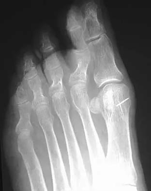
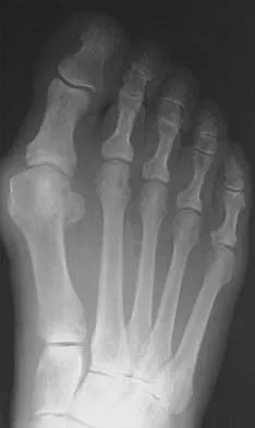
A 64-year-old man with a history of diabetes mellitus underwent open reduction and internal fixation of a displaced ankle fracture 8 weeks ago. Examination now reveals recent onset erythema, warmth, and swelling of the midfoot. Radiographs are shown in Figures 23a through 23d. What is the most likely reason for the swelling of the foot?
Explanation
Question 4
When considering a flexor digitorum longus tendon transfer as part of the surgical treatment in patients with symptomatic flatfoot deformity caused by posterior tibial tendon insufficiency, which of the following patients is the most appropriate candidate?
Explanation
Question 5
What is the most likely cause of recurrent symptoms following excision of a third web space neuroma?
Explanation
Question 6
A 45-year-old woman has had radiating pain in the medial ankle for the past 3 months. Examination reveals a small mass in the retromedial ankle region and a positive Tinel's sign. An intraoperative photograph and a hematoxylin/eosin biopsy specimen are shown in Figures 24a and 24b. Treatment should consist of
Explanation
Question 7
An 83-year-old woman with a long history of her foot slowly and progressively "turning out" now reports significant ankle pain. History reveals that she has significant cardiac disease and exercise-induced angina. Examination reveals a deficiency in the posterior tibial tendon; however, the hindfoot remains moderately supple. Radiographs reveal a valgus tilt of the tibiotalar joint and early arthrosis. What is the most appropriate orthotic management?
Explanation
Question 8
What complication is frequently associated with the Weil lesser metatarsal osteotomy (distal, oblique) in the treatment of claw toe deformities?
Explanation
Question 9
What are the five major compartments of the foot?
Explanation
Question 10
Figures 25a and 25b show the radiographs of a 66-year-old man who has had a long history of bilateral painful flatfoot deformities. Examination reveals that his foot is partially correctable passively, albeit with discomfort, and he has an Achilles tendon contracture. An ankle-foot orthosis has failed to provide relief. Treatment should now consist of
Explanation
Question 11
A 77-year-old man with diabetes mellitus has had a nonhealing Wagner grade I ulcer under the medial sesamoid for the past 3 months. He smokes tobacco regularly. He has undergone several debridements and total contact casting. Examination reveals no palpable pulses. He has no erythema or purulence, and he is afebrile. Radiographs reveal no abnormalities. What is the best initial diagnostic test to help determine why the ulcer has failed to heal?
Explanation
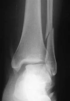
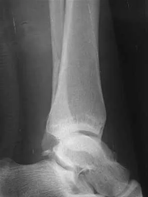
Question 12
A 28-year-old man who sustained an ankle fracture in a motor vehicle accident underwent open reduction and internal fixation 3 months ago. He continues to report significant ankle pain with ambulation. Radiographs are shown in Figure 26. What is the next most appropriate step in management?
Explanation
Question 13
The first branch of the lateral plantar nerve innervates the
Explanation
Question 14
The radiograph shown in Figure 27 shows measurement of what angle?
Explanation
Question 15
Which of the following orthotic features best reduces pain in patients with hallux rigidus?
Explanation
Question 16
An 11-year-old girl sustained an injury to her right foot when a 500-lb headstone fell on it. The headstone was removed after 3 minutes. Radiographs show multiple midfoot fractures. Examination reveals severe pain that is worse with passive toe motion. Clinical photographs are shown in Figure 28. Management should consist of
Explanation
Question 17
A 5-year-old boy has had midfoot pain with activity for the past 3 months. He has no pain at rest. Radiographs are shown in Figures 29a and 29b. Management should consist of
Explanation
Question 18
A 62-year-old man with diabetes mellitus has had a persistent 2-cm ulcer under the third metatarsal head for the past 4 months. He reports that he has had similar ulcers twice before, and both healed with nonsurgical management. He has used multiple types of commercial walking braces, shoes, and commercial dressings without resolution. He is insensate to the Semmes-Weinstein 5.07 monofilament. When the wound is probed with culture swab, there is no communication with the metatarsal head. Radiographs, bone scans, and laboratory studies reveal no evidence of osteomyelitis. What is the most predictable method of accomplishing wound healing without recurrence?
Explanation
Question 19
Figure 30 shows the radiograph of a 38-year-old man who reports persistent pain laterally and plantarly about the fifth metatarsal head. Examination reveals calluses dorsolaterally and plantarly about the fifth metatarsal head. Nonsurgical management has failed to provide relief. Surgical treatment should include
Explanation
Question 20
An 11-year-old boy stepped on a nail and sustained a puncture to the right forefoot 6 days ago. He was wearing tennis shoes at the time of injury. Treatment in the emergency department consisted of local debridement and tetanus prophylaxis; a radiograph was negative for foreign body, chondral defect, or fracture. He was discharged with a 3-day prescription of amoxicillin and clavulanate. The patient now has increasing pain and tenderness at the puncture site. What is the best course of action?
Explanation
Question 21
An 20-year-old elite college football player has ecchymosis, swelling, and pain on the lateral side of his foot after a game. Radiographs are shown in Figures 31a through 31c. Management should consist of
Explanation
Question 22
Which of the following structures are found in the anterior tarsal tunnel?
Explanation
Question 23
A 55-year-old man who runs on the weekends reports a 1-year history of continued pain directly posteriorly in the heel. Management consisting of anti-inflammatory drugs, icing techniques, a heel-counter in his shoe split, and physical therapy consisting of stretching, contrast baths, custom orthotics, and iontophoresis has failed to provide relief. Not only is his lifestyle disrupted with respect to running, but he now has pain with normal ambulation with all forms of shoe wear. He is not necessarily concerned with returning to running; he is primarily seeking pain relief. A lateral radiograph and clinical photograph are shown in Figures 32a and 32b. Treatment should now consist of
Explanation
Question 24
A 45-year-old man who has had recurrent pain and swelling of the left Achilles tendon insertion for the past 10 years reports that physical therapy and activity modification have provided relief in the past. He now has continued pain despite these efforts. He also reports occasional bouts of dysuria that he attributes to a history of prostatitis. He also notes recent eye irritation that he attributes to allergies. A lateral heel radiograph is shown in Figure 33. Which of the following laboratory studies would best aid in diagnosis?
Explanation
Question 25
A 29-year-old man reports severe knee instability and popliteal pain. History reveals that he had polio of the left lower extremity as a child and has been brace-free his entire life. Examination reveals that he walks with 40 degrees of knee hyperextension and has a fixed ankle equinus deformity of 30 degrees. He has no active motors about the knee or ankle. Which of the following methods will provide knee stability and pain relief?
Explanation
Question 26
A 55-year-old female with long-standing poorly controlled type 2 diabetes mellitus presents with a swollen, warm, and erythematous left foot and ankle. She recently started a walking program. Radiographs demonstrate early periarticular fragmentation at the tarsometatarsal joints. Which of the following best describes the underlying neurovascular pathogenesis of this condition?
Explanation
Question 27
When performing a calcaneal exostectomy and Achilles tendon debridement for insertional Achilles tendinopathy, what percentage of the Achilles tendon insertion can typically be detached before structural augmentation or reattachment with bone anchors is biomechanically required?
Explanation
Question 28
A 24-year-old professional football player sustains a hyperplantarflexion injury to his right foot. He has plantar ecchymosis and pain with pronation and abduction of the midfoot. Weight-bearing radiographs show a 2 mm diastasis between the base of the first and second metatarsals. What is the most appropriate definitive management?
Explanation
Question 29
A 60-year-old male presents with dorsal foot pain and stiffness of the great toe. On examination, he has a palpable dorsal osteophyte at the first metatarsophalangeal (MTP) joint and pain primarily with dorsiflexion. Radiographs reveal joint space narrowing with a large dorsal osteophyte but preserved plantar joint space. According to the Coughlin and Shurnas classification, this is Grade 2. What is the most appropriate initial surgical management if conservative measures fail?
Explanation
Question 30
A 28-year-old male sustains an external rotation injury to his ankle. Radiographs show no fracture. An MRI reveals a complete tear of the anterior inferior tibiofibular ligament (AITFL) and interosseous membrane, with an intact posterior inferior tibiofibular ligament (PITFL). Intraoperative stress testing confirms syndesmotic instability. During fixation with suture button devices, what is the correct anatomical trajectory for drilling from the fibula to the tibia?
Explanation
Question 31
A 32-year-old male falls from a height and sustains a Hawkins Type III talar neck fracture. Which of the following vascular structures provides the primary blood supply to the talar body and is at greatest risk of disruption in this injury pattern?
Explanation
Question 32
A 55-year-old female presents with progressive flattening of her left foot, medial-sided pain, and difficulty performing a single-leg heel rise. Examination reveals a flexible flatfoot deformity with forefoot abduction (too-many-toes sign). Which of the following procedures is typically included in the surgical reconstruction for this stage of posterior tibial tendon dysfunction?
Explanation
Question 33
A 45-year-old runner complains of burning pain and tingling radiating into the plantar aspect of his foot, exacerbated by running and prolonged standing. Examination shows a positive Tinel's sign posterior to the medial malleolus. Which of the following structures is located most anteriorly within the tarsal tunnel?
Explanation
Question 34
A 42-year-old obese male undergoes an endoscopic plantar fascia release for recalcitrant plantar fasciitis after 18 months of failed conservative management. The surgeon completely transects the plantar fascia. Which of the following is the most likely biomechanical complication of completely releasing the plantar fascia?
Explanation
Question 35
A 35-year-old recreational basketball player sustains an acute Achilles tendon rupture. He opts for non-operative management with a functional rehabilitation protocol. Compared to traditional cast immobilization, functional rehabilitation for non-operative management of Achilles tendon ruptures has been shown to result in:
Explanation
Question 36
A 25-year-old male is brought to the emergency department after a high-speed motorcycle accident. Radiographs and CT scans of the foot and ankle reveal a displaced fracture of the talar neck. The subtalar joint is dislocated, but the tibiotalar and talonavicular joints remain concentrically reduced. Based on the most appropriate classification system for this injury, what is the estimated risk of developing avascular necrosis (AVN) of the talar body?
Explanation
Question 37
A 55-year-old female presents with a progressive, painful flatfoot deformity of her right foot that has failed 6 months of conservative management with custom orthotics. Clinical examination demonstrates a flexible hindfoot valgus and an inability to perform a single-limb heel rise. Radiographs demonstrate advanced collapse of the medial longitudinal arch with 45% uncoverage of the talonavicular joint on the weight-bearing AP view. Which of the following surgical strategies is most appropriate?
Explanation
Question 38
A 22-year-old collegiate football player sustains an axial loading injury to a plantarflexed foot. Weight-bearing radiographs reveal a 3.5 mm diastasis between the medial and middle cuneiforms, with no obvious fractures visualized. An MRI confirms a complete, purely ligamentous rupture of the Lisfranc ligament complex. According to recent high-level evidence, which of the following treatments provides the best long-term functional outcome and lowest rate of hardware removal?
Explanation
Question 39
A 48-year-old female presents for surgical management of a painful hallux valgus deformity. Clinical examination reveals profound hypermobility of the first tarsometatarsal (TMT) joint in the sagittal plane. Weight-bearing radiographs show a Hallux Valgus Angle (HVA) of 42 degrees and an Intermetatarsal Angle (IMA) of 18 degrees. Which of the following procedures is most strongly indicated?
Explanation
Question 40
Total ankle arthroplasty (TAA) is increasingly utilized for end-stage ankle osteoarthritis. However, stringent patient selection is critical for prosthesis survival. Which of the following is considered an absolute contraindication for primary total ankle arthroplasty?
Explanation
Question 41
A 28-year-old professional skier reports chronic posterolateral ankle pain and a 'snapping' sensation over the lateral malleolus when turning forcefully. Examination reveals subluxation of the peroneal tendons over the distal fibula with active dorsiflexion and eversion. MRI demonstrates a torn superior peroneal retinaculum (SPR) and a convex posterior fibular border. Operative intervention is planned. In addition to primary repair or reconstruction of the SPR, which surgical step is most critical to prevent recurrence?
Explanation
Question 42
A 62-year-old male with poorly controlled type II diabetes and peripheral neuropathy presents with a globally swollen, erythematous, and warm right foot for the past 3 weeks. He denies any trauma or skin ulcerations. His oral temperature is 37.1 °C, WBC is 8.5 x 10^9/L, and CRP is mildly elevated. Radiographs show soft tissue swelling and early fragmentation of the navicular with subtle periarticular debris, but no focal osteopenia. If the limb is elevated for 10 minutes, the erythema significantly diminishes. What is the most appropriate initial management?
Explanation
Question 43
A 20-year-old Division I collegiate basketball player sustains a 5th metatarsal fracture during practice. Radiographs demonstrate a transverse fracture at the metaphyseal-diaphyseal junction extending into the fourth-fifth intermetatarsal facet. There is no evidence of intramedullary sclerosis. To maximize his chances of returning to play this season and minimize the risk of nonunion, which management strategy is most appropriate?
Explanation
Question 44
During open reduction and internal fixation of a pronation-external rotation (PER) ankle fracture, you suspect an associated syndesmotic injury. The medial clear space was widened preoperatively. After rigid fixation of the fibula, you perform an intraoperative 'hook test' which demonstrates 4 mm of lateral translation of the fibula. You decide to fix the syndesmosis. Which of the following modalities has the highest sensitivity for detecting postoperative syndesmotic malreduction?
Explanation
Question 45
A 42-year-old healthy male suffers an acute, complete mid-substance rupture of his Achilles tendon while playing tennis. He opts for non-operative management. Based on recent prospective randomized controlled trials (e.g., Willits et al.), which rehabilitation protocol has been shown to result in re-rupture rates comparable to surgical repair?
Explanation
Question 46
A 45-year-old female presents with a progressive medial deviation of her great toe 1 year after a distal chevron osteotomy with a lateral soft tissue release for hallux valgus. She complains of pain and difficulty with shoe wear. Examination reveals a flexible hallux varus deformity. Radiographs show a congruent first metatarsophalangeal (MTP) joint with a negative intermetatarsal angle and no evidence of MTP joint arthritis. What is the most appropriate surgical management?
Explanation
Question 47
A 55-year-old female presents with a 2-year history of progressive medial foot pain and flattening of her arch. Examination reveals a flexible hindfoot valgus, a positive 'too many toes' sign, and inability to perform a single-leg heel rise. Weight-bearing radiographs demonstrate uncovering of the talonavicular joint of 40%. Which of the following surgical combinations is most appropriate for this patient?
Explanation
Question 48
A 30-year-old male sustains a high-energy motor vehicle collision resulting in a Hawkins Type III talar neck fracture. During the surgical approach for reduction and internal fixation, the surgeon must be meticulous to preserve the remaining blood supply to the talar body. In this specific fracture pattern, which of the following vessels is most likely providing the sole remaining blood supply to the extruded talar body?
Explanation
Question 49
A 62-year-old man presents to the clinic to discuss surgical options for end-stage ankle arthritis. He has a complicated medical history and is weighing the risks and benefits of total ankle arthroplasty (TAA) versus ankle arthrodesis. Which of the following conditions is considered an absolute contraindication to total ankle arthroplasty?
Explanation
Question 50
When managing a displaced intra-articular calcaneus fracture, surgeons often debate between an extensile lateral approach and a limited sinus tarsi approach. Which of the following is the primary advantage of utilizing the sinus tarsi approach compared to the traditional extensile lateral approach?
Explanation
Question 51
A 22-year-old collegiate basketball player sustains an acute Zone 2 proximal fifth metatarsal fracture (Jones fracture). To minimize the risk of nonunion and facilitate an early return to play, intramedullary screw fixation is planned. To prevent an iatrogenic medial cortical breach of the metatarsal shaft during drilling and screw insertion, what is the ideal entry point?
Explanation
Question 52
A 35-year-old male sustains a purely ligamentous Lisfranc injury. After nonoperative management fails to provide a stable arch, surgical intervention is discussed. Compared to primary open reduction and internal fixation (ORIF), recent literature suggests that primary arthrodesis for purely ligamentous Lisfranc injuries provides which of the following advantages?

Explanation
Question 53
A 42-year-old recreational athlete presents with an acute, closed Achilles tendon rupture. He is evaluating the pros and cons of nonoperative management with a functional rehabilitation protocol versus primary surgical repair. Based on current Level I evidence, what is the most accurate statement regarding the comparison of these two treatment strategies?
Explanation
Question 54
A 58-year-old male with long-standing, poorly controlled type 2 diabetes presents with a unilaterally swollen, warm, and erythematous right foot and ankle. He denies any recent trauma or systemic symptoms such as fever. Pedal pulses are bounding. Plain radiographs show no fractures, dislocations, or destructive bone changes. Upon elevating the foot for 10 minutes, the erythema resolves completely. What is the most appropriate initial management?
Explanation
Question 55
A 60-year-old man undergoes surgical debridement of severe non-insertional Achilles tendinosis. Because more than 50% of the tendon requires excision, an augmentation using a flexor hallucis longus (FHL) tendon transfer is performed. Which of the following is a key biomechanical advantage of using the FHL for Achilles augmentation?
Explanation
Question 56
A 24-year-old female presents with a rigid cavovarus foot deformity. Neurological workup confirms Charcot-Marie-Tooth disease. Which of the following muscle imbalances is the primary driver of the plantarflexed first ray in this patient's condition?
Explanation
Question 57
A 35-year-old male sustained a Hawkins Type II talar neck fracture and underwent open reduction and internal fixation. At 8 weeks postoperatively, an AP radiograph of the ankle demonstrates a subchondral radiolucent band in the talar dome. What does this radiographic finding indicate?

Explanation
Question 58
A 55-year-old male presents with progressive pain and stiffness in his right great toe. Examination reveals pain at the extremes of dorsiflexion, which is limited to 20 degrees. Radiographs demonstrate dorsal osteophytes and mild joint space narrowing (Coughlin and Shurnas Grade 2). Conservative measures, including rigid Morton extensions and NSAIDs, have failed. What is the most appropriate surgical intervention?
Explanation
Question 59
A 52-year-old female presents with a progressive flatfoot deformity. Examination shows a flexible hindfoot valgus and inability to perform a single-leg heel raise. Additionally, there is uncovering of the talonavicular joint (more than 40%) on AP weight-bearing radiographs, indicative of severe forefoot abduction. She has failed prolonged brace management. What combination of procedures is most appropriate?
Explanation
Question 60
A 21-year-old collegiate basketball player undergoes intramedullary screw fixation for an acute Zone 2 fifth metatarsal base fracture (Jones fracture) to expedite return to play. Postoperatively, he complains of numbness along the lateral aspect of his foot. Which of the following anatomical structures was most likely injured during the surgical approach?
Explanation
Question 61
A 30-year-old male sustains an ankle fracture-dislocation. Intraoperative stress testing after fixation of the fibula confirms syndesmotic instability. The surgeon opts for dynamic (suture-button) fixation rather than rigid screw fixation. According to current literature, what is the primary advantage of dynamic fixation over static screw fixation for syndesmotic injuries?
Explanation
Question 62
A 60-year-old diabetic patient presents with a warm, swollen, and erythematous right foot. He has a plantar ulcer under the first metatarsal head. Which of the following imaging modalities is the most sensitive and specific for differentiating acute Charcot neuroarthropathy from pedal osteomyelitis?

Explanation
Question 63
A 40-year-old recreational athlete sustains an acute Achilles tendon rupture. After discussing treatment options, the patient opts for nonoperative management utilizing a functional rehabilitation protocol. Compared to traditional nonoperative cast immobilization, what is the primary benefit of utilizing an early functional rehabilitation protocol?
Explanation
Question 64
A professional football player sustains a hyperextension injury to his first metatarsophalangeal (MTP) joint, resulting in a severe turf toe injury. MRI confirms a complete tear of the plantar plate complex. Which of the following is an absolute indication for surgical repair of this injury?
Explanation
Question 65
A 45-year-old female runner complains of burning pain and tingling in the plantar aspect of her right foot that worsens with activity and at night. Examination reveals a positive Tinel's sign posterior to the medial malleolus. Compression of which of the following nerves within the fibro-osseous tunnel is responsible for her symptoms?
Explanation
Question 66
A 22-year-old female presents with a progressive, bilateral cavovarus foot deformity. She reports frequent ankle sprains and difficulty finding comfortable footwear. Neurological examination suggests a diagnosis of Charcot-Marie-Tooth disease. A Coleman block test is performed, and the hindfoot varus corrects to neutral when the first ray is allowed to drop off the block. Which of the following best describes the primary muscular imbalance responsible for the initial development of this deformity?
Explanation
Question 67
A 68-year-old male presents with severe, end-stage post-traumatic osteoarthritis of the right ankle. He is interested in joint preservation and inquires about a Total Ankle Arthroplasty (TAA) instead of an ankle arthrodesis. Which of the following conditions is considered an absolute contraindication to performing a primary Total Ankle Arthroplasty in this patient?
Explanation
Question 68
A 55-year-old woman returns to the clinic 8 months after undergoing a distal chevron osteotomy with a lateral soft-tissue release for a moderate hallux valgus deformity. She now complains of worsening medial forefoot pain and difficulty wearing enclosed shoes. Clinical examination reveals a first metatarsophalangeal (MTP) joint that is deviated medially, and she is unable to actively flex the MTP joint. Which of the following intraoperative technical errors is the most common cause of this specific postoperative complication?
Explanation
Question 69
A 25-year-old professional soccer player sustains an inversion and internal rotation injury to his ankle while pivoting. He has pain over the anterior aspect of the distal tibiofibular syndesmosis. Weight-bearing radiographs of the ankle reveal a normal mortise with no widening of the medial clear space. An MRI demonstrates a complete rupture of the anterior inferior tibiofibular ligament (AITFL) extending 4 cm proximally into the interosseous membrane. The deltoid ligament complex is intact. What is the most appropriate management for this player?
Explanation
Question 70
A 60-year-old female presents with a progressive, painful flatfoot deformity. Clinical examination demonstrates a positive 'too many toes' sign, an inability to perform a single-limb heel rise, and a flexible hindfoot that corrects to neutral passively. Radiographs show significant collapse of the medial longitudinal arch and 45% lateral uncovering of the talonavicular joint on the AP view. What is the most appropriate surgical intervention for this specific stage of adult-acquired flatfoot deformity?
Explanation
Question 71
A 28-year-old male undergoes open reduction and internal fixation for a displaced talar neck fracture sustained in a fall from a height. At his 8-week postoperative follow-up, an AP radiograph of the ankle demonstrates a distinct, continuous subchondral radiolucent band across the dome of the talus. What is the clinical significance of this radiographic finding?
Explanation
Question 72
A 21-year-old Division I collegiate basketball player presents with acute lateral foot pain after landing awkwardly. Radiographs reveal a transverse fracture of the fifth metatarsal located at the metaphyseal-diaphyseal junction, extending into the fourth-fifth intermetatarsal facet articulation. There is no significant sclerosis at the fracture margins. To minimize the risk of nonunion and expedite his return to competitive play, what is the most widely recommended treatment?
Explanation
Question 73
Historically, acute Achilles tendon ruptures were treated surgically to minimize re-rupture rates, despite higher rates of soft-tissue complications. Based on high-quality randomized controlled trials (e.g., Willits et al.) utilizing modern early functional rehabilitation protocols, which of the following statements most accurately reflects the current understanding of operative versus non-operative management?
Explanation
Question 74
A 38-year-old warehouse worker sustains a crush injury to his foot. Radiographs and a subsequent CT scan demonstrate a highly comminuted, intra-articular fracture-dislocation involving the first, second, and third tarsometatarsal joints (Lisfranc injury). The articular surfaces of the medial and middle cuneiforms are extensively fragmented and impacted. What is the most appropriate definitive surgical management to minimize the need for future procedures?
Explanation
Question 75
A 12-year-old boy presents with a history of recurrent ankle sprains and a painful, rigid flatfoot. Radiographs demonstrate an 'anteater nose' sign on the lateral view. A CT scan confirms the diagnosis of a calcaneonavicular coalition. After 6 months of failed conservative management including casting, surgical resection is planned. To minimize the risk of coalition recurrence post-resection, which of the following autologous structures is most commonly utilized as interpositional material?
Explanation
Question 76
A 58-year-old male with a 15-year history of poorly controlled type 2 diabetes presents with a unilaterally swollen, red, and warm right foot. He reports no preceding trauma and denies fevers or chills. On examination, he has bounding pedal pulses and loss of protective sensation to the 5.07 Semmes-Weinstein monofilament. The erythema improves significantly when the leg is elevated for 10 minutes. Radiographs demonstrate early fragmentation and subluxation at the tarsometatarsal joints.
What is the most appropriate initial management for this condition?
Explanation
Question 77
A 32-year-old female sustains a purely ligamentous Lisfranc injury following a twisting event while horseback riding. Weight-bearing radiographs reveal 4 mm of diastasis between the medial cuneiform and the base of the second metatarsal, without evidence of osseous avulsions. She undergoes primary arthrodesis of the first, second, and third tarsometatarsal joints. Compared to open reduction and internal fixation (ORIF), what is the expected clinical advantage of primary arthrodesis for this specific injury pattern?
Explanation
Question 78
A 21-year-old elite collegiate basketball player sustains a fracture of the fifth metatarsal. Radiographs demonstrate a transverse fracture line located at the metaphyseal-diaphyseal junction, which extends into the fourth-fifth intermetatarsal articulation. To optimize the patient's safe return to play and minimize the risk of nonunion, what is the most appropriate management?
Explanation
Question 79
A 62-year-old man with end-stage post-traumatic ankle osteoarthritis is being evaluated for a total ankle arthroplasty (TAA). Which of the following patient factors is considered an absolute contraindication to performing a TAA?
Explanation
Question 80
A 45-year-old female presents with a progressive and painful bunion deformity. Clinical examination demonstrates notable hypermobility in the sagittal plane at the first tarsometatarsal (TMT) joint. Weight-bearing radiographs reveal a Hallux Valgus Angle (HVA) of 42 degrees and an Intermetatarsal Angle (IMA) of 18 degrees. Based on these findings, which surgical procedure is most appropriate to address her deformity and prevent recurrence?
Explanation
Question 81
A 28-year-old male is involved in a high-speed motor vehicle collision and sustains a Hawkins Type III talar neck fracture. Which of the following best describes the typical disruption of the blood supply to the talar body in this specific injury pattern?
Explanation
Question 82
A 40-year-old recreational tennis player sustains an acute Achilles tendon rupture. After a thorough discussion, he elects to undergo non-operative management incorporating an early functional rehabilitation protocol. Compared to acute open surgical repair, which of the following is true regarding his expected clinical outcome?
Explanation
Question 83
A 14-year-old boy presents with bilateral rigid flatfeet and a history of recurrent ankle sprains. Examination demonstrates severe restriction of subtalar motion and peroneal spasticity. On the lateral weight-bearing radiograph of the foot, a distinct 'C-sign' is identified. What is the most likely diagnosis, and which anatomical structures are fused?
Explanation
Question 84
A 55-year-old female presents with a painful, progressive flatfoot deformity. On examination, she is completely unable to perform a single-leg heel raise on the affected side. Her hindfoot rests in valgus but is manually correctable to neutral. Weight-bearing anteroposterior radiographs demonstrate >40% lateral subluxation (uncovering) of the talonavicular joint. In addition to a flexor digitorum longus (FDL) transfer to the navicular, which of the following osseous procedures is most appropriate?
Explanation
Question 85
A 25-year-old professional soccer player undergoes operative fixation for a syndesmotic injury utilizing a flexible suture-button construct. Compared to traditional rigid syndesmotic screw fixation, what is a recognized clinical or biomechanical advantage of the suture-button construct?
Explanation
Question 86
A 32-year-old male sustained a displaced talar neck fracture treated with open reduction and internal fixation. At 8 weeks postoperatively, an anteroposterior radiograph of the ankle shows a subchondral lucency in the talar dome.
What does this radiographic finding indicate?
Explanation
Question 87
A 19-year-old male presents with bilateral progressive foot deformities characterized by high arches, claw toes, and varus hindfeet. He reports frequent lateral ankle sprains. The Coleman block test demonstrates a flexible hindfoot. Which of the following best describes the classic muscle imbalance contributing to this patient's deformity?
Explanation
Question 88
A 65-year-old female with severe post-traumatic ankle osteoarthritis is considering surgical options. She has a history of poorly controlled type 2 diabetes and peripheral neuropathy, lacking protective sensation in her distal extremities. In evaluating her for a total ankle arthroplasty (TAA) versus ankle arthrodesis, which of the following is an absolute contraindication to TAA in this patient?
Explanation
Question 89
A 55-year-old female presents with a painful, progressive flatfoot deformity. Examination shows a valgus hindfoot, prominent medial eminence, and 'too many toes' sign laterally. She has 45% uncovering of the talonavicular joint on weight-bearing AP radiographs. She is unable to perform a single-leg heel raise. The hindfoot remains flexible to manual reduction.
Which of the following surgical procedures is most appropriate for this patient?
Explanation
Question 90
A 22-year-old professional football player sustains a hyperextension injury to his right great toe. MRI demonstrates a complete disruption of the plantar plate with proximal retraction of the sesamoids of 5 mm compared to the contralateral side. What is the most appropriate management for this athlete?
Explanation
Question 91
A 28-year-old female sustains an axial load to a plantarflexed foot. Weight-bearing radiographs show a 4 mm diastasis between the medial and middle cuneiforms and bases of the 1st and 2nd metatarsals. No fractures are identified on computed tomography.
According to current literature, which of the following treatments provides the most predictable long-term functional outcome with the lowest rate of reoperation for this specific injury pattern?

Explanation
Question 92
During open reduction and internal fixation of a severe pronation-external rotation (PER-4) ankle fracture, a syndesmotic screw is planned. To optimize anatomic reduction, the surgeon must be aware of the normal anatomy and biomechanics of the distal tibiofibular joint. Which of the following statements is true regarding the distal tibiofibular syndesmosis?
Explanation
Question 93
A 19-year-old collegiate basketball player sustains an acute fifth metatarsal fracture. Radiographs show a transverse fracture at the metaphyseal-diaphyseal junction involving the fourth-fifth intermetatarsal articulation. Given the patient's athletic status and the fracture location, which of the following is the most appropriate management?
Explanation
Question 94
A 52-year-old male presents with dorsal foot pain and stiffness of the big toe. Physical examination reveals a palpable dorsal exostosis and restricted dorsiflexion of the first MTP joint. Radiographs confirm joint space narrowing, subchondral sclerosis, and a large dorsal osteophyte (Coughlin and Shurnas Grade 2). Conservative measures have failed. Which of the following surgical interventions is most appropriate for preserving joint motion while relieving symptoms?
Explanation
Question 95
A 58-year-old male with long-standing, poorly controlled diabetes mellitus presents with a swollen, warm, erythematous left foot. He denies trauma and has no open ulcers. Radiographs reveal fragmentation and periosteal reaction around the midfoot.
What is the most definitive imaging modality to differentiate an acute Charcot neuroarthropathy from osteomyelitis in the absence of a skin ulcer?
Explanation
Question 96
A 42-year-old woman presents with progressive medial deviation and pain of her great toe 8 months following a bunionectomy. Physical examination reveals a hallux valgus angle of -15 degrees. She actively plantarflexes the interphalangeal joint to compensate for the inability of the metatarsophalangeal (MTP) joint to purchase the ground. Radiographs reveal medial subluxation of the proximal phalanx on the metatarsal head. Which of the following technical errors during the index procedure is the most common cause of this complication?
Explanation
Question 97
A 48-year-old warehouse worker sustains a hyperplantarflexion injury to his midfoot. Weight-bearing radiographs reveal a 4 mm diastasis between the first and second metatarsal bases without evidence of fracture. MRI confirms a complete tear of the Lisfranc ligament complex. Based on prospective randomized studies comparing operative treatments for purely ligamentous Lisfranc injuries, which of the following is the most significant advantage of primary arthrodesis over open reduction and internal fixation (ORIF)?
Explanation
Question 98
A 55-year-old woman undergoes surgical reconstruction for Stage IIB adult acquired flatfoot deformity. The procedure includes a medializing calcaneal osteotomy, lateral column lengthening, flexor digitorum longus (FDL) transfer, and spring ligament repair. Following fixation of the hindfoot, intraoperative assessment using a simulated weight-bearing view reveals a residual forefoot varus with a clinically elevated first ray. Which of the following is the most appropriate next surgical step to achieve a plantigrade foot?
Explanation
Question 99
A 62-year-old man presents with debilitating end-stage ankle osteoarthritis. He has exhausted all non-operative management options and is inquiring about a total ankle arthroplasty (TAA). Which of the following patient factors is considered an absolute contraindication to TAA?
Explanation
Question 100
A 32-year-old male sustains an acute, closed Achilles tendon rupture while playing tennis. He elects non-operative management. He is placed in a functional rehabilitation protocol incorporating early weight-bearing in a brace. According to current evidence-based literature, how do the outcomes of early functional rehabilitation compare to surgical repair for acute Achilles tendon ruptures?
Explanation
None
