العربية
Part of the Master Guide

Orthopedic Board Review Set 675: 100 MCQs for ABOS, OITE, FRCS – Hip Focus

AAOS & ABOS Orthopedic Trauma MCQs (Set 4): Pelvic, Acetabular & Lower Extremity Fractures

27 Apr 2026 61 min read 102 Views
Trauma 2006 MCQs - Part 4

Key Takeaway

This high-yield question set (Set 4) for AAOS and ABOS exams focuses on critical orthopedic trauma. It includes MCQs on diagnosis and management of pelvic and acetabular fractures, plus lower extremity long bone fractures. Enhance board preparation with essential concepts covering key surgical and non-surgical considerations.

AAOS & ABOS Orthopedic Trauma MCQs (Set 4): Pelvic, Acetabular & Lower Extremity Fractures

Comprehensive 100-Question Exam


00:00

Start Quiz

Question 1

A 30-year-old woman sustains a transverse amputation of the distal phalanx of the index finger, leaving exposed bone. What is the most appropriate management of the soft-tissue defect?


Explanation

V-Y advancement flaps are ideal for fingertip amputations that are transverse or dorsal oblique in nature. Healing by secondary intention is contraindicated with exposed bone. Shortening of exposed bone to allow primary skin closure is a possible alternative, as long as significant shortening of the index finger is avoided. A Moberg flap is useful only for distal amputations of the thumb. The first dorsal metacarpal artery-island pedicled flap uses tissue from the dorsum of the proximal index finger, and is typically used to resurface defects of the thumb. Fassler PR: Fingertip injuries: Evaluation and treatment. J Am Acad Orthop Surg 1996;4:84-92.

Question 2

What is the best approach to reduce and stabilize a displaced volar lunate facet fracture of the wrist?


Explanation

A volar lunate fragment of a distal radial fracture is considered a critical component to overall joint stability and function. Obtaining a reduction is difficult through a standard volar approach to the radius between the flexor carpi radialis and radial artery. Visualization and reduction of the ulnar volar facet is not possible from this approach. An extended carpal tunnel incision provides access to the entire articular surface, except for the distal radial styloid component. Hanel DP, Jones MD, Trumble TE: Wrist fractures. Orthop Clin North Am 2002;33:35-57.

Question 3

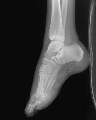
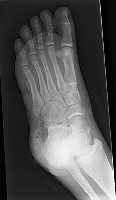
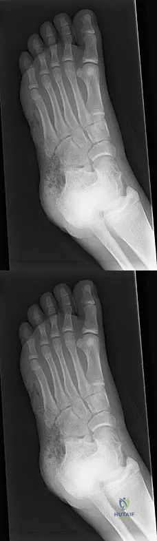
A 17-year-old man sustained a 5-mm laceration on the lateral aspect of the hindfoot while working on a farm. Examination in the emergency department revealed no fractures. Twenty-four hours later, he returns to the emergency department with increasing foot pain. Thin brown drainage is seen emanating from the wound. He has a temperature of 102.0 degrees F (38.9 degrees C), a pulse rate of 120, and a blood pressure of 80/40 mm Hg. Examination of the foot reveals diffuse swelling, ecchymosis, tenderness, and crepitus with palpation. Current radiographs are shown in Figures 40a and 40b. Management should now consist of


Explanation

The mechanism and environment in which the injury occurred, the clinical picture, and the radiographic findings of gas in the tissues suggest an anaerobic Gram-positive bacterial infection. This can be a life- and limb-threatening infection. Treatment should consist of wide debridement of all devitalized tissue, and intravenous antibiotics should be started. Wounds should be left open to allow bacterial effluent and increase oxygen tension in the wound. Hyperbaric oxygen may be used as an adjuvant but is no substitute for debridement. Pellegrini VD, Reid JS, Evarts CM: Complications, in Rockwood CA, Green DP, Bucholz RW, et al (eds): Rockwood and Green's Fractures in Adults, ed 4. Philadelphia, PA, Lippincott-Raven, 1996, vol 1, pp 458-463.


Question 4

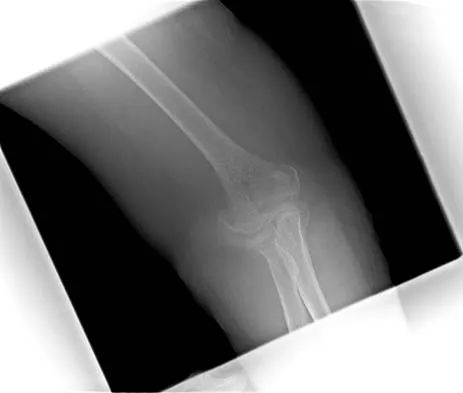
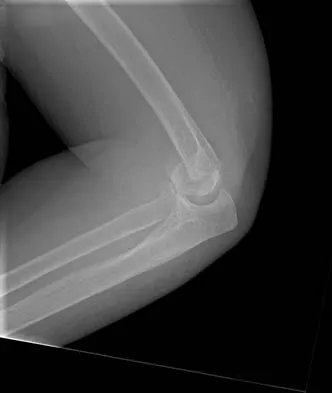
A healthy, active, independent 74-year-old woman fell and sustained the elbow injury shown in Figures 41a and 41b. Management should consist of


Explanation

Open reduction and internal fixation of distal humeral fractures in elderly patients often fails. These fractures characteristically have a very small distal segment and poor bone quality, resulting in failure of fixation and nonunion. Nonunion is often painful and functionally debilitating. Total elbow arthroplasty provides good results when used for distal humeral fractures in elderly patients with osteopenic bone and fracture patterns thought to be irreconstructable. Long arm casting may result in union, but the resulting stiffness is unacceptable for an active patient. Elbow arthrodesis has few indications. A sling and range-of-motion exercises will often result in a painful and debilitating nonunion at the fracture site. Frankle MA, Herscovici D Jr, DiPasquale TG, et al: A comparison of open reduction and internal fixation and primary total elbow arthroplasty in the treatment of intra-articular distal humerus fractures in women older than 65. J Orthop Trauma 2003;17:473-480. Cobb TK, Morrey BF: Total elbow arthroplasty as primary treatment for distal humerus fractures in elderly patients. J Bone Joint Surg Am 1997;79:826-832.


Question 5

A 35-year-old man sustains a closed Monteggia fracture. Examination reveals that sensation, vascular status, and finger flexion are normal. When he extends his wrist, it deviates radially, and he is unable to extend his fingers or thumb. After reduction of the fracture, what is the next step in treatment for the extensor deficits of the thumb and fingers?


Explanation

The posterior interosseous nerve is located adjacent to the radial neck, placing it at risk for a traction injury with a dislocation of the proximal radius. The typical neurapraxia that results can be expected to resolve with observation within the first 6 to 12 weeks. If recovery is not clinically evident by 3 months, neurophysiologic studies are indicated. Jessing P: Monteggia lesions and their complicating nerve damage. Acta Orthop Scand 1975;46:601-609.

Question 6

A 25-year-old man is brought to the emergency department following a motor vehicle accident. Extrication time was 2 hours, and in the field he had a systolic blood pressure by palpation of 90 mm Hg. Intravenous therapy was started, and on arrival in the emergency department he has a systolic blood pressure of 90 mm Hg with a pulse rate of 130. Examination reveals a flail chest and a femoral diaphyseal fracture. Ultrasound of the abdomen is positive. The trauma surgeons take him to the operating room for an exploratory laparotomy. At the conclusion of the procedure, he has a systolic pressure of 100 mm Hg with a pulse rate of 110. Oxygen saturation is 90% on 100% oxygen, and he has a temperature of 95.0 degrees F (35 degrees C). What is the recommended treatment of the femoral fracture at this time?


Explanation

This is a "borderline trauma" patient where serious consideration for damage control orthopaedic surgery is required. His prolonged hypotension, abdominal injury, and chest injury put him at higher risk for serious postinjury complications. Further surgery, such as definitive fracture fixation, adds metabolic load and injury to his system. It is prudent to consider femoral fracture stabilization with an external fixator until he is physiologically recovered as evidenced by a normal base excess and/or lactate acid levels, as well as all other parameters of resuscitation. A borderline patient has been described as polytrauma with an ISS > 20 and thoracic trauma (AIS > 2); polytrauma and abdominal/pelvic trauma (Moore > 3) and hemodynamic shock (initial BP < 90 mm Hg); ISS > 40; bilateral lung contusions on radiographs; initial mean pulmonary arterial pressure > 24 mm Hg; pulmonary artery pressure increase during intramedullary nailing > 6 mm Hg. Factors that worsen the situation following surgery include multiple long bones and truncal injury (AIS > 2), estimated surgery time of more than 6 hours, arterial injury and hemodynamic instability, and exaggerated inflammatory response (eg, Il-6 > 800 pg/mL). It is incumbent on the orthopaedic surgeon who is a member of the trauma team to make sure that he or she is aware of these factors and guides the team to the best patient care. Pape HC, Hildebrand F, Pertschy S, et al: Changes in the management of femoral shaft fractures in polytrauma patients: From early total care to damage control orthopaedic surgery. J Trauma 2002;53:452-461.

Question 7

A 10-year-old girl has a right elbow deformity that is the result of trauma 5 years ago. She has no pain despite the arm deformity. The radiographs in Figures 42a and 42b show complete healing. This radiographic appearance demonstrates what complication?


Explanation

Cubitus varus is a common complication of displaced supracondylar humeral fractures that are treated with closed reduction and cast immobilization. Treatment with closed reduction and percutaneous pinning decreases the incidence of this complication. Cubitus varus also can occur in minimally displaced fractures when unrecognized collapse of the medial column of the distal humerus is not corrected with manipulation. This can be detected on physical examination of the carrying angle or on radiographs measuring Baumann's angle, both in comparison to the opposite side. Cubitus varus may result in unacceptable cosmesis and may predispose the patient to fractures of the lateral condyle. The lateral radiograph demonstrates the crescent sign from overlap of the distal humerus with the olecranon seen in patients with cubitus varus. Patients with growth arrest to the medial trochlear physis would have atrophy of the trochlea on radiographs. Flynn JM, Sarwark JF, Waters PM, et al: The surgical management of pediatric fractures of the upper extremity. Instr Course Lect 2003;52:635-45. Papandrea R, Waters PM: Posttraumatic reconstruction of the elbow in the pediatric patient. Clin Orthop 2000;370:115-126.


Question 8

A 64-year-old woman has left wrist pain and deformity after falling on her hand. Examination shows intact skin and no neurologic or vascular injuries. Radiographs are shown in Figures 43a and 43b. What is the most appropriate management for the injury?


Explanation

The patient has a volar displaced two-part intra-articular distal radial fracture-dislocation of the wrist. Although a closed reduction is usually easily obtained, it is very difficult to maintain the reduction without internal fixation. The approach is determined by the direction of the dislocation, in this case volar. Stabilization with a buttress plate neutralizes the axial loading forces on the fractured fragment. A dorsal placed angular stable plate will not provide this buttress effect and will make the reduction difficult.


Question 9

A 26-year-old man was thrown from a car and sustained the injury seen in Figures 44a and 44b. Nonsurgical management of this injury is recommended. Which of the following factors increases the risk of nonunion?


Explanation

The patient has a displaced comminuted clavicle middle one third fracture from a high-energy mechanism. Recent literature on high-energy clavicular fractures suggests a higher rate of nonunion than previously reported. A nonunion rate of 30% has been reported by Hill and associates when the fracture fragments are displaced more than 1.5 cm. In addition, several patients had neurologic symptoms related to the injury. Robinson and associates reported an increased risk of nonunion in women, elderly patients, comminuted fractures, and injuries with a lack of cortical contact. Hill JM, McGuire MH, Crosby LA: Closed treatment of displaced middle-third fractures of the clavicle gives poor results. J Bone Joint Surg Br 1997;79:537-539. Wick M, Muller EJ, Kollig E: Midshaft fractures of the clavicle with a shortening of more than 2 cm predispose to nonunion. Arch Orthop Trauma Surg 2001;121:207-211.


Question 10

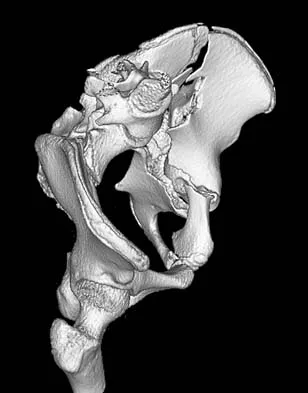
A 13-year-old girl was riding on an all-terrain vehicle when the driver struck a tree. She sustained the injury shown in Figures 45a through 45d. This injury is best described as what type of acetabular fracture pattern?


Explanation

The fracture is a both-column fracture in the Judet/Letournel classification and a C3 in the AO classification. There is extension into the sacroiliac joint along the pelvic brim and comminution along the posterior column above the sciatic notch. Both the anterior and posterior columns are separately broken and displaced. However, the defining feature of a both-column pattern, as seen in this patient, is that all articular fragments are on fracture fragments and no joint surface is left intact to the axial skeleton above. The use of three-dimensional images makes it easier to view the location of the fracture fragments and the amount and direction of displacement. Helfet DL, Beck M, Gautier E, et al: Surgical techniques for acetabular fractures, in Tile M, Helfet DL, Kellam JF (eds): Fractures of the Pelvis and Acetabulum. Philadelphia, PA, Lippincott Williams & Wilkins, 2003, pp 533-603. Tile M: Describing the injury: Classification of acetabular fractures, in Tile M, Helfet DL, Kellam JF (eds): Fractures of the Pelvis and Acetabulum, ed 3. Philadelphia, PA, Lippincott Williams & Wilkins, 2003, pp 427-475.


Question 11

A woman injures the metacarpophalangeal (MCP) joint of her thumb while skiing. Examination reveals tenderness along the ulnar aspect of the MCP joint. Radially directed stress of the joint in full extension produces 5 degrees of angulation. When the MCP joint is flexed 30 degrees, a radially directed stress produces 45 degrees of angulation. Radiographs are otherwise normal. Management should consist of


Explanation

Injuries to the ulnar collateral ligament of the MCP joint of the thumb commonly occur in recreational skiers. Historically, this injury has been referred to as "gamekeeper's thumb." The ligament consists of the proper collateral ligament and the more volar accessory collateral ligament. In extension, the accessory ligament is taut, and in flexion, the proper ligament is taut. For a complete tear of the ligament complex to occur, there must be laxity in full extension. Incomplete tears respond well to thumb spica splinting or casting for 2 to 3 weeks and gradual resumption of range of motion. Prolonged immobilization of incomplete injuries leads to higher rates of MCP joint stiffness. Stener B: Displacement of the ruptured ulnar collateral ligament of the metacarpo-phalangeal joint of the thumb: A clinical and anatomical study. J Bone Joint Surg Br 1971;44:869.

Question 12

A 37-year-old laborer falls 12 feet and sustains a comminuted tibial plafond fracture. Three years after treatment using standard techniques, what will be the most likely outcome?


Explanation

Two recent studies by Pollak and associates and Marsh and associates have focused on function after high-energy tibial plafond fractures. Findings are unfavorable even when anatomic reduction is performed in the best centers and patients are provided excellent rehabilitation. Function improves up to 2 years after injury, but even basic walking skills remain adversely affected. Virtually all patients have long-term adverse general health effects compared to their gender and age-matched peers. Posttraumatic degenerative arthritis is present in most ankles. Patients should be told early about the long-term prognosis, and early vocational/psychological counseling should be given. Despite these adverse outcomes, only a minority of patients require fusion or arthroplasty. Pollak AN, McCarthy ML, Bess RS, et al: Outcomes after treatment of high-energy tibial plafond fractures. J Bone Joint Surg Am 2003;85:1893-1900.

Question 13

A 45-year-old woman sustains an injury to her lower leg. Examination reveals that there is a deformity with no neurologic or vascular problems. The skin is intact. Radiographs are shown in Figures 46a and 46b. Which of the following factors would make closed management the least appropriate choice for this injury?


Explanation

All the factors listed, with the exception of an ipsilateral femoral fracture, are representative of a low-energy stable tibial shaft fracture that will do well with closed reduction and immobilization in a long leg cast, followed by weight bearing as tolerated and then a functional brace or patellar tendon bearing cast until union is achieved. Shortening will not increase from that seen on these initial radiographs. The spiral fracture provides a broad surface for healing, and the fibular fracture at another level indicates a stable soft-tissue envelope which, with the immobilization device, will stabilize the fracture reduction. An ipsilateral femoral fracture is a strong indication to surgically stabilize both fractures. Trafton PG: Tibial shaft fractures, in Browner BD (ed): Skeletal Trauma, ed 3. Philadelphia, PA, WB Saunders, 2003, pp 2153-2169.


Question 14

Which of the following medications may have a negative effect on bone healing following fracture?


Explanation

Nonsteroidal anti-inflammatory drugs that are COX-1 primary inhibitors have been shown in animal studies to delay or inhibit fracture healing. COX-2 inhibitors also delay healing but to a lesser extent than COX-1 inhibitors. The other medications listed do not alter fracture callus formation. Gerstenfeld LC, Thiede M, Seibert K, et al: Differential inhibition of fracture healing by non-selective and cyclooxygenase-2 selective non-steroidal anti-inflammatory drugs. J Orthop Res 2003;21:670-675.

Question 15

A 16-year-old boy has abdominal and back pain after being involved in a high-velocity head-on motor vehicle accident. He was restrained in the rear of the automobile by a lap belt only. A radiograph and CT scan are shown in Figure 47. The patient has no other injuries. Optimal management should include


Explanation

Pediatric bony Chance fractures occur following severe flexion injuries as seen after motor vehicle accidents with lap belt restraints. There is a high rate of associated intra-abdominal injuries. In the absence of associated injuries, these fractures are best treated with immobilization. Bed rest is not necessary. Surgical fixation usually is not needed. Surgical stabilization and two-level fusion may be indicated in select individuals with progressive kyphosis of more than 25 degrees or other conditions that preclude cast or brace immobilization. Greenwald TA, Mann DC: Pediatric seatbelt injuries: Diagnosis and treatment of lumbar flexion-distraction injuries. Paraplegia 1994;32:743-751. Glassman SD, Johnson JR, Holt RT: Seatbelt injuries in children. J Trauma 1992;33:882-886.


Question 16

What inflammatory mediator has been most closely associated with the magnitude of the systemic inflammatory response to trauma and with the development of multiple organ dysfunction syndrome (MODS)?


Explanation

Multiple cytokines (inflammatory mediators) are released following trauma, and their levels can be measured in serum. Persistent elevated levels of IL-6 (> 800 pg/mL) indicate an exaggerated systemic inflammatory response to trauma and have been associated with the development of MODS. Recent work has shown that extensive surgical procedures should be avoided when IL-6 levels remain elevated to prevent the precipitation of MODS. In the future, it is likely that this mediator and possibly others will be used to determine timing and techniques of future treatment. Patrick DA, Moore FA, Moore EE, et al: Jack A. Barney Resident Research Award winner: The inflammatory profile of interleukin-6, interleukin-8, and soluble intercellular adhesion molecule-1 in postinjury multiple organ failure. Am J Surg 1996;172:425-429. Pape HC, van Griesven M, Rice J, et al: Major secondary surgery in blunt trauma patients and perioperative cytokine liberation: Determination of the clinical relevance of biochemical markers. J Trauma 2001;50:989-1000.

Question 17

An 8-year-old girl sustained a displaced fracture at the base of the femoral neck in a motor vehicle accident. Management should consist of


Explanation

Pediatric intracapsular hip fractures are challenging because of the high rates of complications, including osteonecrosis and varus malunion. These patients should be treated as emergencies. Principles of treatment include anatomic reduction with internal fixation. Screw fixation short of the physis is preferred and may need to be supplemented with spica cast immobilization. Fixation may be achieved with smooth pins across the physis when little metaphyseal bone is available. Fixation across the physis with threaded screws is acceptable only when the patient is close to skeletal maturity. Pediatric hip screws are appropriate if immediately available. Emergent open reduction, capsulotomy, or joint aspiration may decrease the rate of osteonecrosis. Cheng JC, Tang N: Decompression and stable internal fixation of femoral neck fractures in children can affect the outcome. J Pediatr Orthop 1999;19:338-343. Azouz EM, Karamitsos C, Reed MH, et al: Types and complications of femoral neck fractures in children. Pediatr Radiol 1993;23:415-420. Song KS, Kim YS, Sohn SW, et al: Arthrotomy and open reduction of the displaced fracture of the femoral neck in children. J Pediatr Orthop B 2001;10:205-210.

Question 18

The plate seen in Figure 48a was applied to the fracture seen in Figure 48b, and is functioning in what capacity?


Explanation

A Weber type B ankle fracture occurs with a supination external rotation mechanism of injury. The fibula generally fails with a spiral fracture pattern. The lag screws provide compression, and the plate acts to neutralize rotational and angular bending forces. A buttress plate resists vertical shear forces. A tension band is used over areas that may fail in tension, such as an olecranon fracture. Compression is provided by the lag screws, and distraction is again resisted by the lag screws.


Question 19

Which of the following findings is considered the strongest indication for surgical treatment of a mallet fracture of the distal phalanx?


Explanation

The majority of mallet fractures can be treated nonsurgically with a distal interphalangeal joint extension splint. Excellent results can be obtained in most patients with splinting alone. The fragment size, amount of displacement, and degree of articular incongruity usually do not affect final outcome, as long as the joint is reduced. Surgical fixation takes on several forms but is fraught with complications including skin/wound problems, loss of fixation, nonunion, and stiffness of the distal interphalangeal joint. Volar subluxation of the distal phalanx remains the primary indication for surgical treatment. Green DP, Butler TE Jr: Fractures and dislocations in the hand, in Rockwood CA, Green DP, Bucholz RW, Heckman JD (eds): Rockwood and Green's Fractures in Adults, ed 4. Philadelphia, PA, Lippincott-Raven, 1996, pp 621-623.

Question 20

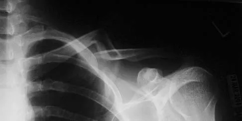
A 20-year-old woman sustained the closed injury shown in Figures 49a and 49b in a motor vehicle accident. Examination reveals that this is an isolated injury; however, she has a complete radial nerve palsy. Management should consist of


Explanation

Lacerated radial nerves are associated with open humeral fractures. All open humeral fractures with radial nerve palsy should be managed with radial nerve exploration and skeletal stabilization. Closed humeral fractures with associated radial nerve palsy usually have an intact nerve with neurapraxia. Most of these patients recover without surgical treatment. If the patient has multiple injuries, skeletal stabilization may be indicated to improve mobilization. For an isolated closed humeral fracture with a radial nerve palsy, the treatment of choice is splinting for 1 to 2 weeks, followed by a humeral fracture brace. Ring D, Chin K, Jupiter JB: Radial nerve palsy associated with high-energy humeral shaft fractures. J Hand Surg Am 2004;29:144-147.


Question 21

The fracture shown in Figure 50 is most reliably treated with what form of fixation?


Explanation

The radiograph shows a comminuted proximal ulnar fracture. The most reliable fixation is a posterior plate, acting as a tension band plate. The fracture involves the proximal shaft of the ulna; therefore, a 3.5-mm compression plate or one of similar size should be used to provide adequate stability. Kirschner wires and tension band wires do not provide axial stability of the comminution of the ulna. Compression screws alone will most likely fail and will not provide axial rotational stability to the construct. A medial plate will not resist the distraction forces across this fracture. McKee MD, Seiler JG, Jupiter JB: The application of the limited contact dynamic compression plate in the upper extremity: An analysis of 114 consecutive cases. Injury 1995;26:661-666.


Question 22

A man sustained the injury shown in Figures 51a and 51b. He underwent closed reduction of the radial head dislocation and open reduction and internal fixation of the ulnar fracture. What is the most common cause of persistent radial head subluxation?


Explanation

The radiographs reveal a Monteggia injury, with a proximal ulnar shaft fracture and a radial head dislocation. Treatment involves open reduction and internal fixation of the ulnar fracture. With correct reduction of the ulna, the radial head is reducible and remains stable, despite an obvious soft-tissue injury around the elbow. Problems with persistent radial head subluxation are almost always attributed to malreduction of the ulnar fracture. Rare causes of persistent radial head subluxation are interposition of soft tissues in the joint and lateral ligamentous injuries. Jupiter JB, Kellam JF: Diaphyseal fractures of the forearm, in Browner B, Jupiter J, Levine A, Trafton P (eds): Skeletal Trauma, ed 2. Philadelphia, PA, WB Saunders, 1992, pp 1421-1454.


Question 23

Which of the following is an indication for surgical management of a Weber type B distal fibular fracture?


Explanation

A widened medial clear space indicates instability caused by an associated tear of the deltoid ligament; therefore, nonsurgical management is not warranted. Weber type B distal fibular fractures without a deltoid tear have a medial clear space of < 4 mm, even under stress, and may be successfully treated nonsurgically. The presence of medial hindfoot ecchymosis and medial ankle tenderness is not fully indicative of medial soft-tissue instability; however, these findings may indicate a deltoid injury and should raise suspicion of an unstable fracture injury pattern. Stress testing is necessary to demonstrate the presence or absence of instability. A small amount of comminution is also cause for increased suspicion of an unstable pattern; however, it is not a direct contraindication when considering nonsurgical management. Two millimeters of fibular displacement without lateral shift of the talus is an acceptable position when considering nonsurgical management of Weber type B distal fibular fractures. Michelson JD, Magid D, Ney DR, et al, Examination of the pathologic anatomy of ankle fractures. J Trauma 1992;32:65-70.

Question 24

Locked plating techniques have been shown to have biomechanical advantages over standard plating in which of the following scenarios?


Explanation

Locked plating is becoming more common. Some biomechanical data comparing locked plating to standard plating have been reported for osteoporotic distal femoral fractures and humeral shaft fractures. Significant differences were seen mainly for osteoporotic fractures without cortical contact. Not all osteoporotic fractures and all comminuted fractures have been shown to demonstrate significant mechanical improvement with locked plating compared to standard plating. Spiral fractures often can be repaired with a lag screw, obtaining adequate cortical contact. Osteoporotic fractures with a torsion mode of failure failed earlier with locked plating systems than with standard plating systems. Zlowodzki M, Williamson S, Cole PA, et al: Biomechanical evaluation of the less invasive stabilization system, angled blade plate, and retrograde intramedullary nail for the internal fixation of distal femur fractures. J Orthop Trauma 2004;18:494-502.

Question 25

A 25-year-old man sustained a head injury after being ejected from his car. Examination reveals a Glasgow Coma Scale score of 7 and a swollen right knee. Clinical examination shows that the knee is very unstable, suggesting tears of the medial collateral and anterior and posterior cruciate ligaments, as well as the posterior lateral corner. What is the most appropriate first step to rule out a vascular injury?


Explanation

A knee dislocation carries the potential for an arterial injury and has always brought up the question of need for arteriography to rule out this limb-threatening injury. However, arteriography has an inherent complication rate that may compromise the general care of the patient. In over 240 published cases with documented knee dislocations that were evaluated for vascular injury by physical examination (without imaging studies), not a single missed injury was reported, for a 100% negative predictive value (0% false-negative rate). This degree of accuracy at excluding major vascular injury is unsurpassed by the results obtained with arteriography but with no risk involved and a marked savings in time, equipment, and costs. Therefore, the most appropriate first step to rule out vascular injury is examination of the pedal pulses. If there is any doubt about an arterial injury, another option is the ankle-brachial index (ABI). If the ABI is greater than 0.9, the chance of arterial injury is again nonexistent. However, a positive physical examination or an ABI of less than 0.9 is not 100% predictive of an arterial injury; therefore, arteriography is recommended. Miranda FE, Dennis JW, Veldenz HC, et al: Confirmation of the safety and accuracy of physical examination in the evaluation of knee dislocation for injury of the popliteal artery: A prospective study. J Trauma 2002;52:247-252.

Question 26

A 45-year-old man is brought to the emergency department after a high-speed motor vehicle collision. He is hypotensive with a blood pressure of 80/40 mm Hg. A pelvic radiograph shows a severely displaced 'open-book' pelvic ring injury. A commercial pelvic binder is ordered. To be most effective, the binder should be centered over which of the following anatomic landmarks?


Explanation

Pelvic binders should be centered over the greater trochanters to effectively reduce pelvic volume and control hemorrhage. Placement over the iliac crests or ASIS is less effective and can paradoxically widen the pelvic ring.

Question 27

A 32-year-old construction worker falls from a scaffolding, sustaining an acetabular fracture. Radiographs and CT imaging reveal a fracture pattern where no portion of the articular surface remains attached to the axial skeleton. Which of the following radiographic findings is pathognomonic for this fracture type?


Explanation

A 'Spur sign' on an obturator oblique radiograph represents the intact ilium projecting posterior to the displaced acetabulum. It is pathognomonic for a both-column acetabular fracture, signifying complete articular detachment.

Question 28

A 28-year-old man sustains a displaced, completely off-ended, trans-cervical femoral neck fracture (Pauwels type III) after a fall from a height. Open reduction and internal fixation are planned. Which of the following constructs provides the most biomechanically stable fixation for this vertically oriented fracture pattern?


Explanation

Pauwels type III fractures are highly vertically oriented and subject to severe shear forces. A sliding hip screw (DHS) with a supplemental anti-rotation screw provides superior biomechanical stability against vertical shear compared to multiple cannulated screws.

Question 29

A 22-year-old athlete sustains a high-energy knee dislocation (KD-III). The knee is successfully reduced in the emergency department. Pulses are symmetric with the uninjured limb, and the Ankle-Brachial Index (ABI) is 0.85. What is the most appropriate next step in management?


Explanation

An ABI less than 0.9 in the setting of a knee dislocation is highly suggestive of an occult vascular injury. CT angiography is strictly indicated to evaluate for a popliteal artery intimal tear or occlusion.

Question 30

A 40-year-old woman is scheduled for open reduction and internal fixation of a severe posterolateral tibial plateau fracture. An isolated posterolateral surgical approach without fibular osteotomy is chosen. Which of the following structures is at greatest risk of iatrogenic injury during this approach?


Explanation

The posterolateral approach to the tibial plateau requires careful dissection to avoid injury to the common peroneal nerve. The nerve runs directly posterior to the fibular head before wrapping anteriorly around the fibular neck.

Question 31

A 75-year-old woman sustains a reverse-oblique intertrochanteric femur fracture (AO/OTA 31-A3). She is medically stable for surgery. Which of the following is the most appropriate fixation implant?


Explanation

Reverse-oblique fractures lack lateral wall support, making sliding hip screws prone to medial displacement and early cutout. A cephalomedullary nail (preferably long) acts as an internal buttress, providing the most stable fixation.

Question 32

A 35-year-old man sustained a Hawkins type II talar neck fracture and underwent open reduction and internal fixation 8 weeks ago. A plain radiograph of the ankle today demonstrates a subchondral radiolucent band in the dome of the talus. What does this finding indicate?


Explanation

The Hawkins sign is a subchondral radiolucency seen 6-8 weeks post-injury, indicating active bone resorption and subchondral osteopenia. This physiological process requires an intact blood supply, thereby essentially ruling out avascular necrosis of the talar body.

Question 33

A 45-year-old man requires ORIF of a displaced posterior wall acetabular fracture via a Kocher-Langenbeck approach. To minimize iatrogenic injury to the sciatic nerve during retraction, how should the ipsilateral lower extremity be positioned intraoperatively?


Explanation

During a Kocher-Langenbeck approach, the sciatic nerve is at high risk from retractor tension. Extending the hip and flexing the knee minimizes tension on the nerve, reducing the risk of a stretch injury.

Question 34

A 26-year-old motorcyclist sustains a coronal plane fracture of the lateral femoral condyle (Hoffa fracture). Open reduction and internal fixation are planned. Which of the following screw configurations provides the most appropriate interfragmentary compression for this specific injury pattern?


Explanation

Hoffa fractures are coronal plane shear fractures of the femoral condyle. Interfragmentary lag screw fixation must be directed orthogonally to the fracture plane, which requires an anterior-to-posterior (or posterior-to-anterior) trajectory.

Question 35

A 42-year-old man presents with a closed, highly comminuted tibial pilon fracture with severe soft tissue swelling and fracture blisters. A spanning external fixator is applied. To avoid tethering of the anterior soft tissues and facilitate future open reduction and internal fixation, how should the foot be positioned during fixator placement?


Explanation

When placing a spanning external fixator for a pilon fracture, the ankle should be held in neutral (0 degrees) to prevent equinus contracture. This optimal position facilitates subsequent definitive fixation and proper soft tissue healing.

Question 36

A 24-year-old soccer player sustains a closed, midshaft tibia fracture treated with intramedullary nailing. Postoperatively, he develops severe, unrelenting leg pain exacerbated by passive stretch of the hallux. If the involved compartment is not rapidly decompressed, which sensory deficit is most likely to develop first?


Explanation

Passive stretch of the hallux exacerbates pain in anterior compartment syndrome. The deep peroneal nerve resides in this compartment, and severe ischemia will lead to sensory loss in its autonomous zone, the first dorsal web space.

Question 37

A 30-year-old woman presents with midfoot pain after falling from a horse with her foot caught in the stirrup. Radiographs show a small bony avulsion in the space between the medial and middle cuneiforms. This 'fleck sign' represents an avulsion of a ligament that connects the medial cuneiform to which structure?


Explanation

The Lisfranc ligament is a stout intra-articular ligament that connects the lateral aspect of the medial cuneiform to the medial aspect of the base of the second metatarsal. A 'fleck sign' indicates an avulsion of this critical stabilizing ligament.

Question 38

A 45-year-old man undergoes tension band wiring for a transverse patella fracture. According to the tension band principle, the wire construct works by converting what type of force at the anterior patellar surface into a compressive force at the articular surface during knee flexion?


Explanation

The tension band principle relies on converting tensile forces (which occur on the convex, anterior surface of the patella during knee flexion) into dynamic compressive forces at the concave articular surface.

Question 39

A 45-year-old man is brought to the trauma bay in hemorrhagic shock following a motorcycle crash. A pelvic radiograph demonstrates an open-book pelvic ring injury.

What is the most appropriate anatomical landmark for the optimal placement of a circumferential pelvic sheet or binder?


Explanation

A pelvic binder should be centered directly over the greater trochanters to effectively close the pelvic ring and reduce intrapelvic volume. Placement over the iliac crests is a common error that can fail to reduce the diastasis or paradoxically widen the pelvis.

Question 40

During an ilioinguinal approach for the fixation of an anterior column acetabular fracture, the surgeon operates in the middle window. Which of the following structures defines the medial border of this surgical window?


Explanation

The middle window of the ilioinguinal approach is bordered laterally by the iliopectineal fascia and medially by the external iliac vessels. This window provides access to the pelvic brim, quadrilateral plate, and superior pubic ramus.

Question 41

A 28-year-old man sustains a displaced, high-shear vertical (Pauwels type III) femoral neck fracture. Open reduction and internal fixation is planned. What is the most common mechanical mode of failure for this specific fracture pattern when treated with three parallel cancellous lag screws?


Explanation

Pauwels type III fractures experience high vertical shear forces across the fracture site. Fixation with parallel cancellous screws alone often fails to resist these shear forces, predictably leading to varus collapse, limb shortening, and subsequent nonunion.

Question 42

A surgeon is treating a proximal third tibial shaft fracture with an intramedullary nail. Apex anterior (procurvatum) and valgus deformities are anticipated during nail passage. Where should blocking (Poller) screws be placed in the proximal fragment to prevent this malalignment?


Explanation

Blocking screws should be placed on the concave side of the anticipated deformity to centralize the nail. To prevent procurvatum (nail goes posterior) and valgus (nail goes lateral) in the proximal segment, screws are placed posterior and lateral to the nail track.

Question 43

A 35-year-old man sustains a subtrochanteric femur fracture. Without specific reduction maneuvers, what is the predictable deformity of the proximal fragment due to the muscular deforming forces?


Explanation

The proximal fragment in a subtrochanteric fracture is classically flexed by the iliopsoas, abducted by the gluteus medius and minimus, and externally rotated by the short external rotators.

Question 44

A 25-year-old man falls from a height of 20 feet. Imaging reveals a Zone 3 sacral fracture according to the Denis classification. What is the most likely neurologic deficit associated with this specific injury pattern?


Explanation

Zone 3 sacral fractures involve the central sacral canal. These injuries have the highest rate of neurologic compromise (up to 60%), most commonly presenting as cauda equina syndrome with bowel and bladder dysfunction.

Question 45

A 35-year-old man sustains a posterior wall acetabular fracture with severe marginal impaction of the articular cartilage after a motor vehicle collision. He is planned for open reduction and internal fixation via a Kocher-Langenbeck approach. What is the most critical intraoperative step for managing the marginal impaction to prevent early osteoarthritis?


Explanation

Marginal impaction in posterior wall acetabular fractures represents osteochondral fragments driven into the underlying cancellous bone. These must be carefully elevated to restore joint congruity, and the resulting void must be filled with bone graft prior to definitive wall fixation.

Question 46

During an ilioinguinal approach for an anterior column acetabular fracture, brisk arterial bleeding is encountered near the superior pubic ramus approximately 5 cm from the pubic symphysis. This bleeding is most likely arising from an anastomosis between which of the following vessel pairs?


Explanation

The corona mortis is a critical vascular anastomosis between the external iliac (or inferior epigastric) system and the obturator vessels. It is found traversing the superior pubic ramus and is highly vulnerable during anterior pelvic and acetabular approaches.

Question 47

A 28-year-old man sustains a highly vertical (Pauwels type III) basicervical femoral neck fracture. To maximize biomechanical stability and minimize the risk of shear failure, which of the following constructs is most appropriate?


Explanation

Vertical Pauwels type III fractures experience high shear forces. In a young patient, a sliding hip screw with a derotation screw offers superior biomechanical stability against shear and varus collapse compared to multiple cancellous screws.

Question 48

An orthopedic trauma surgeon is performing antegrade intramedullary nailing of a femoral shaft fracture. The starting point is chosen at the piriformis fossa. If the guide wire is inadvertently placed too anteriorly on the femoral neck, which of the following iatrogenic complications is most likely to occur?


Explanation

An anterior starting point for a piriformis entry nail misses the central axis of the medullary canal and places excessive stress on the anterior femoral neck during reaming and nail insertion, highly risking an iatrogenic femoral neck fracture.

Question 49

A 45-year-old woman presents with a medial tibial plateau fracture with extension into the intercondylar eminence (Schatzker type IV). The fracture includes a significantly displaced posteromedial fragment. What is the most appropriate surgical approach and fixation strategy?


Explanation

Schatzker IV fractures involve the medial plateau and commonly feature a posteromedial shear fragment. A posteromedial approach allows for direct reduction and placement of an anti-glide plate to effectively neutralize the deforming shear forces.

Question 50

A 32-year-old man undergoes intramedullary nailing for a closed transverse tibial shaft fracture. In the recovery room, he requires increasing doses of opioids and exhibits severe pain with passive stretch of the hallux. His diastolic blood pressure is 70 mm Hg, and an intracompartmental pressure reading of the anterior compartment is 55 mm Hg. What is the most appropriate immediate management?


Explanation

The patient has clinical acute compartment syndrome confirmed by a delta P (diastolic pressure minus compartment pressure) of less than 30 mm Hg. The standard of care is emergent four-compartment fasciotomy to prevent irreversible muscle and nerve necrosis.

Question 51

A 25-year-old man sustained a Hawkins type II talar neck fracture and underwent open reduction and internal fixation. At his 6-week follow-up, a mortise radiograph reveals a subchondral radiolucent band across the dome of the talus (Hawkins sign). What does this radiographic finding indicate?


Explanation

The Hawkins sign represents subchondral osteopenia resulting from disuse hyperemia. Its presence indicates that the blood supply to the talar body is intact, predicting a very low risk of subsequent avascular necrosis.

Question 52

A 50-year-old male smoker with poorly controlled diabetes undergoes open reduction and internal fixation of a displaced intra-articular calcaneus fracture via an extensile lateral approach. Which of the following is the most common complication associated with this specific approach?


Explanation

The extensile lateral approach for calcaneus fractures relies on a full-thickness fasciocutaneous flap supplied by the lateral calcaneal artery. Wound complications, such as edge necrosis and dehiscence, occur in up to 25% of cases, particularly in smokers and diabetics.

Question 53

A 22-year-old construction worker falls from a height and sustains a vertical shear pelvic fracture. Radiographs show superior displacement of the right hemipelvis and avulsion of the right L5 transverse process. Which of the following ligamentous complexes are completely disrupted in this injury pattern?


Explanation

A vertical shear fracture pattern involves global instability of the hemipelvis. It necessitates the complete disruption of the anterior and posterior pelvic ring, including both the anterior and posterior sacroiliac ligaments, as well as the pelvic floor (sacrotuberous and sacrospinous ligaments).

Question 54

An anteroposterior (AP) radiograph of the pelvis is obtained for a 60-year-old woman who fell from a ladder. The film demonstrates an isolated disruption of the iliopectineal line with a completely intact ilioischial line. Which acetabular structure is fractured?


Explanation

On an AP radiograph of the pelvis, the iliopectineal line correlates anatomically with the anterior column of the acetabulum, while the ilioischial line correlates with the posterior column.

Question 55

A 68-year-old woman on long-term alendronate therapy presents with chronic left thigh pain. Radiographs demonstrate lateral cortical thickening and a transverse radiolucent line in the subtrochanteric region of the femur. Which of the following describes the fundamental pathophysiology of this specific fracture type?


Explanation

Bisphosphonate-related atypical femoral fractures are stress fractures that occur due to prolonged, severe suppression of normal bone turnover. This prevents the remodeling of normal microdamage, eventually leading to structural failure on the tension (lateral) side of the femur.

Question 56

A 32-year-old man is involved in a high-speed motor vehicle collision and sustains a displaced posterior wall acetabular fracture with a posterior hip dislocation.

Following emergent closed reduction of the hip, CT imaging confirms a large, single-piece posterior wall fragment. Which of the following surgical approaches is most appropriate for definitive fixation?


Explanation

The Kocher-Langenbeck approach is the standard approach for isolated posterior wall and posterior column acetabular fractures. It allows direct visualization and buttress plating of the posterior wall.

Question 57

A hemodynamically unstable 45-year-old woman is brought to the trauma bay after a crush injury. Radiographs show an anterior-posterior compression type III (APC-III) pelvic ring injury with complete disruption of the sacroiliac joints.

The initial management to stabilize the pelvic volume should be placement of a pelvic binder centered over which of the following anatomic landmarks?


Explanation

A pelvic binder must be centered over the greater trochanters to effectively close the pelvic ring and reduce pelvic volume. Placement over the iliac crests is incorrect and can paradoxically worsen the deformity.

Question 58

A 28-year-old man sustains a completely displaced, highly vertical (Pauwels type III) femoral neck fracture. Which of the following fixation constructs provides the highest biomechanical stability for this specific fracture pattern?


Explanation

Pauwels type III femoral neck fractures in young adults experience high shear forces. A fixed-angle device, such as a sliding hip screw with a derotational screw, provides superior biomechanical stability compared to multiple cancellous screws.

Question 59

An 80-year-old woman presents with a reverse obliquity intertrochanteric femur fracture after a ground-level fall. Which of the following implants is most appropriate to prevent medial shaft displacement and failure?


Explanation

Reverse obliquity and fractures with lateral wall incompetence are highly unstable and do poorly with sliding hip screws due to lateral translation. A cephalomedullary nail provides the necessary intramedullary support to prevent this displacement.

Question 60

A 24-year-old polytrauma patient presents with bilateral closed femoral shaft fractures, severe pulmonary contusions, and an initial lactate of 5.0 mmol/L.

What is the most appropriate initial management of the femoral fractures?


Explanation

In a borderline or unstable polytrauma patient with significant pulmonary injury and elevated lactate, damage control orthopedics (temporary external fixation) is indicated. Early total care (intramedullary nailing) increases the risk of ARDS and systemic inflammatory response syndrome (SIRS) in this population.

Question 61

During open reduction and internal fixation of a distal femur fracture, you identify a separate coronal plane fracture of the lateral femoral condyle (Hoffa fragment). What is the optimal fixation strategy for this specific fragment?


Explanation

A Hoffa fracture is a coronal shear fracture of the femoral condyle. It requires fixation perpendicular to the fracture plane, typically using anterior-to-posterior (AP) or posterior-to-anterior (PA) lag screws.

Question 62

A 35-year-old man undergoes intramedullary nailing for a proximal-third tibial shaft fracture. Intraoperatively, the fracture demonstrates an apex anterior (procurvatum) and valgus deformity. Which of the following techniques is most effective in preventing this deformity?


Explanation

Proximal-third tibia fractures are notorious for apex anterior and valgus deformities during nailing. Blocking screws placed posterior to the nail (to prevent procurvatum) and lateral to the nail (to prevent valgus) in the proximal fragment help guide the nail and maintain reduction.

Question 63

A 45-year-old man sustains a Schatzker IV tibial plateau fracture (medial plateau) from a high-energy varus force.

Which of the following approaches is most appropriate for direct visualization and buttressing of this fracture?


Explanation

Schatzker IV fractures typically involve a posteromedial shear fragment. A posteromedial approach allows for an anti-glide or buttress plate to be applied directly over the apex of the fracture, providing stable fixation.

Question 64

A 30-year-old construction worker falls from a roof, sustaining a closed, highly comminuted tibial pilon fracture with massive soft tissue swelling and fracture blisters. What is the most appropriate definitive management sequence?


Explanation

High-energy pilon fractures with severe soft tissue compromise are best managed with staged protocols. Initial spanning external fixation protects the soft tissues, allowing swelling to subside (wrinkle sign) before definitive internal fixation is safely performed.

Question 65

A 28-year-old male sustains a Hawkins type III fracture of the talar neck. Which of the following accurately describes the associated dislocations and the approximate risk of avascular necrosis (AVN)?


Explanation

A Hawkins type III talar neck fracture involves displacement with dislocation of the subtalar, tibiotalar, and often talonavicular joints. Because all major blood supplies to the talar body are disrupted, the risk of AVN is exceptionally high, approaching 90-100%.

Question 66

During the extensile lateral approach for open reduction and internal fixation of a displaced intra-articular calcaneus fracture, which nerve is at greatest risk of iatrogenic injury in the distal portion of the incision?


Explanation

The sural nerve crosses the lateral border of the foot and is at risk during the distal extent of the extensile lateral approach to the calcaneus. Careful full-thickness flap elevation is required to protect it.

Question 67

A 22-year-old college football player sustains a purely ligamentous Lisfranc injury. Weight-bearing radiographs show a 4 mm diastasis between the first and second metatarsal bases. What is the most widely supported definitive surgical treatment for this specific injury pattern?


Explanation

Evidence suggests that primary arthrodesis yields better long-term functional outcomes and lower reoperation rates compared to ORIF for purely ligamentous Lisfranc injuries. ORIF is generally preferred if there are large, stable bony avulsions.

Question 68

When utilizing the ilioinguinal approach for an anterior column acetabular fracture, the surgeon must identify and ligate the 'corona mortis'. This structure represents an anastomosis between the obturator vessels and the:


Explanation

The corona mortis is an important vascular anastomosis crossing the superior pubic ramus. It connects the obturator vessels with the external iliac or deep inferior epigastric vessels and can cause massive hemorrhage if torn.

Question 69

A 50-year-old man is brought to the emergency department after a crushing injury to his pelvis.

He has blood at the urethral meatus and a high-riding prostate on digital rectal exam. What is the most appropriate next step in his urologic evaluation?


Explanation

Blood at the meatus and a high-riding prostate indicate a potential urethral injury. A retrograde urethrogram (RUG) must be performed before any attempt at Foley catheter placement to avoid converting a partial tear into a complete transection.

Question 70

During internal fixation of a bimalleolar ankle fracture, the surgeon performs a 'Cotton test' pulling the fibula laterally. Which specific anatomic structure is this test primarily designed to evaluate?


Explanation

The Cotton test (or hook test) involves applying lateral traction to the fibula using a bone hook. It is performed intraoperatively to assess the stability of the distal tibiofibular syndesmosis after medial and lateral bony fixation.

Question 71

A 45-year-old man is brought to the trauma bay following a motorcycle collision. He is hypotensive with a blood pressure of 75/40 mm Hg. Radiographs reveal an anteroposterior compression type III (APC-III) pelvic ring injury. A pelvic binder is applied, and he receives 2 units of uncrossmatched packed red blood cells, but remains hemodynamically unstable. A FAST exam is negative. What is the most appropriate next step in management?


Explanation

In a hemodynamically unstable patient with a pelvic fracture and negative FAST, the source of bleeding is presumed to be the pelvis. Immediate preperitoneal pelvic packing or angiography is indicated to control hemorrhage.

Question 72

A 32-year-old man sustains a posterior hip dislocation and an associated posterior wall acetabular fracture. Following closed reduction, a CT scan of the pelvis is obtained, which demonstrates a 15-mm area of marginal impaction of the articular cartilage. What is the most critical step during the operative management of this fracture?


Explanation

Marginal impaction involves osteochondral fragments pushed into the underlying cancellous bone. Successful ORIF requires elevation of these fragments to restore joint congruity, followed by bone grafting of the underlying void.

Question 73

A 65-year-old woman sustains a subtrochanteric femur fracture. During closed reduction prior to intramedullary nailing, the proximal fragment is noted to be severely displaced. Which of the following best describes the typical deforming forces acting on the proximal fragment?


Explanation

The proximal fragment in a subtrochanteric fracture is flexed by the iliopsoas, abducted by the gluteus medius and minimus, and externally rotated by the short external rotators.

Question 74

A 40-year-old man presents with a high-energy bicondylar tibial plateau fracture (Schatzker VI). CT imaging reveals a large, displaced posteromedial fragment. Biomechanically, what is the most appropriate surgical strategy for addressing this specific fragment?


Explanation

The posteromedial fragment in bicondylar tibial plateau fractures typically has a vertical shear pattern. It is best managed via a posteromedial approach using an apex-distal plate in a buttress mode.

Question 75

A 24-year-old man undergoes closed reduction and percutaneous pinning for a Hawkins type II talar neck fracture. Radiographs obtained 8 weeks postoperatively demonstrate a distinct subchondral radiolucent band in the dome of the talus. What does this radiographic finding indicate?


Explanation

A subchondral radiolucent band in the talar dome (Hawkins sign) represents subchondral atrophy from disuse in the setting of an intact blood supply, indicating that the talar body remains viable.

Question 76

A 38-year-old woman is evaluated for a closed pelvic ring injury after a pedestrian-versus-auto accident. Examination reveals a large, fluctuant soft-tissue swelling over the greater trochanter with overlying skin ecchymosis and decreased sensation. What is the pathophysiology of this associated soft-tissue injury?


Explanation

This describes a Morel-Lavallée lesion, a closed degloving injury where subcutaneous tissue is sheared off the underlying fascia. This disrupts perforating vessels, creating a cavity that fills with blood, lymph, and necrotic fat.

Question 77

During the surgical fixation of an anterior column acetabular fracture via an ilioinguinal approach, significant arterial hemorrhage is encountered posterior to the superior pubic ramus. This bleeding is most likely originating from an anastomosis between the obturator vessels and which of the following?


Explanation

The corona mortis is a vascular anastomosis between the obturator vessels and the external iliac or inferior epigastric vessels. It crosses the superior pubic ramus and is at high risk during the ilioinguinal or Stoppa approaches.

Question 78

A 26-year-old man sustains a completely displaced, vertically oriented femoral neck fracture (Pauwels type III). Given his young age, joint-preserving surgery is planned. Biomechanically, which of the following construct choices offers the greatest resistance to vertical shear forces?


Explanation

Pauwels III (vertical) fractures experience high shear forces. A sliding hip screw (fixed-angle device) combined with a derotation screw provides superior biomechanical stability against vertical shear compared to multiple cancellous screws.

Question 79

A 35-year-old man presents with an anteroposterior compression type III (APC-III) pelvic ring injury following a motorcycle collision. He arrives hypotensive, and a pelvic binder is applied. After receiving 2 units of packed red blood cells, his blood pressure remains 75/40 mm Hg. A Focused Assessment with Sonography for Trauma (FAST) exam is negative. What is the most appropriate next step in management?


Explanation

In a hemodynamically unstable patient with a pelvic ring injury and a negative FAST exam, the source of bleeding is likely retroperitoneal. Preperitoneal pelvic packing or pelvic angiography with embolization are the interventions of choice. Exploratory laparotomy is contraindicated for isolated extraperitoneal bleeding.

Question 80

During the surgical management of an anterior column acetabular fracture via the modified Stoppa approach, a retropubic vascular anastomosis is encountered. Injury to this structure, often called the "corona mortis," can cause massive hemorrhage. This structure typically represents an anastomosis between which of the following vessels?


Explanation

The corona mortis is a critical retropubic vascular anastomosis connecting the external iliac system (typically the inferior epigastric vessels) and the internal iliac system (obturator vessels). It crosses the superior pubic ramus and is highly vulnerable to iatrogenic injury during the ilioinguinal or modified Stoppa approaches.

Question 81

A 40-year-old man falls from a height and sustains a displaced acetabular fracture. Plain radiographs demonstrate the "spur sign" on the obturator oblique view. Which of the following acetabular fracture patterns is pathognomonic for this radiographic finding?


Explanation

The "spur sign" represents the fractured inferior aspect of the intact iliac wing (the strut) protruding outward and is classically seen on the obturator oblique radiograph. It is pathognomonic for a both-column acetabular fracture, indicating complete dissociation of all articular segments from the axial skeleton.

Question 82

A 24-year-old male sustains a vertically oriented, displaced femoral neck fracture (Pauwels type III) after a fall from a roof. To minimize shear forces and maximize biomechanical stability, which of the following constructs is most appropriate for definitive fixation?


Explanation

Pauwels type III femoral neck fractures have a vertical fracture line that subjects the repair to high shear forces, increasing the risk of varus collapse and nonunion. A fixed-angle device such as a sliding hip screw, supplemented with a derotational screw, provides superior biomechanical stability compared to multiple parallel cancellous screws.

Question 83

A 38-year-old male is brought to the emergency department after a severe crush injury to his right leg. Radiographs reveal a highly comminuted Schatzker VI tibial plateau fracture. He complains of severe, unrelenting leg pain despite intravenous narcotics. Compartment pressure testing reveals an absolute anterior compartment pressure of 42 mm Hg. His current blood pressure is 110/65 mm Hg. What is the most appropriate management?


Explanation

The patient has a delta pressure (Diastolic BP - Compartment Pressure) of 23 mm Hg (65 - 42). A delta pressure of 30 mm Hg or less is the threshold for diagnosing acute compartment syndrome, necessitating emergent four-compartment fasciotomy. A bridging external fixator provides necessary skeletal stability while deferring definitive fixation until soft tissues heal.

Question 84

A 28-year-old female presents with a closed Hawkins type III fracture of the talar neck (associated with tibiotalar and subtalar dislocations). Despite prompt and anatomic open reduction and internal fixation, the surgeon counsels her regarding the high risk of a specific complication. What is the approximate risk of avascular necrosis (AVN) of the talar body in this injury pattern?


Explanation

Hawkins type III fractures involve disruption of the three major blood supplies to the talar body (artery of the tarsal canal, deltoid branch, and branches from the dorsalis pedis). Consequently, the incidence of avascular necrosis in these high-energy injuries approaches 75% to 100%.

Question 85

A 50-year-old construction worker presents with a severe, displaced OTA/AO type 43C3 (pilon) fracture of the distal tibia. Examination reveals marked soft tissue swelling, hemorrhagic fracture blisters, and threatened skin over the medial malleolus. What is the current standard of care for the initial orthopedic management of this injury?


Explanation

High-energy pilon fractures are associated with profound soft-tissue compromise. The standard of care is a two-staged approach: immediate application of a spanning external fixator to restore length and alignment, followed by definitive internal fixation 10-21 days later once the soft tissue swelling and blisters have resolved.

Question 86

A 26-year-old man sustains bilateral closed femoral shaft fractures, severe pulmonary contusions, and a severe closed head injury (GCS 6) following a motor vehicle accident. He requires aggressive resuscitation and is chemically paralyzed. According to the principles of Damage Control Orthopedics (DCO), what is the most appropriate initial management of his femoral fractures?


Explanation

In a polytrauma patient who is unstable, borderline, or "in extremis" (e.g., severe head injury, lung injury, or shock), the Damage Control Orthopedics (DCO) protocol dictates rapid stabilization to minimize the "second hit." Application of temporary spanning external fixators is the safest and most effective initial treatment.

Question 87

During the extensile lateral approach for open reduction and internal fixation of a displaced intra-articular calcaneus fracture, the surgeon elevates a full-thickness "no-touch" subperiosteal flap. Which of the following nerves is located within this flap and is at highest risk for iatrogenic injury or entrapment during retraction?


Explanation

The sural nerve courses along the lateral aspect of the hindfoot and is intimately involved with the soft tissues elevated during an extensile lateral approach to the calcaneus. A full-thickness subperiosteal flap must be elevated carefully to keep the sural nerve protected within the flap and avoid direct trauma or retraction injury.

Question 88

A 68-year-old woman sustains a lateral compression type 1 (LC-1) pelvic ring injury after a ground-level fall, presenting with a sacral ala fracture and ipsilateral rami fractures. She is hemodynamically stable. Radiographs show less than 1 cm of posterior displacement. What is the most appropriate initial management strategy?


Explanation

LC-1 pelvic ring fractures are inherently stable, both rotationally and vertically, due to intact posterior tension band ligaments. In the absence of extreme pain or dynamic instability, nonoperative management with protected weight-bearing and early mobilization is the standard of care.

None

Clinic OS
Medically Verified Content by
Prof. Clinic OS
Consultant Orthopedic & Spine Surgeon
Chapter Index