العربية
Part of the Master Guide

Orthopedic Board Review Set 675: 100 MCQs for ABOS, OITE, FRCS – Hip Focus

AAOS Pediatric Orthopedics (Set 2): DDH, SCFE & Femur Fractures | Board Review

23 Apr 2026 63 min read 96 Views
Pediatrics 2004 MCQs - Part 2

Key Takeaway

This high-yield question set for the AAOS/ABOS/OITE exams focuses on crucial pediatric orthopedic topics. It covers the diagnosis and management of Developmental Dysplasia of the Hip (DDH), Slipped Capital Femoral Epiphysis (SCFE), and common pediatric femur fractures, essential for board review.

AAOS Pediatric Orthopedics (Set 2): DDH, SCFE & Femur Fractures | Board Review

Comprehensive 100-Question Exam


00:00

Start Quiz

Question 1

Figures 11a and 11b show the clinical photograph and radiograph of a newborn. Based on these findings, what is the best course of action?


Explanation

The newborn has posteromedial bowing of the tibia and calcaneal valgus deformity of the foot. Both are thought to be caused by abnormal intrauterine positioning. The foot deformity typically responds to stretching. The tibial bowing straightens with growth. The long-term problem is limb-length discrepancy. Heyman CH, Herndon CH, Heiple KG: Congenital posterior angulation of the tibia with talipes calcaneus. J Bone Joint Surg Am 1959;41:476-488.

Question 2

Figure 12 shows the radiograph of a patient who has anterior knee pain. History reveals a femoral fracture at age 5 years. What is the most likely cause of the deformity?


Explanation

The radiograph shows a recurvatum deformity of the proximal tibia with growth arrest of the tibial tubercle apophysis. This deformity has been described in association with femoral shaft fractures in children and has been attributed to a clinically silent, concommitant injury to the proximal tibial physes and also to iatrogenic injury associated with a proximal tibial traction pin. Overlengthened hamstrings and rupture of the posterior cruciate ligament may lead to knee hyperextension; however, these problems should not cause bone deformity. Osgood-Schlatter disease occurs when growth is nearly complete and usually leads to prominence of the tibial tubercle. Patellar tendon rupture is rare in children and would not cause this deformity unless the repair was performed with screws across the apophysis. Hresko MT, Kasser JR: Physeal arrest about the knee associated with non-physeal injuries of the lower extremity. J Bone Joint Surg Am 1989;71:698-703.


Question 3

An 11-year-old boy has had a fever and pain and swelling over the lateral aspect of his right ankle for the past 3 days. Examination reveals warmth, swelling, and tenderness over the lateral malleolus, and he has a temperature of 103.2 degrees F (39.5 degrees C). Laboratory studies show a WBC count of 13,200/mm3 with 61% neutrophils, an erythocyte sedimentation rate of 112 mm/h, and a C-reactive protein of 15.7. Radiographs and a T2-weighted MRI scan are shown in Figures 13a through 13c. Aspiration yields 1 mL of purulent fluid. Management should now consist of


Explanation

The initial signs and symptoms of acute hematogenous osteomyelitis vary widely but usually include fever, bone pain, and impaired use of the involved extremity. In lower extremity infections, the child may limp or refuse to walk. Examination often reveals bone tenderness. In more advanced cases, erythema, warmth, and swelling may be present. The WBC and neutrophil counts are not always elevated, but the erythocyte sedimentation rate will be abnormal in more than 90% of patients. When the infection is diagnosed early, before a subperiosteal abscess has formed, antibiotics alone may be adequate to treat the infection. This patient has a more advanced infection, however, with the MRI scan revealing a subperiosteal abscess that was confirmed by aspiration. When an abscess is present, surgical drainage is generally indicated to remove devitalized tissue and to enhance the efficacy of the antibiotics. Further studies, such as bone or indium scans, are not necessary and will delay definitive treatment. Scott RJ, Christofersen MR, Robertson WW Jr, et al: Acute osteomyelitis in children: A review of 116 cases. J Pediatr Orthop 1990;10:649-652.


Question 4

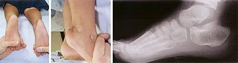
Figure 14 shows the clinical photographs and radiograph of an 8-year-old girl who has a progressive equinus deformity of the right ankle. There is no history of trauma or infection. What is the most likely diagnosis?


Explanation

Focal scleroderma is characterized by the formation of patches of sclerotic skin, also known as morphea, or streaks of sclerosis (linear scleroderma). Systemic involvement in focal scleroderma is unusual; however, progression during childhood is common. Contracture of underlying tissues is common, often resulting in serious joint contractures. Bony changes similar to those seen in melorheostosis can be seen. This patient has characteristic skin changes, atrophy of the soft tissues, Achilles tendon contractures, and calcaneal deformities. There are no signs of arthrogryposis, which usually presents with bilateral congenital deformities, including equinovarus. Klippel-Trenaunay-Weber syndrome is characterized by venous malformation in association with focal overgrowth.


Question 5

Which of the following patients is considered the most appropriate candidate for an isolated split posterior tendon transfer?


Explanation

Isolated split posterior tendon transfer alone is best performed in a patient with cerebral palsy who is between the ages of 4 and 7 years and has a flexible equinovarus foot. Rigid deformities often must be managed with a combination of soft-tissue and bony procedures. Patients with out-of-phase activity may be best managed with a transfer of the posterior tibialis to the dorsum of the foot, while those with continuous activity are better candidates for an isolated split posterior tendon transfer. Drennan JC (ed): The Child's Foot and Ankle. New York, NY, Raven Press, 1992, pp 291-294. Green NE, Griffin PP, Shiavi R: Split posterior tibial-tendon transfer in cerebral palsy. J Bone Joint Surg Am 1983;65:748-754.


Question 6

The mother of a 2-year-old boy reports that he had pain in the right hip all night and refuses to walk on the leg this morning. He is afebrile. Examination reveals pain on hip extension and adduction, but he is able to internally and externally rotate the hip approximately 20 degrees in each direction without pain. Laboratory studies reveal a WBC count of 7,400/mm3, with 62% polymorphonuclear neutrophil leukocytes. The AP radiograph shown in Figure 15 reveals a left teardrop distance of 8 mm, while the right side measures 10 mm. Which of the following diagnostic studies will best help confirm the diagnosis?


Explanation

The differential diagnosis includes septic hip and transient synovitis. Both disorders may present with a joint effusion. The increased teardrop distance and loss of range of motion implies that there is excess fluid in the joint. While the other tests can confirm this, only aspiration can characterize the fluid further, thereby indicating the etiology of the effusion. Provided there is enough fluid obtained at aspiration, the joint fluid should be sent for Gram stain, culture, cell count, glucose, and protein studies.


Question 7

A senior resident is scheduled to perform a posterior medial release on a 10-month-old infant who has a congenital clubfoot deformity. Informed consent is obtained for the procedure. The supervising surgeon is obligated to give the parents what information?


Explanation

Informed consent is generally considered to be a process of mutual decision making between the physician and patient. The physician is required to provide to the patient all material information that is needed for the patient to make an informed decision. The courts have held that a patient's choice of surgeon is as important to the consent as the procedure itself. Assistance by a surgical trainee with adequate supervision is permissible when there has been adequate disclosure. Adequate supervision may be defined as active participation by the attending during the essential parts of the procedure. Allowing a substitute surgeon to operate on a patient without the patient's knowledge "ghost surgery" may result in charges of battery against the substitute surgeon and malpractice against the surgeon to whom the patient gave consent. Kocher MS: Ghost surgery: The ethical and legal implications of who does the operation. J Bone Joint Surg Am 2002;84:148-150.


Question 8

Figure 16 shows the clinical photograph of a 3-month-old infant with a foot deformity that has been nonprogressive since birth. Examination reveals that the deformity corrects actively and with passive manipulation. There is no associated equinus. Management should consist of


Explanation

The patient has bilateral metatarsus adductus deformities. In a long-term follow-up study by Farsetti and associates, deformities that were passively correctable spontaneously resolved and no treatment was required. More rigid deformities were successfully treated with serial manipulation, with good results in 90%. There were no poor results. Therefore, observation is the management of choice for passively correctable deformities. In feet that are more rigid, serial manipulation and casting is the management of choice.


Question 9

Figure 17 shows the radiograph of an 11-year-old boy with Duchenne muscular dystrophy who has been nonambulatory for the past 2 years. Management of the spinal deformity should consist of


Explanation

The presence of any curve greater than 20 degrees in a nonambulatory patient with Duchenne muscular dystrophy is an indication for posterior fusion with instrumentation. Because of progressive cardiomyopathy and pulmonary deficiency, waiting until the curve is larger can increase the risk of pulmonary or cardiac complications during or following surgery. There is some disagreement as to whether all such fusions must extend to the pelvis. Bracing or other nonsurgical management is ineffective and is not indicated in this situation. Sussman M: Duchenne muscular dystrophy. J Am Acad Orthop Surg 2002;10:138-151.


Question 10

A 13-year-old girl with Down syndrome has bilateral chronic patellar dislocations. She denies knee pain. She is able to straighten her knees and walks with a symmetric but awkward gait. She does not flex her knees in midstance. Examination reveals that the patellae cannot be brought into a reduced position. Management should consist of


Explanation

Chronic dislocation of the patella is occasionally seen in patients with Down syndrome. In early childhood, patellar realignment may restore stability of the patellae. In later childhood, bony changes in the patellar groove interfere with stability, even if surgical realignment is performed. Realignment can also lead to increased knee pain postoperatively. In asymptomatic patients who are able to extend their knees, continued observation is the management of choice. Dugdale TW, Renshaw TS: Instability of the patellofemoral joint in Down syndrome. J Bone Joint Surg Am 1986;68:405-413.


Question 11

A 3-year-old patient with L3 myelomeningocele has bilateral dislocated hips. Management should consist of


Explanation

In patients with myelomeningocele, the presence of bilateral hip dislocation does not affect ambulation, bracing requirements, sitting ability, degree of scoliosis, or level of comfort. There is little evidence to support active treatment of bilateral hip dislocations in patients with myelomeningocele proximal to L4. Fraser RK, Hoffman EB, Sparks LT, et al: The unstable hip and mid-lumbar myelomeningocele. J Bone Joint Surg Br 1992;74:143-146.


Question 12

A 4-year-old child sustains a spiral fracture to the tibia in an unwitnessed fall. History reveals three other fractures to long bones, and the parents are vague about the etiology of each. There is no family history of bone disease. The parents ask if the child has osteogenesis imperfecta (OI); however, there are no clinical or radiographic indications of this diagnosis. In addition to fracture care, management should include


Explanation

Child abuse and OI are frequently both in the differential diagnosis of a child with multiple fractures. If OI is suspected, testing is appropriate to confirm this diagnosis. This may include skull radiographs to look for wormian bones and/or fibroblast culturing and collagen analysis of a punch biopsy. Unfortunately, because of the large number of mutations that can yield the disease, DNA testing is not commercially available for OI. In this patient, however, the physician suspects nonaccidental trauma and is legally obligated in most states to notify child protective services. Because the child may be at considerable risk of further injury, hospitalization is indicated to protect the child until child protective services can complete a home investigation and assess the degree of risk. Work-up for both OI and child abuse can be done during the hospitalization. Rockwood CA, Wilkins KE, King RE (eds): Fractures in Children. Philadelphia, PA, JB Lippincott, 1984, vol 3, pp 173-175. Kempe CH, Silverman FN, Stelle BF, Droegemueller W, Silver HK: The battered-child syndrome. JAMA 1962;181:17-24.


Question 13

A 10-year-old girl has been unable to walk for the past 5 days because of bilateral hip pain. Administration of IV morphine has provided some pain relief. She is afebrile. History reveals that she had an upper respiratory tract infection 3 weeks ago that resolved uneventfully. Examination reveals moderate pain with internal rotation and abduction, while log rolling maneuvers do not cause significant pain. An MRI scan shows a small effusion of one hip; however, a bone scan and plain radiographs are normal. Initial laboratory studies showed a markedly elevated WBC count, which subsequently declined to normal levels with IV antibiotics. Current studies show an erythrocyte sedimentation rate (ESR) of 100 mm/h. Aspiration of the hip obtains 3 mL of fluid; Gram stain is negative for bacteria, but a cell count shows a WBC count of 16,500/mm3. Streptozyme titer of the peripheral blood is 200 units (normal is less than 100 units). Management should now consist of


Explanation

This clinical situation requires careful analysis because some data suggest infection and some a noninfectious inflammatory process. Bilateral hip involvement, the absence of significant fluid collection or fever, the streptozyme level, and the history of upper respiratory infection all suggest poststreptococcal toxic synovitis as the most likely cause for the clinical presentation. In the first 24 hours, this type of presentation might warrant incision and drainage given uncertainty of the diagnosis and the risks associated with missing an infection. However, 5 days after onset, surgery is not warranted, especially given that the patient remains afebrile and her symptoms are improving. Cardiology consultation would be appropriate. There is no evidence to suggest slipped capital femoral epiphysis. Treatment with antibiotics is not advised because there is no bacteriologic data on which to base treatment. De Cunto CL, Giannini EH, Fink CW, et al: Prognosis of children with poststreptococcal reactive arthritis. J Pediatr Infect Dis 1988;7:683-686.


Question 14

What is the best initial screening test for a patient with a limb-length discrepancy?


Explanation

With the patient standing, add blocks under the short leg until the pelvis is level, then measure the blocks to determine the discrepancy. This method is an accurate, simple, and inexpensive way to assess limb-length discrepancy. Differences of less than 2 cm need no treatment. Increasing discrepancy in a growing child should be followed clinically. Radiographic examination can include scanography, CT scanography, or a standing pelvic radiograph with the pelvis leveled. CT scanography is the most accurate diagnostic test when hip, knee, or ankle contractures are present. Herring JA: Tachdjian's Pediatric Orthopedics, ed 4. Philadelphia, PA, WB Saunders, 2002, pp 1041-1045. Schoenecker PL, Rich MM: The lower extremity, in Morrissy RT, Weinstein SL (eds): Lovell and Winter's Pediatric Orthopaedics, ed 5. Philadelphia, PA, Lippincott Williams and Wilkins, 2001, pp 1120-1122. Stanitski DF: Limb-length inequality: Assessment and treatment options. J Am Acad Orthop Surg 1999;7:143-153.


Question 15

A 6-year-old girl has never been able to crawl or walk and can sit only when propped. History reveals no complications during pregnancy or delivery. Examination reveals a 30-degree scoliosis from T4 to L3. Deep tendon reflexes are absent, but fasciculations are present. The most likely genetic defect is the result of an abnormality in


Explanation

The patient's findings are consistent with an intermediate form of spinal muscular atrophy. Children with this condition appear normal at birth but are not able to walk. The disorder affects anterior horn cells. Fasciculations may be present, but deep tendon reflexes are typically absent. The development of scoliosis is almost universal with this type of spinal muscular atrophy. More than 90% of patients with spinal muscular atrophy have deletions in the telomeric survival motor neuron gene. Peripheral myelin protein 22 is abnormal in Charcot-Marie-Tooth type IA. Connexin 32 is abnormal in the X-linked type of Charcot-Marie-Tooth disease. Neurofibromin is affected in neurofibromatosis type 1. Friedreich's ataxia is secondary to a disorder of frataxin. Buckwalter JA, Einhorn TA, Simon SR (eds): Orthopaedic Basic Science, ed 2. Rosemont, IL, American Academy of Orthopaedic Surgeons, 2000, pp 111-131.


Question 16

Figure 18a shows the clinical photograph of a 2-year old boy who has a deformity of the right leg. Examination reveals eight cutaneous markings similar to those shown in Figure 18b. Radiographs are shown in Figure 18c. Management should consist of


Explanation

The diagnosis of neurofibromatosis may be based on the presence of at least six cafe-au-lait spots larger than 5 mm in diameter and the osseous lesion shown in Figure 18c. Neurofibromatosis occurs in 50% of patients who have an anterolateral bowing deformity of the tibia, and this bowing may be the first clinical manifestation of this disorder. The patient has anterolateral bowing of the tibia and fibula that warrants concern for a possible fracture and pseudarthrosis; therefore, the limb should be protected in a total contact orthosis to prevent fracture. In contradistinction to posteromedial bowing of the tibia and fibula, spontaneous remodeling of an anterolateral bowing deformity is not expected. Intramedullary nailing or the use of a vascularized fibula is reserved for the treatment of a congenital pseudarthrosis of the tibia. Crawford AH Jr, Bagamery N: Osseous manifestations of neurofibromatosis in childhood. J Pediatr Orthop 1986;6:72-88.


Question 17

What is the most common problem seen following epiphysiodesis for limb-length discrepancy?


Explanation

Errors in timing are by far the most common in this technically safe procedure. Incomplete growth arrest has been reported in up to 15% of patients versus timing errors in 61%. Fracture through the site has been reported rarely. Neurovascular and cartilaginous injury are extremely uncommon but always need to be considered when performing surgery in the vicinity of these structures. Blair VP III, Walker SJ, Sheridan JJ, Schoenecker PL: Epiphysiodesis: A problem of timing. J Pediatr Orthop 1982;2:281-284.


Question 18

Figure 19 shows the radiograph of a 6-month-old infant who has limited hip motion. History reveals no complications during pregnancy or delivery. Examination reveals that hip abduction is 45 degrees in flexion bilaterally. The neurologic examination is normal. What is the best course of action?


Explanation

Diminished hip abduction can occur in normal children and is not always associated with hip pathology; therefore, initial management should consist of observation.


Question 19

A 6-year-old boy with spastic diplegic cerebral palsy has a crouched gait. Examination reveals hip flexion contractures of 15 degrees and popliteal angles of 70 degrees. Equinus contractures measure 10 degrees with the knees extended. Which of the following surgical procedures, if performed alone, will worsen the crouching?


Explanation

Children with spastic diplegic cerebral palsy often have contractures of multiple joints. Because the gait abnormalities can be complex, isolated surgery is rarely indicated. To avoid compensatory deformities at other joints, it is preferable to correct all deformities in a single operation. Isolated heel cord lengthening in the presence of tight hamstrings and hip flexors will lead to progressive flexion at the hips and knees, thus worsening a crouched gait. Split posterior tibial tendon transfer is used for patients with hindfoot varus, which is not present in this patient. Gage JR: Distal hamstring lengthening/release and rectus femoris transfer, in Sussman MD (ed): The Diplegic Child. Rosemont, IL, American Academy of Orthopaedic Surgeons, 1992, pp 324-326.


Question 20

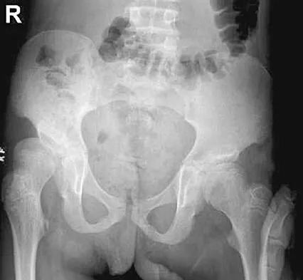
The parents of a 13-year-old boy with Down syndrome report that he has an increasing limp and decreased endurance with activities. Lateral flexion-extension radiographs of the cervical spine show no evidence of instability. Examination reveals a right Trendelenburg limp and an obvious limb-length discrepancy. Hip motion is symmetric except for some decreased abduction on the right side. A standing AP radiograph is shown in Figure 20. Management should consist of


Explanation

Ligamentous laxity and muscle hypotonia seen in Down syndrome contribute to the incidence of hip subluxation and dislocation. These factors can be progressive and lead to degenerative arthritis in adults with Down syndrome. Because this patient has a progressive limp and decreased endurance, observation and a shoe lift are not options. Bracing may be an option in the younger child before significant bony changes occur. Surgical intervention is the treatment of choice in this patient; however, all components of the deformity need to be addressed. Because of the increased capsular laxity, there is a high likelihood of recurrence if capsulorrhaphy is not included with the pelvic and femoral osteotomies. Surgery in these patients is associated with a high rate of complications. Shaw ED, Beals RK: The hip joint in Down's syndrome: A study of its structure and associated disease. Clin Orthop 1992;278:101-107. Aprin H, Zinc WP, Hall JE: Management of dislocation of the hip in Down's syndrome. J Pediatr Orthop 1985;5:428-431.


Question 21

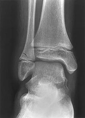
A 9-year-old girl reports the immediate onset of severe groin pain and the inability to walk after tripping on a curb. Examination reveals marked hip pain with passive range of motion. A radiograph is shown in Figure 21. Regardless of treatment, what is the most common complication following this injury?


Explanation

The patient has an unstable slipped capital femoral epiphysis (SCFE). According to the classification system based on physeal stability, an unstable SCFE is one in which the patient is unable to walk, even with crutches. Ishemic necrosis, or osteonecrosis, of the femoral head is the most devastating complication of SCFE. One study found a 47% incidence of ischemic necrosis following unstable slips. This complication is most likely the result of vascular injury associated with initial femoral head displacement rather than the result of either tamponade from joint effusion or gentle repositioning prior to stabilization. Chondrolysis is a relatively uncommon complication following treatment of SCFE. This complication has been associated with persistent penetration of the hip joint with screws or pins used to stabilize the femoral head or with spica cast immobilization. There are no reports to suggest that osteochondritis dissecans, nonunion, or coxa magna follows treatment of SCFE. Loder RT, Richards BS, Shapiro PS, et al: Acute slipped capital epiphysis: The importance of physeal stability. J Bone Joint Surg Am 1993;75:1134-1140.


Question 22

An 8-year-old boy reports progressive difficulty with walking. Examination reveals muscle weakness, with proximal groups more affected than distal muscles. Deep tendon reflexes are within normal limits. Laboratory studies show a creatine kinase level of 7,200 IU. Based on these findings, what is the most likely diagnosis?


Explanation

Patients with Becker muscular dystrophy have an abnormality in dystrophin, but unlike patients with Duchenne muscular dystrophy, some dystrophin is present. As a result, the progression of muscle weakness is slower, with the diagnosis typically made after age 8 years. Similar to patients with Duchenne muscular dystrophy, patients with Becker muscular dystrophy have pseudohypertrophy of the calves, markedly increased creatine kinase levels, and X-linked transmission of the condition. In addition, these patients are more prone to cardiomyopathy; a condition that should be carefully evaluated if any surgery is required. Patients with spinal muscular atrophy also have proximal muscle weakness, but the onset of weakness occurs earlier in childhood. These patients also have absent deep tendon reflexes and fasciculations, but pseudohypertrophy is absent and creatine kinase levels are normal. Patients with Emery-Dreifuss dystrophy may have a similar clinical picture to Becker's muscular dystrophy, but pseudohypertrophy is absent and creatine kinase levels are only mildly elevated. In addition, neck extension, elbow flexion, and ankle equinus contractures develop at an early age. Limb girdle dystrophy is a group of progressive muscular dystrophies that is not associated with pseudohypertrophy or a significant elevation of creatine kinase levels. Guillain-Barre syndrome is a condition associated with results from postinfectious demyelination of the peripheral nerve. These patients have the acute onset of weakness, hypotonia, and areflexia; creatine kinase levels are normal. Sussman MD: Muscular dystrophy, in Fitzgerald RH, Kaufer H, Malkani AL (eds): Orthopaedics. St Louis, MO, Mosby, 2002, pp 1573-1583.


Question 23

A 7-year-old boy sustained a 2-cm laceration to the anterior aspect of his left knee after falling on a rock. Examination reveals that the joint surface is not visible through the wound. Radiographs show no evidence of a foreign body or free air in the joint. Management should consist of


Explanation

The possibility of an open joint injury should be considered in any patient who has a small periarticular laceration. Failure to promptly diagnose and treat such injuries may lead to septic arthritis. The diagnosis of an open joint is easily made when there is visible communication of the joint through the traumatic wound, or when intra-articular air is present on a radiograph. In the absence of these findings, the diagnosis of an open joint may be established by the saline load test, in which a volume of saline is injected into the joint under sterile conditions. If fluid extravasates through the traumatic wound, the diagnosis of an open joint is established. Voit and associates used a saline load test in 50 patients with periarticular lacerations suggestive of joint penetration. When they compared the clinical prediction of whether or not the laceration had penetrated the joint and the test results, the authors reported a false-positive clinical result in 39% of patients and a false-negative clinical result in 43%. The authors concluded that the saline load test was valuable in evaluating periarticular lacerations. Voit GA, Irvine G, Beals RK: Saline load test for penetration of periarticular lacerations. J Bone Joint Surg Br 1996;78:732-733.


Question 24

What radiographic measurement is best used to assess the adequacy of deformity correction for the patient shown in Figure 22?


Explanation

Developmental coxa vara develops in early childhood and results in a progressive decrease in the proximal femoral neck-shaft angle with growth. The characteristic radiographic features are seen in this patient and include a decreased neck-shaft angle, a more vertical position of the physeal plate, and a triangular metaphyseal fragment in the inferior femoral neck, surrounded by an inverted radiolucent Y pattern. The main goal of surgery is to correct the varus angulation into a more normal range. Valgus overcorrection is preferred. A recent study emphasized the importance of adequately correcting the Hilgenreiner physeal angle to less than 38 degrees to minimize the risk of recurrent angulation. No study has documented the use of any of the other listed radiographic measurements to the outcome of treating developmental coxa vara. Carroll K, Coleman S, Stevens PM: Coxa vara: Surgical outcomes of valgus osteotomies. J Pediatr Orthop 1997;17:220-224.


Question 25

Figure 23 shows the radiograph of a 7 year-old girl with a low thoracic-level myelomeningocele. She has a history of skin ulcers over the apex of the deformity, but her current skin condition is good. Management of the spinal deformity should consist of


Explanation

This form of severe kyphosis results in intractable difficulties with sitting position, compression of internal organs, and chronic skin breakdown. Kyphectomy and posterior fusion with instrumentation, while associated with a high rate of complications, provides one of the best solutions to this clinical dilemma. The other choices are either completely ineffective or inadequate in managing this degree of deformity. Lindseth RE: Spine deformity in myelomeningocele. Instr Course Lect 1991;40:273-279.


Question 26

A 6-week-old female is being treated with a Pavlik harness for developmental dysplasia of the hip (DDH). At her 1-week follow-up, the mother notes that the child has stopped extending the knee on the affected side. On examination, the quadriceps are flaccid. What is the most appropriate next step in management?


Explanation

Femoral nerve palsy is a known complication of excessive hip flexion in a Pavlik harness. The harness must be discontinued to allow for nerve recovery before resuming any abduction treatment.

Question 27

A 9-month-old child with DDH is undergoing closed reduction and spica casting. According to Ramsey, the safe zone for positioning the hip to minimize the risk of avascular necrosis while maintaining reduction is defined by the arc between which two positions?


Explanation

Ramsey's safe zone is the arc between maximum abduction and the angle at which the hip redislocates in adduction. Forcing the hip into extreme abduction outside this zone significantly increases the risk of avascular necrosis.

Question 28

An 18-month-old requires open reduction for a chronically dislocated hip due to DDH. Which of the following anatomical structures is considered an intra-articular obstacle to successful closed reduction?


Explanation

Intra-articular obstacles to reduction in DDH include the transverse acetabular ligament, ligamentum teres, pulvinar, and an inverted limbus. Extra-articular obstacles include the iliopsoas, adductors, and an hourglass constriction of the capsule.

Question 29

A 5-year-old female with residual acetabular dysplasia requires a pelvic osteotomy to improve anterolateral femoral head coverage. The surgeon plans an incomplete pericapsular osteotomy that hinges on the triradiate cartilage. Which procedure is being described?


Explanation

The Pemberton osteotomy is an incomplete pericapsular cut that hinges on the triradiate cartilage, effectively reducing acetabular volume and improving anterolateral coverage. In contrast, the Salter osteotomy hinges at the pubic symphysis.

Question 30

A 4-week-old female born breech undergoes an ultrasound screening for DDH. The report notes an alpha angle of 45 degrees and a beta angle of 65 degrees. According to Graf's classification, what does the alpha angle represent?


Explanation

In the Graf ultrasound method, the alpha angle measures the bony roof of the acetabulum (normal is > 60 degrees). The beta angle measures the cartilaginous roof.

Question 31

A 6-month-old infant was treated with a Pavlik harness for 3 months. Recent radiographs reveal fragmentation and delayed ossification of the left femoral head. Excessive positioning in which direction during treatment is most strongly associated with this complication?


Explanation

Avascular necrosis (AVN) of the femoral head in DDH treatment is most commonly caused by excessive abduction. This position compresses the medial circumflex femoral artery, compromising blood flow to the epiphysis.

Question 32

A 4-year-old child undergoes a Salter innominate osteotomy for the treatment of DDH. Which of the following biomechanical changes occurs as a direct result of this specific osteotomy?


Explanation

The Salter osteotomy is a complete innominate cut that redirects the entire acetabulum to provide anterolateral coverage. Because it hinges at the pubic symphysis, it biomechanically lateralizes and distalizes the joint center, often lengthening the limb slightly.

Question 33

A 13-year-old obese male presents with left groin and knee pain. On physical examination, which finding is considered the classic pathognomonic sign during passive flexion of the affected hip?


Explanation

Obligate external rotation during passive hip flexion is the classic physical examination finding in Slipped Capital Femoral Epiphysis (SCFE). This occurs due to the anterior and superior translation of the femoral neck relative to the epiphysis.

Question 34

A 9-year-old female presents with bilateral Slipped Capital Femoral Epiphysis (SCFE). She is in the 10th percentile for height and weight.

Which of the following laboratory investigations is most indicated for this patient?


Explanation

Patients presenting with SCFE under the age of 10 years or with bilateral disease have a high incidence of underlying endocrine disorders, most commonly hypothyroidism. TSH and Free T4 are the primary screening tests indicated.

Question 35

A 14-year-old male is unable to bear weight on his right leg after a minor fall. Radiographs demonstrate a severe, acute unstable SCFE. What is the primary advantage of the modified Dunn procedure via a surgical dislocation approach compared to in-situ pinning for this patient?


Explanation

The modified Dunn procedure involves a surgical hip dislocation to anatomically reduce the slipped epiphysis. Its main advantage is the direct visualization and protection of the posterior retinacular vessels, though it is technically demanding and AVN risk remains.

Question 36

An 11-year-old boy undergoes in-situ pinning for a symptomatic left SCFE. Which of the following is the strongest clinical indication for prophylactic pinning of the asymptomatic right hip?


Explanation

Prophylactic pinning of the contralateral hip is highly recommended in patients with underlying endocrine or metabolic disorders (such as renal osteodystrophy or hypothyroidism) because their risk of developing a subsequent contralateral slip is exceedingly high.

Question 37

A 14-year-old male treated with in-situ pinning for a stable SCFE 6 months ago now presents with severe hip stiffness and worsening pain. Radiographs reveal global joint space narrowing to 2 mm. What is the most likely cause of this complication?


Explanation

Chondrolysis presents with severe stiffness and diffuse joint space narrowing (< 3 mm). The most common iatrogenic cause is unrecognized pin penetration into the articular space during SCFE fixation.

Question 38

A 12-year-old female presents with hip pain for 3 weeks but is able to ambulate into the clinic with a mild limp.

According to the Loder classification, what is her approximate risk of developing avascular necrosis (AVN) following in-situ pinning?


Explanation

The Loder classification divides SCFE into stable (patient can bear weight) and unstable (unable to bear weight). A stable SCFE carries a very low risk of AVN, historically less than 5%.

Question 39

A 13-year-old male presents with vague knee pain. An AP pelvis radiograph is obtained. Which of the following radiographic signs strongly indicates a subtle SCFE?


Explanation

Klein's line is drawn along the superior margin of the femoral neck on an AP radiograph. In a normal hip, it intersects the lateral portion of the epiphysis. Trethowan's sign in SCFE occurs when the line passes completely superior to the epiphysis.

Question 40

A 6-week-old female infant is brought to the clinic for a routine check-up. She was born via cesarean section for a breech presentation. Clinical examination reveals symmetrical thigh creases and negative Barlow and Ortolani maneuvers. What is the most appropriate next step in management?


Explanation

Infants with a history of breech presentation have a higher risk of DDH and should undergo ultrasound screening at 4 to 6 weeks of age, even with a normal clinical examination.

Question 41

A 4-month-old infant with developmental dysplasia of the hip has been treated in a Pavlik harness for 3 weeks. The parents report that the child has stopped kicking the left leg. On examination, the knee lacks active extension but has normal sensation. What is the most likely cause?


Explanation

Femoral nerve palsy is a known complication of Pavlik harness treatment, typically resulting from hyperflexion of the hips. Management involves removing the harness or adjusting the flexion straps.

Question 42

A 14-year-old boy with a BMI in the 95th percentile presents with 3 weeks of vague left knee pain and a slight limp. He is able to bear weight on the affected limb. Radiographs show a widened and irregular left capital femoral physis with a posterior and inferior slip. What is the most appropriate treatment?


Explanation

This is a stable slipped capital femoral epiphysis (SCFE). The standard of care is in situ fixation with a single cannulated screw placed in the center of the epiphysis.

Question 43

In evaluating a patient with a slipped capital femoral epiphysis (SCFE), which of the following is considered an indication for prophylactic pinning of the contralateral asymptomatic hip?


Explanation

Prophylactic pinning of the contralateral hip is generally recommended for patients with endocrine or metabolic disorders (like renal osteodystrophy or hypothyroidism), prior radiation therapy, or age < 10 years.

Question 44

A 4-year-old boy sustains a completely displaced, isolated, midshaft femur fracture after a fall from a playground structure. He weighs 18 kg (40 lbs). What is the most appropriate initial treatment?


Explanation

For children under 5 years old and weighing less than 50 lbs, an early or immediate hip spica cast is the treatment of choice for closed, isolated midshaft femur fractures.

Question 45

A 9-year-old boy presents with an acute, unstable slipped capital femoral epiphysis (SCFE) after a minor fall. He is unable to bear weight. Which of the following complications is most highly associated with this specific type of SCFE compared to a stable SCFE?


Explanation

Unstable SCFE (defined as the inability to bear weight) has a significantly higher rate of avascular necrosis (up to 50%) compared to stable SCFE.

Question 46

A 12-month-old girl is noted to have a painless limp. Examination reveals a positive Galeazzi sign and asymmetric thigh folds. Radiographs demonstrate a superolaterally displaced right femoral head with an acetabular index of 40 degrees. What is the most appropriate initial management?


Explanation

In a child aged 6 to 18 months with DDH, the initial treatment of choice is typically closed reduction and spica casting. A Pavlik harness is generally ineffective after 6 months of age.

Question 47

A 10-year-old boy weighing 40 kg sustains a closed, length-stable transverse midshaft femur fracture. Which of the following is the most appropriate surgical treatment?


Explanation

Flexible intramedullary nailing is the standard of care for length-stable femur fractures in children aged 5 to 11 years who weigh less than 50 kg (110 lbs).

Question 48

During the surgical treatment of a slipped capital femoral epiphysis (SCFE) with in situ pinning, the surgeon must be careful to avoid joint penetration. Which complication is most directly associated with unrecognized pin penetration into the hip joint?


Explanation

Unrecognized pin penetration during in situ fixation of SCFE is classically associated with chondrolysis, leading to severe stiffness and joint space narrowing.

Question 49

A 13-year-old boy who recently underwent intramedullary nailing for a femur fracture via a piriformis fossa entry point presents for a follow-up 2 years later. What is the most likely complication associated with this specific entry point in a skeletally immature patient?


Explanation

Use of a piriformis fossa entry point for rigid intramedullary nailing in skeletally immature patients carries a significant risk of iatrogenic avascular necrosis of the femoral head. A lateral trochanteric entry is strongly preferred.

Question 50

A 3-week-old male infant undergoes ultrasound screening for developmental dysplasia of the hip (DDH). The alpha angle is measured at 40 degrees. According to the Graf classification, what does this alpha angle indicate?


Explanation

In the Graf classification, an alpha angle of less than 43 degrees (Type III) indicates a subluxated hip with poor bony roof coverage. This structural deficiency requires immediate treatment.

Question 51

A 6-year-old boy sustains a spiral midshaft femur fracture. He is managed with a spica cast. Two years later, his parents are concerned about a leg length discrepancy. Which of the following is the most expected outcome regarding limb length after this injury?


Explanation

Children between 2 and 10 years of age typically experience an overgrowth phenomenon following a femur fracture, usually resulting in 1 to 2 cm of overgrowth of the fractured limb within the first 2 years.

Question 52

A 5-month-old infant is being treated with a Pavlik harness for DDH. The ultrasound at 4 weeks of treatment shows failure of reduction of the hip. What is the most appropriate next step in management?


Explanation

If a Pavlik harness fails to achieve reduction within 3 to 4 weeks, it should be discontinued to prevent "Pavlik harness disease" (abrading the posterior acetabulum). The next standard step is closed reduction and spica casting.

Question 53

Which of the following radiographic findings on an AP pelvis is most indicative of developmental dysplasia of the hip (DDH) in an 8-month-old child?


Explanation

In DDH, the femoral head typically migrates superolaterally, causing a break in Shenton's line. A normal hip has the ossific nucleus in the lower inner quadrant formed by Perkin and Hilgenreiner lines.

Question 54

A 6-week-old female is undergoing treatment for developmental dysplasia of the hip with a Pavlik harness. During a follow-up visit, the mother notes the child is no longer kicking her leg on the affected side. On examination, active knee extension is absent, but the hip remains well reduced. What is the most appropriate next step in management?


Explanation

Absent knee extension indicates a femoral nerve palsy, the most common nerve palsy associated with the Pavlik harness. The harness should be removed temporarily to allow the nerve to recover, which usually happens within a few days to weeks.

Question 55

A 5-week-old female infant undergoes hip ultrasound screening due to a breech presentation. The alpha angle is 55 degrees and the beta angle is 60 degrees. Dynamic testing demonstrates a stable hip. What is the most appropriate next step?


Explanation

An alpha angle between 50 and 59 degrees at this age classifies as Graf Type IIa, indicating physiologic immaturity. Because the hip is stable, the correct management is observation and repeat ultrasound in 4 to 6 weeks to ensure normal development.

Question 56

A 3-year-old girl is diagnosed with unilateral DDH. Closed reduction was unsuccessful. During an open reduction, an innominate osteotomy is planned to address acetabular dysplasia. Which of the following osteotomies hinges on the pubic symphysis to provide anterolateral coverage?


Explanation

The Salter innominate osteotomy involves a complete cut through the ilium and hinges on the pubic symphysis to redirect the acetabulum. This provides anterolateral coverage for the femoral head.

Question 57

An 8-year-old boy presents with an acute on chronic slipped capital femoral epiphysis. His height and weight are both at the 25th percentile for his age. Which of the following laboratory studies is most strongly indicated?


Explanation

SCFE in atypical patients (age younger than 10, or weight below the 50th percentile) raises strong suspicion for an underlying endocrine or metabolic disorder. Hypothyroidism is a common cause, making TSH an essential screening test.

Question 58

A 13-year-old obese boy presents to the emergency department with severe right hip pain after a minor fall. He is unable to bear weight on the right leg, even with crutches. Radiographs confirm a severe slipped capital femoral epiphysis (SCFE). Which of the following represents the highest risk of complication for this patient?


Explanation

Inability to bear weight defines an unstable SCFE according to the Loder classification. Unstable SCFE carries a significantly higher risk of osteonecrosis (up to 47%) compared to stable SCFE.

Question 59

A 12-year-old boy is scheduled for in situ pinning of a left-sided slipped capital femoral epiphysis. Prophylactic pinning of the asymptomatic right hip is most strongly recommended if the patient also has:


Explanation

Prophylactic contralateral pinning is highly recommended in patients with endocrine or metabolic disorders, such as renal osteodystrophy or hypothyroidism, due to a very high risk of bilateral involvement.

Question 60

A 9-month-old infant is brought to the emergency department with a swollen and deformed left thigh. Radiographs reveal a spiral fracture of the femoral shaft. Assuming no other injuries, which of the following is the most appropriate initial orthopedic management?


Explanation

Spica casting is the standard of care for isolated femur fractures in children aged 6 months to 5 years. While child abuse must be ruled out, the orthopedic management of the fracture relies on cast immobilization.

Question 61

A 6-year-old boy sustains an isolated diaphyseal femur fracture and is treated with flexible intramedullary nailing. To anticipate the most common complication related to leg length, how much overgrowth is typically expected after a femur fracture in this age group?


Explanation

In children aged 2 to 10 years, a femur fracture typically stimulates overgrowth of 1 to 2 cm due to hyperemia of the physes. Surgeons may intentionally leave the fractured limb slightly short to compensate.

Question 62

When performing a closed reduction and spica casting for a 9-month-old with developmental dysplasia of the hip, a percutaneous adductor tenotomy is often performed. The primary purpose of this tenotomy is to:


Explanation

The adductor tenotomy relieves medial tension, allowing for adequate abduction without excessive pressure on the femoral head. This widens the "safe zone" of Ramsey and minimizes the risk of avascular necrosis.

Question 63

A 3-month-old girl with DDH is treated with a Pavlik harness. During a follow-up visit, she is noted to have decreased active knee extension on the affected side. What is the most appropriate next step in management?


Explanation

Femoral nerve palsy is a known complication of the Pavlik harness caused by excessive hip flexion. Loosening the anterior straps reduces hyperflexion and typically allows for full recovery of the nerve.

Question 64

A 13-year-old obese boy presents to the emergency department with acute left groin pain and an inability to bear weight after a minor fall. Radiographs show a slipped capital femoral epiphysis. According to Loder's classification, what is the primary significance of his inability to bear weight?


Explanation

Loder classified SCFE into stable (able to bear weight) and unstable (unable to bear weight). Unstable SCFE is associated with a significantly higher risk of avascular necrosis (up to 47%) compared to stable SCFE.

Question 65

A 6-year-old boy sustains a completely displaced midshaft femur fracture. If treated with a spica cast, what is the acceptable amount of shortening to aim for during reduction to account for expected overgrowth?


Explanation

In children aged 2 to 10 years, femur fractures often stimulate longitudinal overgrowth due to hyperemia. An initial shortening of 1 to 1.5 cm is accepted and expected to correct over time.

Question 66

A 4-week-old female infant undergoes a screening hip ultrasound for a breech presentation. The alpha angle is measured at 65 degrees and the beta angle at 45 degrees. Which of the following is the most appropriate management?


Explanation

An alpha angle greater than 60 degrees and a beta angle less than 55 degrees represent a Graf type I (normal) hip. No treatment is required, and the parents can be reassured.

Question 67

Prophylactic pinning of the contralateral, asymptomatic hip in a patient presenting with a unilateral slipped capital femoral epiphysis (SCFE) is most strongly indicated in which of the following scenarios?


Explanation

Patients with underlying endocrine disorders (e.g., hyperparathyroidism, hypothyroidism, renal osteodystrophy) have a very high rate of developing bilateral SCFE. Prophylactic pinning of the contralateral hip is strongly recommended in these patients.

Question 68

A 12-year-old boy sustains a transverse midshaft femur fracture. He is treated with a rigid intramedullary nail using a piriformis fossa starting point. Which of the following is the most devastating complication associated with this specific surgical technique in this age group?


Explanation

Using a piriformis fossa entry point for rigid nailing in skeletally immature patients carries a high risk of iatrogenic avascular necrosis. This is due to injury to the ascending branches of the medial femoral circumflex artery.

Question 69

An 18-month-old girl presents with a painless limp. Examination reveals a positive Galeazzi sign and limited abduction of the right hip. Radiographs confirm a dislocated right hip. Which of the following is the most appropriate definitive treatment?


Explanation

In a child older than 18 months with untreated DDH, closed methods generally fail. Open reduction is required, often combined with a pelvic osteotomy (e.g., Salter) to address the secondary acetabular dysplasia.

Question 70

A 12-year-old boy presents with an unstable slipped capital femoral epiphysis (SCFE). The surgeon plans to perform a capsulotomy and percutaneous in situ pinning. What is the primary rationale for performing an anterior capsulotomy in this setting?


Explanation

Unstable SCFE is associated with a hemarthrosis that increases intracapsular pressure, potentially compromising epiphyseal blood flow. An anterior capsulotomy decompresses the joint and is thought to reduce the high risk of AVN.

Question 71

A 6-week-old female infant is being treated with a Pavlik harness for a dislocated left hip. During a follow-up visit, the parents report that the child has stopped actively extending her left knee. On examination, there is decreased active knee extension and a diminished patellar reflex on the left. What is the most likely cause of this finding, and what is the appropriate management?


Explanation

Hyperflexion in a Pavlik harness can compress the femoral nerve against the rim of the pelvis, leading to transient femoral nerve palsy. The appropriate management is to loosen the anterior straps to decrease hip flexion, which typically results in spontaneous resolution.

Question 72

A 4-week-old female infant born in the breech presentation undergoes ultrasound screening for developmental dysplasia of the hip (DDH). The coronal image reveals an alpha angle of 40 degrees and a beta angle of 80 degrees. According to the Graf classification, what is the best initial management for this patient?


Explanation

An alpha angle of less than 60 degrees indicates dysplasia, and an angle of 40 degrees falls into Graf Type III (dislocated but reducible). The standard of care for a newborn to 6-month-old with a dysplastic or dislocated hip is the application of a Pavlik harness.

Question 73

A 13-year-old obese boy presents to the emergency department with acute severe right hip pain after a minor fall. He is completely unable to bear weight on the right leg, even with the use of crutches. Radiographs confirm a severe slipped capital femoral epiphysis (SCFE). Based on the Loder classification, this specific clinical presentation carries the highest risk for which of the following complications?


Explanation

The Loder classification defines an unstable SCFE by the patient's inability to ambulate, with or without crutches. Unstable SCFE is associated with a markedly high rate of avascular necrosis (up to 50%), whereas stable SCFE rarely results in AVN.

Question 74

A 9-month-old infant is brought to the pediatric emergency department with swelling and deformity of the left thigh. Radiographs demonstrate a spiral fracture of the femoral diaphysis. Which of the following factors in this patient's presentation is the strongest indicator of non-accidental trauma (child abuse)?


Explanation

Femur fractures in non-ambulatory infants (typically under 1 year of age) have a high association with non-accidental trauma (up to 80%). While spiral fractures were historically deemed suspicious, recent studies show age and ambulatory capability are the most reliable indicators of abuse.

Question 75

A 3-year-old boy sustains an isolated, closed midshaft femur fracture with 2 cm of shortening after falling from a playground structure. His neurovascular exam is intact. What is the most appropriate definitive management for this patient?


Explanation

For children aged 6 months to 5 years with an isolated femur fracture and acceptable shortening (<2-3 cm), early hip spica casting is the gold standard treatment. Operative interventions like flexible nailing are generally reserved for children older than 5 years.

Question 76

A 12-year-old girl is diagnosed with a unilateral stable slipped capital femoral epiphysis (SCFE). She has a known medical history of panhypopituitarism and renal osteodystrophy. When counseling the parents about surgical management, what is the primary indication for recommending prophylactic in situ pinning of the contralateral hip?


Explanation

Patients with underlying endocrine disorders (e.g., hypothyroidism, renal osteodystrophy, panhypopituitarism) have an exceedingly high risk (up to 100%) of developing bilateral SCFE. Prophylactic pinning of the contralateral asymptomatic hip is strongly recommended in this population.

Question 77

During a closed reduction for developmental dysplasia of the hip (DDH) under general anesthesia in a 9-month-old, the hip reduces at 100 degrees of flexion and 60 degrees of abduction. However, it redislocates when abduction is reduced to 40 degrees. The safe zone of Ramsey is determined to be narrow. Which of the following is the best next step to safely enlarge this zone?


Explanation

A narrow safe zone of Ramsey (< 20 degrees between reduction and redislocation) increases the risk of avascular necrosis if the hip is forced into hyperabduction. A percutaneous adductor longus tenotomy safely increases the maximal abduction, widening the safe zone for a safer spica cast position.

Question 78

An 8-year-old boy, weighing 35 kg, underwent flexible intramedullary nailing for a transverse midshaft femur fracture 6 months ago. The fracture has healed well radiographically, but he now complains of knee pain and a slight limp. What is the most common complication of this procedure causing his current symptoms?


Explanation

The most common complication following titanium elastic nailing for pediatric femur fractures is symptomatic hardware prominence at the distal insertion site, leading to soft tissue irritation and knee pain. This typically resolves after implant removal once the fracture is fully healed.

Question 79

A 14-year-old boy is undergoing in situ single-screw fixation for a stable slipped capital femoral epiphysis (SCFE). To provide optimal stability and minimize the risk of unrecognized joint penetration, where should the screw threads be ideally positioned within the epiphysis?


Explanation

The ideal screw placement for a SCFE is the center-center position (center of the epiphysis on both the AP and lateral radiographs). This trajectory is perpendicular to the physis, provides maximal mechanical stability, and significantly reduces the risk of inadvertent joint penetration.

Question 80

An 11-year-old boy weighing 65 kg (143 lbs) sustains a length-unstable, comminuted midshaft femur fracture. He is planned for rigid intramedullary nailing. To minimize the risk of iatrogenic avascular necrosis (AVN) of the femoral head, which entry point is strictly recommended in this pediatric age group?


Explanation

Piriformis fossa entry in children carries a significant risk of injuring the medial circumflex femoral artery anastomosis, leading to AVN of the femoral head. A lateral greater trochanteric entry point avoids this blood supply and is the standard for rigid nailing in older children.

Question 81

A 13-year-old girl was treated 8 months ago with a single cannulated screw for a SCFE. She now presents with progressive hip stiffness, worsening pain, and a significant loss of hip range of motion. Radiographs demonstrate diffuse joint space narrowing of the affected hip without focal collapse. What is the most likely etiology of this late complication?


Explanation

Chondrolysis is characterized by acute loss of articular cartilage and joint space narrowing, presenting with severe stiffness. In the setting of SCFE treatment, it is most strongly associated with unrecognized intra-articular hardware penetration.

Question 82

A 2-year-old girl is brought to the clinic for a newly noticed painless limp. Radiographs demonstrate a completely dislocated right hip with a false acetabulum and a dysplastic true acetabulum. Based on her age and presentation, what is the most appropriate initial management?


Explanation

Children presenting with DDH after 18-24 months of age typically have significant secondary adaptive changes (acetabular dysplasia and capsular constriction). Successful treatment usually requires an open reduction combined with a pelvic osteotomy and often a femoral shortening osteotomy.

Question 83

A 12-year-old, overweight boy complains of vague left knee pain for the past 3 months. His knee examination is completely unremarkable. When the examiner passively flexes his left hip to 90 degrees, the leg obligately deviates into external rotation. What is the most likely underlying diagnosis?


Explanation

Obligate external rotation of the hip during passive flexion is a pathognomonic physical exam finding for a slipped capital femoral epiphysis (SCFE). Furthermore, SCFE frequently presents as referred knee or thigh pain, leading to potential misdiagnosis if the hip is not examined.

Question 84

A 10-year-old boy is diagnosed with a unilateral stable slipped capital femoral epiphysis. Which of the following radiographic parameters is considered the most reliable predictor for the development of a subsequent contralateral slip?


Explanation

The Modified Oxford bone age score relies on pelvic radiographs to determine skeletal maturity and is the most reliable predictor of a contralateral slip. Patients with a lower score (greater skeletal immaturity) are at the highest risk for sequential SCFE.

Question 85

A 6-week-old female is undergoing treatment with a Pavlik harness for developmental dysplasia of the hip. At a follow-up visit, the parents report the infant is no longer kicking her right leg. Examination reveals an inability to actively extend the knee on the affected side. This complication is most directly caused by which of the following?


Explanation

Femoral nerve palsy in a Pavlik harness is caused by hyperflexion of the hips, typically resulting from overly tight anterior straps. Management consists of loosening the straps to reduce flexion and monitoring for spontaneous recovery.

Question 86

A 4-year-old boy sustains an isolated, closed midshaft femur fracture and is treated with immediate spica casting. To appropriately account for anticipated post-traumatic limb overgrowth, what is the ideal amount of initial fracture shortening accepted in the cast?


Explanation

In children aged 2 to 10 years, femoral shaft fractures often stimulate physeal overgrowth. Accepting 15 to 20 mm (1.5 to 2 cm) of initial shortening in the spica cast helps prevent ultimate leg length discrepancy.

Question 87

According to the Loder classification, an unstable slipped capital femoral epiphysis (SCFE) is associated with a significantly higher rate of which of the following complications when compared to a stable SCFE?


Explanation

The Loder classification defines an unstable SCFE by the patient's inability to ambulate even with crutches. Unstable slips carry a nearly 50% risk of avascular necrosis, whereas the rate in stable slips is close to zero.

Question 88

A 3-year-old girl is diagnosed with a neglected, completely dislocated unilateral developmental dysplasia of the hip. She is scheduled for an open reduction and pelvic osteotomy. What is the primary biomechanical rationale for performing a concurrent femoral shortening osteotomy in this patient?


Explanation

In older children (typically over 2-3 years) with untreated DDH, a femoral shortening osteotomy is often necessary during open reduction. Shortening the femur relieves soft-tissue tension across the joint, significantly reducing the risk of avascular necrosis.

Question 89

A 12-year-old male who weighs 65 kg (143 lbs) presents with a length-unstable, comminuted midshaft femur fracture. His greater trochanteric physis remains open. To minimize the risk of avascular necrosis of the femoral head while providing stable fixation, what is the preferred starting point if a rigid intramedullary nail is selected?


Explanation

In adolescents with open physes, a rigid lateral entry nail utilizing the lateral aspect of the greater trochanter avoids the medial femoral circumflex artery's ascending branch. This effectively minimizes the risk of iatrogenic avascular necrosis compared to piriformis fossa entry.

Question 90

A 14-year-old girl presents with progressive groin pain and marked stiffness 8 months after undergoing an uncomplicated in situ pinning for a stable slipped capital femoral epiphysis. Radiographs reveal diffuse narrowing of the hip joint space with preservation of femoral head sphericity. What is the most likely diagnosis?


Explanation

Chondrolysis is characterized by diffuse joint space narrowing and severe stiffness following SCFE treatment, with the femoral head typically remaining spherical. Risk factors include severe slips, spica casting, and unrecognized intra-articular hardware penetration.

Question 91

When interpreting an anteroposterior pelvic radiograph of a 6-month-old infant for developmental dysplasia of the hip, the normal position of the proximal femoral ossific nucleus (or its expected location) should be in which quadrant formed by Hilgenreiner's and Perkin's lines?


Explanation

Hilgenreiner's line is drawn horizontally through the triradiate cartilages, and Perkin's line is drawn perpendicular to it at the lateral margin of the acetabulum. The normal femoral head must reside in the inferomedial quadrant.

Question 92

When performing in situ pinning for a stable slipped capital femoral epiphysis using a single fully threaded cannulated screw, what is the optimal position of the screw within the epiphysis to achieve maximal biomechanical stability and reduce the risk of joint penetration?


Explanation

The ideal trajectory for an in situ screw in a SCFE is perpendicular to the physis, placing the threads central in both the AP and lateral radiographic views. This center-center position provides maximum hold in the epiphysis while limiting the "in-out-in" phenomenon.

Question 93

An ultrasound is performed on a 4-week-old female infant to evaluate for developmental dysplasia of the hip. According to the Graf classification, a normal, mature hip (Type 1) is defined by which of the following sonographic measurements?


Explanation

In the Graf ultrasound classification, the alpha angle measures the concavity of the bony acetabular roof. An alpha angle greater than 60 degrees indicates a normal, mature hip (Type 1).

None

Clinic OS
Medically Verified Content by
Prof. Clinic OS
Consultant Orthopedic & Spine Surgeon
Chapter Index