العربية
Part of the Master Guide

Orthopedic Board Review Set 675: 100 MCQs for ABOS, OITE, FRCS – Hip Focus

Orthopedic Hip & Knee 2026 MCQs: Board Review Questions & Answers (Part 3)

23 Apr 2026 87 min read 94 Views
Figure for Hpkn 2007 MCQs - Part 3 - Question 51

Key Takeaway

In this comprehensive guide, we discuss everything you need to know about Orthopedic Hip & Knee 2026 MCQs: Board Review Questions & Answers (Part 3). Top-rated Orthopedic Hip & Knee 2026 MCQs bank. Practice with clinical case questions, orthopedic surgery board review, and evidence-based answers updated for 2026.

Orthopedic Hip & Knee 2026 MCQs: Board Review Questions & Answers (Part 3)

Comprehensive 100-Question Exam


00:00

Start Quiz

Question 1

Figure 30 shows the MRI scan of a 68-year-old woman who has left hip pain. What is the most appropriate treatment?


Explanation

The patient has a large zone of osteonecrosis of the left femoral head. The wedge-shaped zone of decreased signal intensity on the T1 image in the subchondral region of the femoral head is typical. Based on these findings, total hip arthroplasty is the most appropriate treatment. Open reduction and internal fixation will not help this condition. Incisional biopsy is indicated only if the MRI scan shows a probable neoplasm. Resection of the proximal femur is indicated only for aggressive malignancy. Arthrodesis may be considered in a younger patient but not in a 68-year-old individual. Other treatments, not listed, such as core decompression, vascularized fibular transplant, and osteotomy may be options in selected patients. Urbaniak JR, Jones JP Jr (eds): Osteonecrosis: Etiology, Diagnosis, and Treatment. Rosemont, IL, American Academy of Orthopaedic Surgeons, 1997, pp 213-223.

Question 2

There is increasing concern about the ethical relationship of orthopaedists to the orthopaedic equipment industry. Which of the following describes the most appropriate relationship?


Explanation

It is appropriate for orthopaedic surgeons to have relationships with industry as long as the relationship is for the good of the patient and no "quid pro quo" intent exists. A grant to cover registration at a CME event is appropriate but travel and hotel for a spouse is not. For orthopaedists who are faculty at a meeting sponsored by industry, it is appropriate for travel and expenses to be covered for that faculty member. Care must be exercised that the faculty member contributes in an amount appropriate for the expenses paid. The faculty member must ensure that information presented is unbiased and based on reasonable data and opinion. Consulting agreements should spell out specifically the duties of the agreement and payment should be appropriate for the time spent. There should be a defined work product for the consulting. Agreements that are thinly veiled payments for use of a company's products must be avoided. In all cases, the agreements must stand up to public scrutiny. Restricted grants for specific industry-sponsored programs aimed at residents are not appropriate. Unrestricted grants intended for attendance at approved CME courses are appropriate. Dinners at which information is presented about topics that can aid in patient care are appropriate as long as the expense is reasonable ($100 or less/person) and the guest list includes individuals who can use the information in a patient case. Clearly a "premium" dinner for office staff to review new surgical instrumentation would not pass this test.

Question 3

A 30-year-old patient has had severe left hip pain and difficulty ambulating, necessitating the use of a cane, for the past 6 months. A photomicrograph of the femoral head sectioned at the time of surgery is shown in Figure 31. What is the most likely diagnosis?


Explanation

The photomicrograph demonstrates a wedge-shaped infarct with femoral head collapse; therefore, the diagnosis is osteonecrosis of the femoral head. Perthes disease and osteoarthritis do not involve a wedge-shaped defect. Tuberculosis of the hip joint results in greater destruction of the articular cartilage. Basset LW, Mirra JM, Cracchiolo A III: Ischemic necrosis of the femoral head: Correlation between magnetic resonance imaging and histologic sections. Clin Orthop 1987;223:181-187.

Question 4

When comparing mobile-bearing total knee arthroplasty (TKA) to fixed-bearing total condylar arthroplasty, the mobile-bearing procedure provides


Explanation

Survivorship is similar in the two groups. In a recent study, mobile-bearing TKAs showed a slightly higher maximum flexion than the total condylar fixed-bearing-type designs (112 degrees versus 108 degrees with no difference in recovery rate). Using a fixed-bearing or a mobile-bearing design did not seem to influence the recovery rate in early results after knee arthroplasty. Mobile-bearing arthroplasties are suggested, in theory, to offer a reduction in polyethylene wear; however, clinical studies have not yet proven this. Recovery rates have yet to be statistically seen as improved with either method. Differences in strength have not been shown. Aglietti P, Baldini A, Buzzi R, et al: Comparison of mobile-bearing and fixed-bearing total knee arthroplasty: A prospective randomized study. J Arthroplasty 2005;20:145-153. Sorrells RB: The rotating platform mobile bearing TKA. Orthopedics 1996;19:793-796.

Question 5

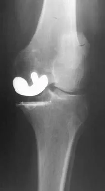
Based on the type of articulation shown in Figure 32, wear is not affected by which of the following factors?


Explanation

Wear in total hip arthroplasty is a very complex phenomenon. The radial mismatch of the femoral head to the acetabular component has been shown in multiple studies to be a significant factor in wear. The mismatch can neither be too small nor too large. When the mismatch is too small, seizing of the implants can occur. When the mismatch is too large, contact stresses increase and produce exceptionally high wear. The ideal radial mismatch should be approximately 50 microns. Surface roughness and ball sphericity are two items that are extremely important with respect to wear. High carbon content has been shown to decrease wear. This device has a very large head-to-neck ratio, so impingement-related wear is unlikely. Amstutz HC, Grigoris P: Metal on metal bearings in hip arthroplasty. Clin Orthop 1996;329:S11-S34. Amstutz HC, Campbell P, McKellop H, et al: Metal on metal total hip replacement workshop consensus document. Clin Orthop 1996;329:S297-S303.

Question 6

Figure 33 shows the venogram of a patient who has a long history of alcohol abuse. Warfarin should be used cautiously because of the interaction with which of the following factors?


Explanation

Warfarin acts by inhibiting clotting factors II, VII, IX, X. The actual mechanism of action is by inhibition of hepatic enzymes, vitamin K epoxide, and perhaps vitamin K reductase. This inhibition results in lack of carboxylation of vitamin K-dependent proteins (II, VII, IX, X). The anticoagulant effect of warfarin can be reversed with vitamin K or fresh-frozen plasma. The use of alcohol may lead to liver dysfunction and an even more limited margin of available factors. Lieberman JR, Wollaeger J, Dorey F, et al: The efficacy of prophylaxis with low-dose warfarin for prevention of pulmonary embolism following total hip arthroplasty. J Bone Joint Surg Am 1997;79:319-325.

Question 7

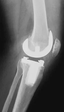
A 78-year-old patient undergoing revision total knee arthroplasty has bone loss throughout the knee at the time of revision. A distal femoral augment is used to restore the joint line. One month after surgery, the patient reports pain and is unable to ambulate. A lateral radiograph is shown in Figure 34. What is the most likely etiology of this problem?


Explanation

Instability is a leading cause of failure following total knee arthroplasty. Instability can present as global instability, extension gap (varus/valgus) instability, or flexion gap (anterior/posterior) instability. Treatment options are numerous based on the exact pathology. The radiograph reveals anterior/posterior instability with dislocation consistent with flexion gap instability. A loose flexion gap can allow the femoral component to ride above the tibial cam post mechanism, resulting in dislocation. Distal femoral augments treat extension gap instability, whereas tibial augments can treat both flexion and extension gap instability. Posterior condyle augments at the distal femur can also be used to treat flexion gap instability. Flexion gap instability is further aggravated by extension mechanism incompetence. Note the excessively thin patella on the lateral radiograph. Pagnano MW, Hanssen AD, Lewallen DG, et al: Flexion instability after primary cruciate retaining total knee arthroplasty. Clin Orthop 1998;356:39-46. McAuley J, Engh GA, Ammeen DJ: Treatment of the unstable total knee arthroplasty. Inst Course Lect 2004;53:237-241.

Question 8

Figure 35 shows the AP radiograph of a patient who underwent a previous upper tibial osteotomy (UTO). The patient may be at risk for which of the following during total knee arthroplasty (TKA)?


Explanation

The results of TKA for patients with a prior UTO are reported to be slightly suboptimal. The major problems are patella baja, difficulty in exposure, and instability. Most of the patients exhibit some degree of instability prior to TKA, and ligamentous balancing may be difficult. Ligamentous structures are at risk of rupture during the difficult exposure. The problem of ligamentous balancing is exacerbated by the change in the joint slope that can occur after UTO. Parvizi J, Hanssen AD, Spangehl MJ: Total knee arthroplasty following proximal tibial osteotomy: Risk factors for failure. J Bone Joint Surg Am 2004;86:474-479.

Question 9

Figure 36 shows the radiograph of a patient who has hip pain and is unable to ambulate. What is the most appropriate management for this patient?


Explanation

The patient has a periprosthetic fracture of the greater trochanter - Vancouver A. The reason for the fracture of the greater trochanter is the extensive periarticular osteolysis that has occurred as a result of polyethylene wear. The latter is demonstrated by eccentric seating of the large femoral head in the acetabulum. The most appropriate management is to reverse the osteolysis process, which involves exchange of the acetabular liner with or without revision of the other components depending on their fixation and position. The greater trochanter can also be fixed during revision surgery. Duncan CP, Masri BA: Fractures of the femur after hip replacement. Instr Course Lect 1995;44:293-304.

Question 10

When polyethylene is exposed to radiation and subsequently heated, certain chemical changes occur in the material. Which of the following statements best describes these changes?


Explanation

Exposure of polyethylene to radiation and then heating it to quench the free radicals leads to a cross-linked material. It converts a high molecular weight polyethylene macromolecule to an interpenetrating network structure of polymer chains. The ductility of the material is decreased, hence the greater risk of fracture. While the wear rate (measured as fewer and smaller particles) against a smooth counterface is markedly reduced, cross-linked polyethylene has shown a larger increase in wear rate when a rougher counterface is used compared to noncross-linked material. Due to reduced mechanical strength, highly cross-linked polyethylene is less resistant to abrasive wear.

Question 11

Familial (Leiden) thrombophilia is of importance in joint arthroplasty because of an abnormality in the clotting cascade. Which of the following statements best describes the condition?


Explanation

Factor V Leiden is a disease caused by an abnormality of factor V in which a single amino acid substitution of glutamine for arginine in the protein C cleavage region leads to decreased inactivation of factor V and thus a greater tendency to form clots. More than half of all individuals with Factor V Leiden will develop deep venous thrombosis in the presence of a single additional risk factor such as long bone fracture or total joint arthroplasty.

Question 12

Figure 37 reveals a periprosthetic fracture around a cemented femoral stem in an 81-year-old patient with Paget's disease and mild coagulopathy. What is the most appropriate reconstructive management on the femoral side?


Explanation

This is an example of a Vancouver B3 periprosthetic fracture that consists of a fracture around a loose femoral stem with poor proximal bone support. Therefore, open reduction and internal fixation is not an option. PFR is an excellent choice for elderly inactive patients with poor femoral bone stock. The surgery can be performed in an expeditious manner, which is very important in a patient with mild coagulopathy. Impaction allografting and APC are both options for younger patients who have bone stock that needs to be restored. The results of revision arthroplasty using proximally coated stems, especially under these circumstances, are poor. Duncan CP, Masri BA: Fractures of the femur after hip replacement. Instr Course Lect 1995;44:293-304. Parvizi J, Sim FH: Proximal femoral replacements with megaprostheses. Clin Orthop 2004;420:169-175.

Question 13

A patient with a documented allergy to nickel requires a total knee arthroplasty. Which of the following prostheses is most likely to provide long-term success in this individual?


Explanation

Nickel allergy is not an infrequent preoperative finding. The ramifications of such allergies in arthroplasty patients are poorly understood at this time. Stainless steel and cobalt-chromium alloys contain relatively high concentrations of nickel. Titanium, oxidized zirconium, and polyethylene do not contain significant amounts of nickel. Titanium is not a good surface for the articulating portion of the femoral component because of its propensity for metallosis. Oxidized zirconium is the only suitable femoral component for patients allergic to nickel. A modular titanium tibial component or an all-polyethylene tibial component would be satisfactory for these patients. Laskin RS: An oxidized Zr ceramic surfaced femoral component for total knee arthroplasty. Clin Orthop 2003;416:191-196.

Question 14

Which of the following is accurate regarding low-molecular-weight heparin used for deep venous thrombosis (DVT) prophylaxis in total joint arthroplasty?


Explanation

Low-molecular-weight heparin is highly bioavailable with a half-life of 3 to 18 hours. This is greater than the 1 hour half-life of unfractionated heparin. Low-molecular-weight heparin offers an advantage over unfractionated heparin by selectively targeting Factor Xa while having a lesser effect on circulating thrombin (Factor IIa). Circulating thrombin Factor IIa is needed for local hemostasis at the site of the surgical wound. Clinical studies have shown a reduction by one third in the incidence of thrombocytopenia with the use of low-molecular-weight heparin. Low-molecular-weight heparin has been shown to demonstrate similar clinical results compared to warfarin with respect to preventing thromboembolic disease after total hip arthroplasty and complications such as bleeding. Zimlich RH, Fulbright BM, Friedman RJ: Current status of anticoagulation therapy after total hip and total knee arthroplasty. J Am Acad Orthop Surg 1996;4:54-62. Colwell CW Jr, Spiro TE, Trowbridge AA, et al: Use of enoxaparin, a low-molecular-weight heparin, and unfractionated heparin for the prevention of deep venous thrombosis after elective hip replacement: A clinical trial comparing efficacy and safety. J Bone Joint Surg Am 1994;76:3-14.

Question 15

A 42-year-old man reports the recent onset of right hip pain. A radiograph and MRI scan are shown in Figures 38a and 38b. A WBC count, erythrocyte sedimentation rate, and hip aspiration are within normal limits. Management should now consist of


Explanation

38b Transient osteoporosis of the hip is an uncommon problem, usually affecting women in the last trimester of pregnancy and middle-aged men. Symptoms include pain in the involved hip with temporary osteopenia; however, there is no joint space involvement. In this patient, the imaging findings are consistent with transient osteoporosis. Short TR/TE (repetition time/echo time) images reveal diffusely decreased signal intensity in the femoral head and intracapsular region of the femoral neck. Increased signal intensity is seen with increased T2-weighting. Within a few months, the pain, as well as the imaging findings, will completely resolve without intervention. Distinguishing the diffuse features of transient osteoporosis of the hip from the segmental findings of osteonecrosis is essential. Unlike transient osteoporosis of the hip, osteonecrosis will have a double-density signal on MRI and may progress radiographically. Surgical intervention and oral corticosteriods are not indicated for treatment. Protected weight bearing until the pain resolves may decrease symptoms while the transient osteoporosis resolves. Potter H, Moran M, Scheider R, et al: Magnetic resonance imaging in diagnosis of transient osteoporosis of the hip. Clin Orthop 1992;280:223-229. Bijl M, van Leeuwen MA, van Rijswijk MH: Transient osteoporosis of the hip: Presentation of typical cases for review of the literature. Clin Exp Rheumatol 1999;17:601-604.

Question 16

During cemented total hip arthroplasty, peak pulmonary embolization of marrow contents occurs when the


Explanation

Peak embolization is observed during femoral stem insertion. Embolization is also observed during acetabular preparation and hip reduction. Lewallen DG, Parvizi J, Ereth MH: Perioperative mortality associated with hip and knee arthroplasty, in Morrey BF (ed): Joint Replacement Arthroplasty, ed 3. Philadelphia, PA, Churchill-Livingstone, 2003, pp 119-127.

Question 17

What are the optimal conditions for leaving the acetabular shell in place, replacing the acetabular liner, and grafting the osteolytic defect shown in Figure 39?


Explanation

Dense pods of ingrowth into the porous coating of cementless ingrowth sockets are seen. Channels through the non-ingrown portion allow access to the trabecular bone of the ilium. Polyethylene wear debris can enter these areas through screw holes. Expansile, lytic lesions can result, which can become large without compromising implant fixation. Loosening is late and results from catastrophic loss of bone. A well-fixed acetabular component with a modular design, a well-designed locking mechanism, and a good survivorship history is a candidate for exchange of the liner and grafting of the osteolytic lesion. Ries MD: Complications in primary total hip arthroplasty: Avoidance and management. Wear. Instr Course Lect 2003;52:257-265. Dumbleton JH, Manley MT, Edidin AA: A literature review of the association between wear rate and osteolysis in total hip arthroplasty. J Arthroplasty 2002;17:649-661.

Question 18

A 53-year-old patient is seen in the emergency department after sustaining a fall onto her left hip. A current radiograph is shown in Figure 40. What is the best treatment option?


Explanation

The patient has sustained a Vancouver B2 periprosthetic femoral fracture (a femoral fracture that occurs around or just distal to a loose stem, with adequate proximal bone stock). The stem is no longer fixed to proximal bone; therefore, retention of the femoral component is not recommended. Nonsurgical management is contraindicated because of the high risk of nonunion and malunion with significant component settling in the distal fragment and leg shortening. Revision femoral arthoplasty must attain distal fixation in adequate host bone, which is usually successful with a porous-coated cylindrical stem. Parvizi J, Rapuri VR, Purtill JJ, et al: Treatment protocol for proximal femoral periprosthetic fractures. J Bone Joint Surg Am 2004;86:8-16.

Question 19

A 67-year-old patient seen in the emergency department reports the acute onset of pain and is unable to ambulate. History reveals that the patient underwent surgical treatment for a periprosthetic femoral fracture 6 months ago. A radiograph is shown in Figure 41. What is the best treatment option at this time?


Explanation

The radiograph reveals a periprosthetic fracture at the tip of the stem with a stable cemented implant. This is classified as a Vancouver type B1 periprosthetic fracture. An attempt at internal fixation has already failed; therefore, the most predictable results would be achieved with distal fixation. After removal of the well-fixed cemented implant, the proximal bone may not be suitable for proximal fixation. Adequate bone stock is available such that an allograft prosthetic composite or a tumor prosthesis is not necessary. The best option is a long stem implant with distal fixation, which serves as an intramedullary device to restore alignment and increase the likelihood of union. Cortical onlay strut grafts are used as an adjunct to definitive fixation. Younger AS, Dunwoody I, Duncan CP: Periprosthetic hip and knee fractures: The scope of the problem. Inst Course Lect 1998;47:251-256.

Question 20

With the increasing availability of total hip arthroplasty (THA) to younger patients with hip osteoarthritis, there has been increased use of alternative bearing surfaces. Compared to a ceramic-on-ceramic articulation, which of the following is a specific advantage of a metal-on-metal bearing surface?


Explanation

Alternative bearing surfaces in THA have received much attention in recent years as more and more hip arthroplasties are being performed on younger patients with hip arthritis. The two most popular nonmetal-on-polyethylene bearing surfaces are metal-on-metal and ceramic-on-ceramic. There are arguments supporting the use of either, but ceramic bearings have been shown to have a theoretic increased risk of fracture compared with cobalt-chromium. This has been shown to be clinically relevant with zirconium ceramics. Newer alumina ceramics are being produced with lower porosity and grain size and with higher density and purity, resulting in lower fracture risk but still greater than that of cobalt-chromium. Heisel C, Silva M, Schmalzried TP: Bearing surface options for total hip replacement in young patients. Instr Course Lect 2004;53:49-65.

Question 21

Which of the following prophylactic regimens for the prevention of deep venous thrombosis after knee arthroplasty has received a grade 1A recommendation in favor of its use from the American College of Chest Physicians (ACCP) in the 2004 guidelines?


Explanation

In the 2004 ACCP guidelines, there were three prophylactic regimens that received a grade 1A favorable recommendation. These included low-molecular-weight heparin, warfarin, or fondaparinux, as long as they are used for at least 10 days. If warfarin is used, the target INR should be 2.0 to 3.0, according to the guidelines. Pneumatic compression sleeves have gained popularity in the orthopaedic community but have not received a grade 1A rating from the ACCP at this time. Use of aspirin by itself is discouraged by the ACCP.

Question 22

In the radiograph shown in Figure 42, the fracture pattern around this well-fixed stem is classified as Vancouver type


Explanation

The Vancouver classifications describes periprosthetic hip fractures in the following way. Type A fractures are in the trochanteric region. Type B1 fractures occur around the stem or at the tip in the face of a well-fixed stem. These are usually treated with open reduction and internal fixation, usually including struts, cable, and/or cable plates. Type B2 fractures occur in the same region with a loose stem. Type B3 fractures occur with a loose stem where the proximal bone is of poor quality and/or severely comminuted. Type C fractures occur well below the stem. Pellicci PM, Tria AJ Jr, Garvin KL (eds): Orthopaedic Knowledge Update: Hip and Knee Reconstruction 2. Rosemont, IL, American Academy of Orthopaedic Surgeons, 2000, pp 149-154. Parvizi J, Rapuri VR, Purtill JJ, et al: Treatment protocol for proximal femoral periprosthetic fractures. J Bone Joint Surg Am 2004;86:8-16.

Question 23

Figures 43a and 43b show the T1- and T2-weighted MRI scans of a 78-year-old woman who reports the sudden atraumatic onset of well-localized medial knee pain. Pain is worse at night and also occurs with weight-bearing activity. What is the most likely diagnosis?


Explanation

43b Osteonecrosis of the tibial plateau occurs infrequently. The symptoms are similar to those of idiopathic osteonecrosis of the medial femoral condyle and include pain and tenderness of the medial aspect of the knee and a slight synovitis. The range of motion of the knee remains within normal limits, and no gross deformity is present. Osteonecrosis of the tibial plateau is easily misdiagnosed as degenerative meniscus or osteoarthritis of the compartment of the knee. Review of lateral radiographs may reveal an osteopenic area in the subchondral bone of the medial tibial plateau. The diagnosis is more easily established with a bone scan where increased uptake of radionucleides is shown over the medial tibial plateau. In osteoarthritic involvement of the medial compartment, uptake is over both the medial femoral condyle and the medial tibial plateau, whereas if osteoarthritis involves the entire knee, uptake is diffuse over the entire joint. Radiographic findings in complex regional pain syndrome are normal as opposed to the findings for osteonecrosis or osteoarthritis. Osteosarcoma has a characteristic radiographic appearance of a bone-forming tumor. Loose bodies can derive from osteochondral fractures; a history of trauma is usually elicited. Osteoarthritis usually presents with joint space narrowing accompanying the weight-bearing pain. Soucacos PN, Berris AE, Xenakis TH, et al: Knee osteonecrosis: Distinguishing features in differential diagnosis, in Urbanik JR, Jones JD (eds): Osteonecrosis. Rosemont, IL, American Academy of Orthopaedic Surgeons, 1997, pp 413-424.

Question 24

Figure 44 shows the radiograph of a 65-year-old man who underwent a revision arthroplasty to remove a loose, cemented femoral stem. When planning the postoperative restrictions, the surgeon should be aware that


Explanation

The transfemoral approach, also known as the extended trochanteric osteotomy, is an important technique to master for revision hip surgery. When performed correctly, it allows excellent exposure of the femoral canal and aids in exposure of the acetabulum. As demonstrated in the study cited, however, it markedly reduces the torque that the composite can withstand without failure. This type of basic science study is important to guide postoperative rehabilitation.

Question 25

A 75-year-old patient returns for follow-up after undergoing bilateral total hip arthroplasty (THA). The right hip is a hybrid THA performed 12 years ago, whereas the left hip is a cementless THA performed 10 years ago. Both acetabular components are the same type, same size, and from the same manufacturer. Both femoral heads are 28-mm cobalt-chromium components. What is the most likely explanation for the advanced polyethylene wear in one hip?


Explanation

Over the past three decades, gamma irradiation and air has been the most common method of sterilizing polyethylene used in total joint arthroplasty. This method of sterilization results in breakage of the chemical bonds within the polymer. While this promotes cross-linking, it also leaves the polyethylene vulnerable to oxidation, especially if packaged in an air environment. Oxidation has been shown to decrease polyethylene's molecular weight, ultimate tensile strength, elongation, and toughness which results in a stiffer, more brittle material that is less resistant to wear. Severity of oxidation and a decrease in mechanical properties have been shown to be related to the length of time that the component is exposed to air (the shelf life). Currier and associates studied the clinical performance of gamma irradiated in air polyethylene components that had been shelf aged. They demonstrated that for the first 5 years of shelf life, polyethylene oxidized rather slowly. However, polyethylene components with a shelf life of more than 5 years would be expected to have minimal mechanical toughness and would likely fail rapidly if implanted. Bohl and associates evaluated 135 patients who had undergone total knee arthroplasty. Survivorship at 5 years was 100% for components with a shelf life of less than 4 years, 89% for components with a shelf life of 4 to 8 years, and 79% for components with a shelf life of more than 8 years. Sychterz and associates reported no correlation between shelf life and true wear rates for components with a shelf life of less than 3 years. In summary, both in vivo and in vitro data suggest that shelf life in excess of 3 to 5 years has a direct effect on wear of polyethylene. Currier BH, Currier JH, Collier JP, et al: Shelf life and in vivo duration: Impacts on performance of tibial bearings. Clin Orthop 1997;342:111-122. Bohl JR, Bohl WR, Postak PD, et al: The Coventry Award: The effects of shelf life on clinical outcome for gamma sterilized polyethylene tibial components. Clin Orthop 1999;367:28-38.

Question 26

During a primary posterior-stabilized total knee arthroplasty, the surgeon inserts the trial components and assesses the gaps. The knee is found to be symmetrically tight in both full extension and 90 degrees of flexion. Which of the following is the most appropriate next step to achieve a balanced knee?


Explanation

A gap that is symmetrically tight in both flexion and extension is best managed by increasing the size of both gaps equally. This is achieved by either decreasing the thickness of the tibial polyethylene insert or resecting more bone from the proximal tibia. Resecting more distal femur would only increase the extension gap. Decreasing the femoral component size primarily increases the flexion gap.

Question 27

A 55-year-old female presents with groin pain 6 years after undergoing a metal-on-metal total hip arthroplasty. Her serum cobalt level is 12 ppb and chromium is 9 ppb. MARS MRI demonstrates a symptomatic 4 cm cystic mass communicating with the joint. Infection has been ruled out. What is the most appropriate definitive management?


Explanation

The patient has an adverse local tissue reaction (ALTR), or pseudotumor, secondary to a metal-on-metal THA. This is indicated by elevated metal ions (>7 ppb) and a symptomatic cystic mass on MRI. The definitive treatment for a symptomatic ALTR with elevated ions and a pseudotumor is revision of the bearing surfaces to a non-metal-on-metal articulation (e.g., ceramic-on-polyethylene), along with excision of the pseudotumor tissue.

Question 28

During a total hip arthroplasty, the surgeon decides to use a high-offset femoral stem instead of a standard-offset stem to optimize abductor mechanics. Assuming the leg length remains completely unchanged, what is the biomechanical effect of this decision?


Explanation

Increasing the femoral offset increases the moment arm of the abductor mechanism. This provides a mechanical advantage, decreasing the force required by the abductor muscles to maintain a level pelvis during single-leg stance. Consequently, the overall joint reaction force across the hip joint is decreased. It also increases the bending moment on the femoral stem and decreases the risk of bony impingement.

Question 29

A 68-year-old male presents with persistent pain and stiffness 18 months after a primary total knee arthroplasty. Serology shows an ESR of 45 mm/hr and a CRP of 25 mg/L. Joint aspiration yields a synovial fluid white blood cell (WBC) count of 4,500 cells/µL with 85% neutrophils. What is the most appropriate next step in management?


Explanation

The patient meets the criteria for a chronic periprosthetic joint infection (PJI). In the chronic setting (more than 4 weeks postoperatively), a synovial fluid WBC > 3,000 cells/µL and PMN% > 80% are highly diagnostic. The gold standard treatment for chronic PJI in North America is a two-stage revision arthroplasty. Debridement, antibiotics, and implant retention (DAIR) is reserved for acute infections.

Question 30

When evaluating a patient for a fixed-bearing medial unicompartmental knee arthroplasty (UKA), which of the following is widely considered an absolute contraindication?


Explanation

Absolute contraindications for unicompartmental knee arthroplasty (UKA) include inflammatory arthropathies (e.g., rheumatoid arthritis), prior septic arthritis, and symptomatic tri-compartmental osteoarthritis. Patient age and weight (BMI) are considered relative contraindications or not contraindications by many modern authors. Asymptomatic patellofemoral arthritis or chondrocalcinosis is not an absolute contraindication.

Question 31

A 62-year-old female presents with a painful 'catch' and a palpable 'pop' at the anterior aspect of her knee when she actively extends her knee from a flexed position. She underwent a posterior-stabilized total knee arthroplasty 14 months ago. Radiographs show well-fixed components with appropriate sizing. What is the most appropriate management?


Explanation

The clinical presentation is classic for patellar clunk syndrome, a recognized complication occurring primarily after posterior-stabilized TKA. It is caused by the formation of a fibrotic nodule at the superior pole of the patella that catches in the intercondylar notch of the femoral component during active knee extension. The definitive treatment is arthroscopic or open excision of the fibrous nodule.

Question 32

A 70-year-old male sustains a posterior dislocation of his total hip arthroplasty while bending to tie his shoes 6 weeks postoperatively. Radiographs demonstrate a well-fixed cup with 45 degrees of inclination and 5 degrees of retroversion. The femoral stem is anteverted by 10 degrees. What is the most likely primary cause of the dislocation?


Explanation

The patient's cup is retroverted by 5 degrees (-5 degrees anteversion) and the stem is anteverted by 10 degrees, resulting in a combined anteversion of only 5 degrees. The normal target for combined anteversion is typically between 25 and 45 degrees to prevent impingement and dislocation. This critically low combined anteversion makes the construct highly prone to anterior impingement in flexion and subsequent posterior dislocation.

Question 33

During the final 20 degrees of active knee extension, the 'screw-home' mechanism occurs to lock the knee in its most stable position. In an open kinetic chain (e.g., seated leg extension), which of the following best describes this obligatory kinematic coupling?


Explanation

The 'screw-home' mechanism is an obligatory rotation that locks the knee during terminal extension. In an open kinetic chain (where the tibia is free to move), the tibia externally rotates on the fixed femur. Conversely, in a closed kinetic chain (where the foot is planted), the femur internally rotates on the fixed tibia.

Question 34

A 65-year-old female undergoes a right total knee arthroplasty for severe valgus osteoarthritis. Postoperatively, she is unable to actively dorsiflex her right ankle or extend her toes, and she has decreased sensation over the dorsum of her foot. Which of the following intraoperative factors is most closely associated with this complication?


Explanation

The patient has a common peroneal nerve palsy, which is a well-documented complication of TKA, particularly following the correction of a severe, long-standing valgus deformity often combined with a flexion contracture. The acute correction stretches the contracted lateral structures, placing tension on the peroneal nerve.

Question 35

During a primary total knee arthroplasty utilizing a measured resection technique, the surgeon aims to establish a balanced rectangular flexion gap. If the femoral component is inadvertently placed in excessive internal rotation relative to the transepicondylar axis, what is the expected effect on the flexion gap?


Explanation

In measured resection TKA, placing the femoral component in internal rotation relative to the transepicondylar axis results in under-resection of the posterior medial femoral condyle and over-resection of the posterior lateral femoral condyle. The thicker retained bone on the medial side causes the medial flexion gap to be tight, while the lateral flexion gap becomes loose.

Question 36

A 68-year-old male presents with a painful total knee arthroplasty 3 years after the index procedure. Radiographs show no component loosening. Joint aspiration yields a synovial white blood cell (WBC) count of 4,200 cells/µL with 88% polymorphonuclear leukocytes (PMNs). Which of the following synovial fluid biomarkers has the highest specificity for diagnosing a periprosthetic joint infection (PJI)?


Explanation

Alpha-defensin is an antimicrobial peptide released by neutrophils in response to infection. It has been shown to have an extremely high specificity (often >95%) and sensitivity for diagnosing periprosthetic joint infections (PJI), outperforming other synovial markers such as CRP, IL-6, and leukocyte esterase, making it highly valuable in diagnostic algorithms.

Question 37

A 79-year-old woman sustains a fall 8 years following a primary total hip arthroplasty. Radiographs demonstrate a displaced periprosthetic fracture of the femur just distal to the tip of the stem. The stem appears radiographically loose, but there is excellent proximal and distal bone stock.

What is the most appropriate surgical management?


Explanation

This is a Vancouver B2 periprosthetic femur fracture (fracture around or just below the stem, loose stem, good bone stock). The standard of care is revision arthroplasty to bypass the fracture and achieve stable diaphyseal fixation. A modular fluted tapered stem is highly successful in achieving diaphyseal fixation and restoring leg length and offset. ORIF alone is contraindicated for a loose stem.

Question 38

Which of the following conditions is considered an absolute contraindication to metal-on-metal hip resurfacing?


Explanation

Impaired renal function is an absolute contraindication to metal-on-metal (MoM) hip resurfacing or total hip arthroplasty. Metal ions (cobalt and chromium) generated by the articulation are primarily excreted by the kidneys. Renal insufficiency leads to toxic systemic accumulation of these ions. Female gender and large subchondral cysts are relative contraindications.

Question 39

During the manufacturing of highly cross-linked polyethylene (HXLPE) for total hip arthroplasty, the material is subjected to gamma irradiation to induce cross-linking. Which of the following subsequent steps is most critical to eliminate free radicals and minimize the risk of in vivo oxidation?


Explanation

Gamma irradiation creates free radicals that can lead to oxidation and subsequent mechanical degradation of the polyethylene. Remelting the polyethylene above its melting point allows the crystalline regions to melt, mobilizing and eliminating residual free radicals, thereby conferring high oxidation resistance. Annealing (heating below the melting point) leaves some residual free radicals.

Question 40

A 58-year-old female presents with isolated medial compartment knee osteoarthritis. She is evaluating options for a unicompartmental knee arthroplasty (UKA). Which of the following is considered an absolute contraindication to performing a UKA?


Explanation

Inflammatory arthropathy (e.g., rheumatoid arthritis) is considered an absolute contraindication to unicompartmental knee arthroplasty due to the systemic, progressive nature of the disease affecting all compartments. Age, weight, and asymptomatic chondrocalcinosis are no longer considered absolute contraindications. A fixed varus deformity of <15 degrees can typically be corrected.

Question 41

A 72-year-old male with a history of a total knee arthroplasty (TKA) 5 years ago presents with a chronic, massive rupture of the patellar tendon following a fall. He is unable to perform a straight leg raise. According to recent literature, which of the following reconstructive techniques demonstrates the best long-term survivorship and prevention of extensor lag?


Explanation

For chronic, massive extensor mechanism disruptions post-TKA, direct repair has a prohibitively high failure rate. Recent literature heavily supports the use of synthetic mesh (such as Marlex mesh) reconstruction over allograft tissue, demonstrating superior long-term durability, fewer infections, and less late stretching (extensor lag).

Question 42

A 69-year-old female experiences recurrent posterior dislocations after a primary total hip arthroplasty performed through a posterior approach. Radiographic evaluation reveals the acetabular component is positioned in 30 degrees of abduction and 5 degrees of retroversion.

What is the most definitive surgical treatment to prevent future dislocations?


Explanation

The patient's acetabular component is severely malpositioned (retroverted and under-abducted). The 'safe zone' for cup placement is typically 40±10 degrees of abduction and 15±10 degrees of anteversion. While a larger head or constrained liner may add transient stability, they do not address the fundamental biomechanical failure and will likely result in impingement or early failure. The most definitive treatment is revising the malpositioned acetabular component.

Question 43

During a posterior-stabilized (PS) total knee arthroplasty, gap assessment reveals a symmetric, well-balanced extension gap, but the flexion gap is unacceptably tight. Which of the following is the most appropriate technical step to resolve this imbalance?


Explanation

A tight flexion gap with a balanced extension gap means the space in flexion must be increased without altering the extension gap. Decreasing the AP size of an anteriorly referenced femoral component decreases the posterior condylar offset, thereby increasing the flexion gap exclusively. Recutting the distal femur would affect only the extension gap. Increasing the poly thickness would tighten both gaps.

Question 44

A 35-year-old male treated with high-dose corticosteroids for a systemic disease presents with right hip pain. Radiographs demonstrate an area of sclerosis with a visible subchondral radiolucent line (crescent sign), but no significant flattening of the femoral head. What is the most appropriate management for this hip?


Explanation

The presence of a 'crescent sign' indicates subchondral fracture/collapse (Steinberg Stage III). Once subchondral collapse has occurred, joint-preserving procedures such as core decompression have unacceptably high failure rates. Total hip arthroplasty (THA) is the most reliable and appropriate treatment for symptomatic AVN with subchondral collapse, yielding the best functional outcomes even in younger patients.

Question 45

When performing a total hip arthroplasty on a 45-year-old female with a Crowe Type IV (high dislocation) developmental dysplasia of the hip (DDH), which of the following intraoperative strategies is most commonly required?


Explanation

In Crowe Type IV DDH, the hip is completely dislocated. Reconstructing the hip at the true anatomic center of rotation is biomechanically superior. However, bringing the femur down risks severe traction injury to the sciatic nerve. Therefore, a subtrochanteric shortening osteotomy is frequently required to place the cup in the true acetabulum while safely reducing the hip without excessive nerve tension. The femoral canal in DDH is typically narrow (stovepipe), requiring specialized stems.

Question 46

A 68-year-old male undergoes a primary total knee arthroplasty. During intraoperative trialing, the knee is found to be tight in flexion but symmetric and well-balanced in extension. What is the most appropriate next step in management to address this gap imbalance?


Explanation

A knee that is tight in flexion and balanced in extension indicates a tight flexion gap. Ways to increase the flexion gap without affecting the extension gap include increasing the posterior slope of the tibial cut, downsizing the femoral component (to a smaller AP dimension, assuming anterior referencing), or translating the femoral component anteriorly. Downsizing the tibial insert would incorrectly increase both the flexion and extension gaps. Releasing the posterior capsule would primarily increase the extension gap.

Question 47

A 72-year-old woman presents with recurrent posterior dislocations following a primary total hip arthroplasty (THA) performed via a posterior approach. She has had three dislocations in the past 4 months. Radiographs demonstrate appropriate component positioning with an acetabular cup anteversion of 15 degrees and abduction of 40 degrees. The femoral stem is stable and in 15 degrees of anteversion. Which of the following is the most appropriate surgical option to minimize the risk of future dislocations?


Explanation

In the setting of recurrent posterior dislocation with well-positioned components and suspected soft tissue laxity or abductor deficiency, converting to a dual mobility bearing is a highly effective and reliable surgical option. Dual mobility bearings significantly increase the jump distance and the impingement-free range of motion. A constrained liner is another option but carries higher rates of wear and mechanical failure, and is typically reserved for severe abductor deficiency when dual mobility is contraindicated. Revising well-positioned components to abnormal versions is incorrect.

Question 48

A 65-year-old male presents with right knee pain 3 years after a total knee arthroplasty. Aspiration yields synovial fluid with a WBC count of 4,500 cells/uL with 85% PMNs. Serum CRP is 25 mg/L, and ESR is 40 mm/hr. Synovial fluid alpha-defensin testing is positive. According to the 2018 International Consensus Meeting (ICM) criteria, what is the correct diagnosis?


Explanation

Based on the 2018 ICM criteria for PJI, major criteria include two positive cultures or the presence of a sinus tract. Minor criteria utilize a scoring system: elevated CRP (>10 mg/L) or D-dimer (2 points), elevated ESR (>30 mm/hr) (1 point), elevated synovial WBC (>3000 cells/uL) or LE (3 points), positive alpha-defensin (3 points), elevated synovial PMN (>80%) (2 points). A score of >= 6 indicates an infection. This patient has: CRP (2) + ESR (1) + WBC (3) + PMN% (2) + alpha-defensin (3) = 11 points. Therefore, the patient is infected and meets the minor criteria score for PJI.

Question 49

A 55-year-old man who underwent a metal-on-metal total hip arthroplasty 10 years ago presents with progressive groin pain and a palpable soft tissue mass. Serum cobalt and chromium levels are significantly elevated (Cobalt 15 ppb, Chromium 12 ppb). MARS MRI demonstrates a large, thick-walled cystic fluid collection compressing the femoral vein. What is the most appropriate management?


Explanation

This patient has an Adverse Local Tissue Reaction (ALTR) or pseudotumor secondary to metal wear debris from a metal-on-metal THA. The presence of a symptomatic pseudotumor, elevated metal ions, and groin pain are absolute indications for revision surgery. The standard treatment is a revision of the bearing surfaces (acetabular cup and femoral head) to a non-metal-on-metal articulation (e.g., ceramic-on-polyethylene) accompanied by extensive debridement of the pseudotumor and necrotic tissues. A well-fixed femoral stem can typically be retained if the trunnion is intact.

Question 50

A 70-year-old woman who underwent a primary total knee arthroplasty 5 years ago presents with an inability to actively extend her knee following a mechanical fall. Radiographs demonstrate a high-riding patella (patella alta) with well-fixed TKA components. Clinical examination reveals a palpable defect inferior to the patella. What is the most appropriate surgical management for this condition?


Explanation

The patient has sustained a complete patellar tendon rupture in the setting of a TKA (indicated by patella alta and inferior defect). Primary repair of a patellar tendon rupture after TKA has an unacceptably high failure rate due to poor tissue quality and compromised blood supply. Extensor mechanism reconstruction using a fresh-frozen full extensor mechanism allograft or Achilles tendon allograft with a bone block is the treatment of choice to restore active extension and optimize survivorship.

Question 51

A 62-year-old man undergoes primary total hip arthroplasty via a direct anterior approach. Intraoperatively, after placing the trial components, the leg lengths are perfectly equal compared to the contralateral side; however, the hip is unstable in extension and external rotation, tending to anteriorly dislocate. Which of the following component changes would most appropriately improve stability without increasing the patient's leg length?


Explanation

The hip is unstable in extension and external rotation, which characterizes anterior instability, a known risk of the direct anterior approach if there is insufficient tension in the anterior structures. Utilizing a high-offset femoral stem increases the global offset (lateralizing the femur), which increases the soft tissue tension of the abductors and short external rotators. This improves stability without lengthening the leg. Increasing the femoral head length would increase both offset and leg length.

Question 52

A 24-year-old male athlete presents with deep anterior groin pain exacerbated by hip flexion and internal rotation. An AP pelvis radiograph demonstrates a crossover sign and a prominent ischial spine sign. The alpha angle on the lateral view is 45 degrees. These radiographic findings are most consistent with which of the following pathomorphologies?


Explanation

The crossover sign (where the anterior wall of the acetabulum crosses the posterior wall) and a prominent ischial spine sign are classic radiographic features of acetabular retroversion. Acetabular retroversion leads to focal anterior overcoverage of the femoral head, causing Pincer-type femoroacetabular impingement (FAI). An alpha angle of 45 degrees is normal (<50-55 degrees), making Cam impingement unlikely. Coxa profunda is characterized by the acetabular fossa medial to the ilioischial line.

Question 53

A 68-year-old female with a severe 25-degree valgus deformity of the right knee undergoes a primary total knee arthroplasty via a lateral parapatellar approach. In the Post-Anesthesia Care Unit, she is unable to actively dorsiflex her right foot or extend her toes, and sensation is decreased over the dorsum of the foot. What is the most appropriate initial step in the management of this complication?


Explanation

Peroneal nerve palsy is a severe complication following TKA for significant valgus deformities, often occurring due to traction or stretching of the nerve when the valgus alignment is acutely corrected to neutral. The immediate initial management includes removing all constrictive dressings and flexing the knee to 20-30 degrees to relieve tension on the peroneal nerve. Immediate surgical exploration is generally not indicated unless there is high suspicion of direct intraoperative transection or entrapment.

Question 54

When comparing posterior-stabilized (PS) to cruciate-retaining (CR) total knee arthroplasty designs, which of the following kinematics or complications is most uniquely characteristic of a PS design?


Explanation

Posterior-stabilized (PS) TKA designs replace the function of the PCL by utilizing a cam and post mechanism to enforce femoral rollback during flexion. This mechanism can lead to specific complications such as patellar clunk syndrome, which is caused by the formation of a fibrous nodule at the superior pole of the patella that catches within the intercondylar box during active extension. PS knees prevent paradoxical anterior sliding (which can occur in CR knees) and require more bone resection to accommodate the intercondylar box.

Question 55

A 60-year-old man presents with a painful right hip 6 years following a primary total hip arthroplasty. He has a metal-on-polyethylene bearing with a titanium femoral stem and a large diameter (36 mm) cobalt-chromium femoral head. Serum cobalt levels are markedly elevated at 12 ppb, while chromium levels are normal. An MRI reveals a solid tissue mass adjacent to the hip joint. What is the most likely etiology of this patient's presentation?


Explanation

The patient's clinical presentation, featuring an Adverse Local Tissue Reaction (ALTR) mass, elevated cobalt, and normal chromium in the setting of a metal-on-polyethylene THA, is characteristic of trunnionosis. This condition is driven by mechanically assisted crevice corrosion (MACC) and fretting at the modular head-neck junction (the trunnion) between a titanium stem and a cobalt-chromium head. The use of large-diameter cobalt-chromium heads increases torque at the trunnion, exacerbating this specific mode of failure.

Question 56

A 64-year-old man presents with progressive left groin pain 6 years after a primary total hip arthroplasty. The implant utilizes a titanium cementless stem, a cobalt-chromium femoral head, and a highly cross-linked polyethylene liner in a titanium shell. Radiographs show no evidence of component loosening. Laboratory workup reveals an erythrocyte sedimentation rate of 12 mm/hr, a C-reactive protein of 0.4 mg/L, a serum cobalt level of 16.5 mcg/L, and a serum chromium level of 1.2 mcg/L. An MRI demonstrates a solid-cystic pseudotumor in the joint space. What is the most likely etiology of his presentation?


Explanation

The patient has elevated metal ions with a classic disproportionate elevation of serum cobalt compared to chromium (Co >> Cr). In the presence of a metal head on a polyethylene liner, this indicates mechanically assisted crevice corrosion (MACC), also known as trunnionosis, at the modular head-neck junction. A metal-on-metal bearing surface wear pattern typically presents with more equally elevated cobalt and chromium levels.

Question 57

A 68-year-old woman complains of recurrent knee swelling and a sensation of her knee 'giving way' particularly when descending stairs, 1 year after a primary posterior-stabilized total knee arthroplasty. On examination, the knee is completely stable to varus and valgus stress in full extension. At 90 degrees of flexion, there is 12 mm of joint opening with both varus and valgus stress, and a positive anterior drawer test. Which intraoperative technical error most likely caused this specific complication?


Explanation

The patient presents with isolated flexion instability, characterized by a stable extension gap and a loose flexion gap. During a measured resection TKA, undersizing the femoral component in the anteroposterior (AP) dimension increases the flexion gap without altering the extension gap, leading to flexion instability. Excessive distal femoral resection would affect only the extension gap, causing extension instability. Excessive proximal tibial resection affects both gaps equally.

Question 58

A 79-year-old woman sustains a fall and presents with severe thigh pain. She underwent a total hip arthroplasty 12 years ago with a polished taper-slip cemented stem. Radiographs demonstrate a periprosthetic spiral fracture of the femur located around the tip of the stem. The stem has subsided 3 cm compared to prior films, and the cement mantle is extensively fractured. Proximal bone stock is adequate.

According to the Vancouver classification, what is the most appropriate management?


Explanation

This is a Vancouver B2 periprosthetic fracture. The fracture is located around or just below the tip of the stem (Type B), the implant is radiographically loose as evidenced by subsidence and a fractured cement mantle (B2), and the proximal bone stock is adequate. The standard of care for a Vancouver B2 fracture is revision of the femoral component to a long cementless diaphyseal-engaging stem that bypasses the fracture by at least two cortical diameters, often accompanied by cerclage wiring of the fracture.

Question 59

A 28-year-old professional hockey player reports deep anterior groin pain that is exacerbated by hip flexion and internal rotation. An anteroposterior radiograph of the pelvis demonstrates a 'crossover sign'.

What is the primary pathophysiologic mechanism responsible for this patient's condition?


Explanation

A 'crossover sign' on an AP pelvis radiograph indicates that the anterior wall of the acetabulum crosses over the posterior wall before reaching the lateral edge of the acetabular roof. This radiographic finding is pathognomonic for focal or global acetabular retroversion, which leads to pincer-type femoroacetabular impingement (FAI) due to anterior overcoverage of the femoral head.

Question 60

A 62-year-old woman is 18 months post-op from a posterior-stabilized total knee arthroplasty. She reports a painful popping and catching sensation at the anterior aspect of her knee when extending her leg from a flexed seated position. Physical exam reveals a palpable, painful clunk at approximately 35-40 degrees of flexion as the knee extends. Which of the following implant design features or surgical factors is most strongly associated with this complication?


Explanation

The patient is presenting with patellar clunk syndrome, a well-recognized complication associated primarily with posterior-stabilized (PS) knee designs. It occurs when a fibrous nodule forms on the deep surface of the quadriceps tendon just proximal to the superior pole of the patella. During extension, this nodule gets caught in the intercondylar notch (box) of the femoral component and then 'clunks' out as the knee extends further. It is most strongly associated with older PS implant designs that feature a sharp, high, or anteriorly positioned intercondylar box.

Question 61

A 55-year-old active man complains of a high-pitched squeaking sound originating from his right hip when walking or bending, 3 years after receiving a primary total hip arthroplasty with a ceramic-on-ceramic bearing. He reports no pain, and radiographs show well-fixed components. Which of the following factors is most strongly associated with the development of this phenomenon?


Explanation

Squeaking in ceramic-on-ceramic total hip arthroplasty is a well-documented phenomenon. While its exact etiology can be multifactorial, it is most strongly associated with edge loading caused by component malposition (such as excessive cup inclination or version). Edge loading leads to stripe wear, loss of fluid film lubrication, and altered tribology, resulting in the high-pitched audible squeak.

Question 62

A 72-year-old woman sustains a complete spontaneous rupture of her patellar tendon 4 years after a primary total knee arthroplasty. The implant components are clinically and radiographically well-fixed, and infection has been ruled out. She is scheduled for an extensor mechanism reconstruction using a whole extensor mechanism allograft. To optimize the functional outcome and minimize postoperative extensor lag, how should the allograft be tensioned during the reconstruction?


Explanation

Extensor mechanism allografts used for post-TKA ruptures are notorious for stretching out over time, leading to significant and debilitating extensor lags. To counteract this expected biological creep, the current standard of care dictates that the allograft must be tensioned 'bar-string' tight with the knee in full, absolute extension (0 degrees) during the surgical reconstruction.

Question 63

A surgeon is performing a primary total hip arthroplasty using the direct anterior approach. The superficial internervous plane is established between the sartorius and the tensor fasciae latae. During this specific stage of the superficial dissection, which of the following neurologic structures is at the highest risk of iatrogenic injury?


Explanation

The direct anterior (Smith-Petersen) approach for total hip arthroplasty utilizes the internervous plane between the sartorius (femoral nerve) and the tensor fasciae latae (superior gluteal nerve). The lateral femoral cutaneous nerve (LFCN) typically crosses anterior to the sartorius and exits the fascia lata near the anterior superior iliac spine, placing it at significant risk during the initial superficial incision and retraction. Injury can lead to meralgia paresthetica.

Question 64

A 38-year-old man presents with an 8-month history of debilitating right groin pain. He has a history of severe asthma managed with frequent bursts of oral corticosteroids. Anteroposterior and lateral radiographs of the hip demonstrate a dense sclerotic rim and a subchondral radiolucent line (crescent sign) in the anterosuperior aspect of the femoral head. The joint space is well-preserved, and there is no flattening of the articular surface. According to the Ficat and Arlet classification, what is the most appropriate definitive management?


Explanation

The presence of a 'crescent sign' on radiographs indicates a subchondral fracture, which classifies the osteonecrosis as Ficat Stage III. While joint-preserving procedures like core decompression are often indicated for pre-collapse stages (Ficat I and II), they have unacceptably high failure rates once subchondral fracture (Stage III) or articular collapse/arthritis (Stage IV) has occurred. Therefore, Total Hip Arthroplasty (THA) is the most reliable and definitive treatment for Ficat Stage III osteonecrosis.

Question 65

A 66-year-old male with a painful total knee arthroplasty 3 years post-operatively undergoes a joint aspiration. The synovial fluid analysis reveals a white blood cell count of 4,200 cells/μL with 88% polymorphonuclear leukocytes. Gram stain is negative. Which of the following synovial fluid biomarkers is known to be an antimicrobial peptide released by neutrophils and is highly specific for diagnosing a periprosthetic joint infection (PJI)?


Explanation

Alpha-defensin is an antimicrobial peptide released by neutrophils in response to pathogens. It has been incorporated into the newer MSIS/ICM criteria for diagnosing periprosthetic joint infection (PJI) due to its high sensitivity and specificity. Unlike systemic markers, synovial alpha-defensin is a direct local marker of infection in the joint fluid and remains highly accurate even in the presence of systemic inflammatory conditions.

Question 66

A surgeon is performing a crucial step in a posterior-stabilized total knee arthroplasty (TKA). After making the initial bone cuts, the trial components are placed. The knee is symmetric and balanced in extension but is too tight in flexion, preventing full range of motion. What is the most appropriate next step?


Explanation

A tight flexion gap with a balanced extension gap requires reducing the posterior condylar offset to loosen the flexion space. Downsizing the femoral component achieves this without altering the extension gap. Resecting more proximal tibia or using a thinner insert would loosen both gaps symmetrically. Resecting more distal femur would loosen the extension gap only.

Question 67

A 65-year-old woman undergoes primary total hip arthroplasty via a posterior approach. Six weeks postoperatively, she sustains a posterior dislocation while picking an item off the floor. CT scan demonstrates the acetabular component is placed in 45 degrees of inclination and 5 degrees of retroversion. The femoral stem is anteverted 15 degrees. What is the most appropriate definitive management if recurrent instability occurs?


Explanation

The target for safe acetabular anteversion is typically 15-20 degrees. Retroversion of the cup strongly predisposes to posterior dislocation. The definitive management for recurrent instability in the setting of an excessively retroverted cup is revision of the acetabular component to correct the version.

Question 68

A 72-year-old man presents with acute onset of severe right knee pain and swelling. He underwent primary TKA 5 years ago and had excellent function. Three days ago, he developed fever and chills following a routine dental cleaning without prophylactic antibiotics. Synovial fluid aspiration yields a WBC count of 85,000 cells/µL with 95% neutrophils. What is the most appropriate surgical management?


Explanation

Acute hematogenous periprosthetic joint infections (characterized by symptom onset < 3 weeks in a previously well-functioning joint) are best managed with Debridement, Antibiotics, and Implant Retention (DAIR), which must include exchange of the modular polyethylene insert to access and debride the posterior joint spaces adequately.

Question 69

An 80-year-old man sustains a periprosthetic femur fracture around a cemented polished taper-slip femoral stem placed 10 years ago. Radiographs demonstrate a spiral fracture at the tip of the stem. The stem is radiographically loose with a fractured cement mantle, but there is adequate cortical bone stock both proximally and distally. According to the Vancouver classification, what is the most appropriate treatment?


Explanation

This describes a Vancouver B2 fracture (fracture around the stem, loose stem, good bone stock). The standard of care is revision arthroplasty to bypass the fracture and achieve stability distal to the fracture site, typically utilizing a cementless long diaphyseal-engaging stem (e.g., fluted, tapered design).

Question 70

A 58-year-old man presents with progressive groin pain 4 years after a primary metal-on-polyethylene THA utilizing a 36mm femoral head and a titanium alloy stem. Inflammatory markers are normal, and joint aspiration is negative for infection. Serum cobalt levels are markedly elevated (15 ppb) while chromium levels are mildly elevated (3 ppb). MARS MRI demonstrates a cystic mass communicating with the joint space. What is the most likely source of the elevated metal ions?


Explanation

Trunnionosis (mechanically assisted crevice corrosion at the modular head-neck junction) typically presents with an elevated Cobalt-to-Chromium ratio and local adverse tissue reactions (ALTR/pseudotumor). It is associated with large femoral heads on titanium stems, which increases torque and micro-motion at the taper junction, even with metal-on-polyethylene bearing surfaces.

Question 71

A 68-year-old woman complains of a painful 'catching' sensation in her knee when rising from a chair, 1 year after a posterior-stabilized TKA. Physical exam reveals a palpable pop at the superior pole of the patella as the knee actively extends from 40 degrees of flexion to full extension. What is the most likely pathogenesis of this condition?


Explanation

Patellar clunk syndrome is caused by a proliferative fibrous nodule forming at the superior pole of the patella (under the quadriceps tendon). During flexion, the nodule enters the intercondylar box of a posterior-stabilized femoral component; during active extension, it catches and 'clunks' as it pops out. Management is typically arthroscopic excision of the nodule.

Question 72

A 62-year-old woman presents with acute onset of severe medial knee pain that began abruptly while walking. Radiographs show minimal joint space narrowing and no obvious fractures. MRI of the knee demonstrates localized bone marrow edema in the medial femoral condyle with a subchondral crescent sign, but no cortical collapse. What is the most appropriate initial management?


Explanation

This presentation is characteristic of spontaneous osteonecrosis of the knee (SONK), increasingly referred to as a subchondral insufficiency fracture of the knee (SIFK). Because there is no subchondral collapse, the initial treatment should be nonoperative with protected weight-bearing and analgesics, as a significant portion will resolve without surgical intervention.

Question 73

During preoperative templating for a total hip arthroplasty, a surgeon notes that the planned femoral component will increase the femoral neck offset by 8 mm compared to the contralateral native hip, without altering the leg length. Which of the following biomechanical effects will this change have?


Explanation

Increasing femoral offset extends the greater trochanter further laterally, thereby increasing the moment arm of the abductor muscles. This mechanically advantageous position decreases the required abductor muscle force needed to maintain a level pelvis during single-leg stance. Consequently, this also decreases the overall joint reactive force across the hip, though it increases the bending moment at the stem-neck junction.

Question 74

A 70-year-old man complains of persistent anterior knee pain and a feeling of instability 2 years after a primary TKA. CT scan evaluation demonstrates that the femoral component is internally rotated 6 degrees relative to the surgical transepicondylar axis, and the tibial component is internally rotated 9 degrees relative to the medial third of the tibial tubercle. What is the most likely clinical consequence of this combined component positioning?


Explanation

Internal rotation of the femoral component medially translates the trochlear groove, and internal rotation of the tibial component externalizes the tibial tubercle. Both errors functionally increase the Q-angle effect, pulling the extensor mechanism laterally. This combined malrotation predictably leads to lateral patellar maltracking, subluxation, and severe anterior knee pain.

Question 75

A 55-year-old active man underwent THA with a ceramic-on-ceramic bearing surface. Three years postoperatively, he complains of an audible squeaking sound from his hip during ambulation, though he denies any pain. Radiographs show well-fixed components with the acetabular cup placed in 65 degrees of inclination and 35 degrees of anteversion. What is the most likely underlying cause of the squeaking?


Explanation

Squeaking in ceramic-on-ceramic THA is heavily associated with component malposition, specifically excessive cup inclination and anteversion. This abnormal biomechanics leads to edge loading, which disrupts fluid film lubrication and causes localized 'stripe wear' on the ceramic head, generating the characteristic squeaking sound.

Question 76

A 68-year-old woman who underwent a posterior-stabilized total knee arthroplasty 18 months ago presents with a painful catching sensation and an audible 'pop' when extending her knee from a flexed position. The range of motion is 0 to 120 degrees. Radiographs show well-fixed components with no evidence of loosening. Which of the following is the most likely cause of her symptoms?


Explanation

This presentation is highly characteristic of 'patellar clunk syndrome,' a complication seen most frequently following posterior-stabilized (PS) total knee arthroplasty. It is caused by the formation of a fibrous nodule at the superior pole of the patella. As the knee extends from a flexed position, this nodule catches in the intercondylar box of the PS femoral component and then pops out, producing a painful clunk. Treatment typically involves arthroscopic or open debridement of the nodule.

Question 77

A 55-year-old man with a metal-on-metal total hip arthroplasty presents with progressive groin pain and swelling 6 years after his index surgery. MRI with metal artifact reduction sequence (MARS) demonstrates a large, thick-walled cystic mass communicating with the joint space. Serum cobalt and chromium levels are elevated. If a biopsy of the periprosthetic tissue is performed, which of the following histologic findings is most characteristic of this patient's pathology?


Explanation

This patient has an adverse local tissue reaction (ALTR) or adverse reaction to metal debris (ARMD) associated with a metal-on-metal bearing. Histologically, this is often characterized by an aseptic lymphocyte-dominated vasculitis-associated lesion (ALVAL), which features extensive perivascular lymphocytic infiltration. Giant cells with birefringent particles are seen in polyethylene wear disease.

Question 78

A 24-year-old collegiate hockey player presents with chronic anterior groin pain exacerbated by hip flexion and internal rotation. Anteroposterior pelvis radiographs reveal a prominent crossover sign and an ischial spine sign. Which of the following best describes the pathomorphology contributing to this patient's impingement?


Explanation

The crossover sign (where the anterior rim of the acetabulum crosses the posterior rim on an AP pelvis radiograph) and the ischial spine sign (visibility of the ischial spines medial to the pelvic brim) are classic radiographic indicators of acetabular retroversion. This structural abnormality causes pincer-type femoroacetabular impingement (FAI). An alpha angle >55 degrees and decreased head-neck offset are indicative of cam-type impingement.

Question 79

A 28-year-old male sustains a direct blow to the anteromedial aspect of his proximal tibia while his knee is flexed. Physical examination reveals increased external rotation of the tibia compared to the contralateral side when tested at 30 degrees of knee flexion, but symmetric external rotation when tested at 90 degrees of knee flexion. Which of the following structures is most likely injured?


Explanation

The dial test is used to evaluate the posterolateral corner (PLC) and the posterior cruciate ligament (PCL). Increased external rotation of more than 10 degrees compared to the uninjured knee at 30 degrees of flexion, but symmetric external rotation at 90 degrees of flexion, is indicative of an isolated PLC injury. If the external rotation was increased at both 30 and 90 degrees, it would suggest a combined PLC and PCL injury.

Question 80

During a direct anterior approach for a total hip arthroplasty, the surgeon dissects through the superficial internervous plane between the tensor fasciae latae and the sartorius. In the distal extent of this field, a leash of vessels is encountered crossing the surgical field transversely, requiring ligation. These vessels are branches of which of the following arteries?


Explanation

The direct anterior (Smith-Petersen) approach uses the internervous plane between the sartorius (femoral nerve) and the tensor fasciae latae (superior gluteal nerve). Ascending branches of the lateral femoral circumflex artery consistently cross this surgical interval transversely in its mid-to-distal portion. These vessels must be identified and ligated or cauterized to prevent postoperative hematoma.

Question 81

A surgeon performs a total knee arthroplasty using a measured resection technique. Postoperatively, the patient develops anterior knee pain and recurrent lateral patellar subluxation. A CT scan of the lower extremity is obtained to evaluate component rotation. Which of the following malpositions of the femoral component is most likely responsible for this complication?


Explanation

Internal rotation of the femoral component in total knee arthroplasty displaces the trochlear groove medially relative to the extensor mechanism, leading to a functional increase in the Q-angle. This promotes lateral patellar tilt, lateral tracking, and potential subluxation or dislocation of the patella.

Question 82

In planning a total hip arthroplasty for a patient with severe osteoarthritis, the surgeon templates to medialize the center of rotation of the acetabular component relative to the native anatomy, without changing its superior-inferior position. What is the primary biomechanical effect of this medialization?


Explanation

Medializing the center of rotation of the hip moves the fulcrum closer to the body's center of gravity. This horizontal shift decreases the body weight moment arm. As a result, the abductor muscles must generate less force to maintain a level pelvis during single-leg stance, which ultimately decreases the total joint reactive force across the hip. Medialization alone does not significantly change the abductor moment arm unless combined with changes in femoral offset.

Question 83

A 45-year-old active male with isolated medial compartment knee osteoarthritis and a varus deformity undergoes a medial opening-wedge high tibial osteotomy. To prevent an unintended increase in the posterior tibial slope during the procedure, how should the osteotomy gap be managed?


Explanation

Due to the triangular shape of the proximal tibia, the anteroposterior dimension of the medial plateau is greater posteriorly than anteriorly. Opening a medial wedge equally in the anterior and posterior aspects will inadvertently increase the posterior tibial slope. To maintain the native sagittal slope, the anterior opening should be approximately one-half to two-thirds the size of the posteromedial opening.

Question 84

A 72-year-old female presents to the emergency department with a posterior dislocation of her total hip arthroplasty, which was performed via a posterior approach 6 weeks ago. After a successful closed reduction, component position is evaluated. Which of the following combinations of component positions would place her at the highest risk for recurrent posterior instability?


Explanation

Posterior instability in total hip arthroplasty is strongly associated with component retroversion. A combination of acetabular retroversion and femoral retroversion results in a severe lack of anterior coverage and early impingement in flexion and internal rotation, which leverages the femoral head posteriorly out of the socket.

Question 85

A 68-year-old male presents with increasing pain and swelling in his total knee arthroplasty 3 years after the index surgery. Laboratory studies show an Erythrocyte Sedimentation Rate (ESR) of 55 mm/hr and a C-Reactive Protein (CRP) of 3.2 mg/dL. Knee aspiration yields a synovial fluid white blood cell (WBC) count of 5,500 cells/µL with 88% neutrophils. According to the 2018 International Consensus Meeting (ICM) criteria, what is the most appropriate definitive management for this patient?


Explanation

The patient has a chronic periprosthetic joint infection (PJI), indicated by symptom onset years after surgery, elevated inflammatory markers (ESR >30 mm/hr, CRP >1.0 mg/dL), and a synovial WBC >3,000 cells/µL with >80% PMNs. The standard of care for chronic PJI in the United States is a two-stage exchange arthroplasty. DAIR is reserved for acute postoperative or acute hematogenous infections (typically within 4 weeks of symptom onset or surgery).

Question 86

Compared to conventional ultra-high molecular weight polyethylene (UHMWPE), highly cross-linked polyethylene (HXLPE) used in total hip arthroplasty has which of the following mechanical characteristics?


Explanation

Highly cross-linked polyethylene (HXLPE) is manufactured by exposing UHMWPE to radiation (gamma or electron beam), which creates free radicals that form cross-links. While this significantly improves wear resistance (reducing volumetric wear and osteolysis), it alters the bulk mechanical properties of the material. Specifically, cross-linking decreases ultimate tensile strength, yield strength, elongation at break (ductility), and fatigue crack propagation resistance. To mitigate free radical oxidation, it is subsequently remelted, annealed, or doped with Vitamin E.

Question 87

A 65-year-old woman presents with anterior knee pain and a sensation of giving way 1 year after a primary total knee arthroplasty. Radiographs reveal lateral patellar subluxation. A CT scan is performed to evaluate component rotation. Which of the following component malpositions is the most likely cause of this complication?


Explanation

Patellar maltracking and lateral subluxation or dislocation after total knee arthroplasty are most frequently caused by internal rotation of the femoral and/or tibial components. Internal rotation of the femoral component translates the trochlear groove medially, increasing the Q angle. Internal rotation of the tibial component lateralizes the tibial tubercle, further increasing the Q angle and the lateral vector on the patella. Combined internal rotation of both components synergistically exacerbates patellar maltracking.

Question 88

A 72-year-old man presents with a painful total hip arthroplasty 4 years after his index procedure. His ESR is 45 mm/hr and CRP is 22 mg/L. Joint aspiration is performed. According to the 2018 International Consensus Meeting (ICM) criteria, which of the following synovial fluid results strongly supports the diagnosis of a chronic periprosthetic joint infection?


Explanation

The 2018 International Consensus Meeting (ICM) criteria for chronic periprosthetic joint infection (PJI) rely on a scoring system based on major and minor criteria. For minor criteria in chronic PJI (>90 days postoperatively), a synovial fluid WBC count greater than 3,000 cells/µL and a polymorphonuclear (PMN) percentage greater than 80% are strongly indicative of infection. Only option C meets both established thresholds to yield the maximum score for these biomarkers.

Question 89

A 55-year-old man presents with progressive groin pain and swelling 6 years after a metal-on-metal total hip arthroplasty. A MARS MRI demonstrates a large cystic mass adjacent to the greater trochanter. Aspiration reveals sterile, cloudy fluid. If a tissue biopsy of the pseudotumor were analyzed, what would be the most characteristic histological finding?


Explanation

Adverse local tissue reaction (ALTR) or aseptic lymphocyte-dominated vasculitis-associated lesion (ALVAL) is a well-recognized complication of metal-on-metal (MoM) hip arthroplasty. Histologically, ALVAL is characterized by a perivascular lymphocytic infiltrate (primarily T cells), macrophage infiltration, and varying degrees of tissue necrosis. This is considered a delayed-type (Type IV) hypersensitivity reaction to metal ions, specifically cobalt and chromium.

Question 90

Which of the following is widely considered an absolute contraindication for a medial unicompartmental knee arthroplasty (UKA)?


Explanation

Inflammatory arthritis (e.g., rheumatoid arthritis) is universally considered an absolute contraindication for unicompartmental knee arthroplasty (UKA) due to the pan-articular nature of the disease, which will predictably progress to involve the unresurfaced compartments. Age > 80 and weight > 90 kg were historically considered contraindications but are now considered relative or non-contraindications. ACL deficiency is a relative contraindication (some fixed-bearing UKAs are performed in this setting). A flexion contracture of < 15 degrees is generally acceptable.

Question 91

A 68-year-old woman undergoes a posterior approach total hip arthroplasty. Six weeks postoperatively, she sustains a posterior dislocation while bending over to tie her shoes. Radiographs demonstrate the acetabular component is placed in 55 degrees of inclination and 5 degrees of retroversion. Which of the following most accurately describes the biomechanical etiology of her instability?


Explanation

The 'safe zone' for acetabular component positioning is historically described as 40 ± 10 degrees of inclination and 15 ± 10 degrees of anteversion. This patient's cup is retroverted (5 degrees of retroversion). Retroversion of the acetabular component leads to anterior impingement of the femoral neck against the anterior rim of the socket during hip flexion and internal rotation (such as bending over to tie shoes). This anterior impingement acts as a fulcrum, levering the femoral head out posteriorly and resulting in a posterior dislocation.

Question 92

A 45-year-old physically active man is considering a hip resurfacing arthroplasty for severe osteoarthritis. Which of the following is a recognized surgical risk factor for early femoral neck fracture following this procedure?


Explanation

Femoral neck fracture is a devastating early complication of hip resurfacing arthroplasty. Surgical risk factors include varus placement of the femoral component and notching of the superior femoral neck during cylindrical reaming. Superior neck notching creates a significant stress riser, predisposing the neck to fracture under load. Patient-specific risk factors include female gender, small femoral head size (<50 mm), and the presence of large cystic lesions in the femoral head/neck. Valgus placement is actually protective as it reduces shear stresses across the neck.

Question 93

During the cementation of the femoral component in an 82-year-old patient undergoing a cemented hemiarthroplasty for a femoral neck fracture, the patient's end-tidal CO2 abruptly drops, followed by severe hypotension and hypoxia. Which of the following is the most likely pathophysiological mechanism of this intraoperative event?


Explanation

Bone Cement Implantation Syndrome (BCIS) is characterized by hypoxia, hypotension, cardiac arrhythmias, increased pulmonary vascular resistance, and potentially cardiac arrest occurring around the time of cementation and prosthesis insertion. The most widely accepted pathophysiological mechanism is the embolization of marrow contents (fat, marrow, air, and bone particles) into the pulmonary circulation. The high intramedullary pressure generated during cement pressurization and prosthesis insertion forces these contents into the venous system. Although monomer toxicity was historically theorized, embolic showers are now known to be the primary cause.

Question 94

In a posterior-stabilized (PS) total knee arthroplasty, the cam-post mechanism is designed to mechanically substitute for the resected posterior cruciate ligament (PCL). What is the primary kinematic function of this mechanism during deep knee flexion?


Explanation

In a native knee, the PCL causes the femur to roll back posteriorly on the tibia during flexion, which prevents anterior translation of the femur and maximizes the lever arm of the extensor mechanism, aiding in deep flexion. In a posterior-stabilized (PS) TKA, the PCL is excised. To replicate this kinematic function, a cam on the femoral component engages a post on the tibial polyethylene insert during mid-to-deep flexion. This engagement forces mandatory femoral rollback and prevents the femur from translating anteriorly (paradoxical anterior sliding), a phenomenon that can occur in cruciate-retaining knees with a non-functional PCL.

Question 95

A 65-year-old woman presents with an inability to actively extend her knee 3 years after a primary total knee arthroplasty. Clinical examination and ultrasound confirm a chronic, retracted patellar tendon rupture. Infection has been definitively ruled out, and the remaining prosthetic components are well-fixed. What is the most appropriate and durable reconstructive option?


Explanation

Chronic patellar tendon rupture following TKA is a severely debilitating complication. Primary repair, even when augmented with local autograft, is associated with unacceptably high failure rates (often exceeding 70%) due to compromised tissue quality and the high mechanical demands placed over a relatively avascular prosthetic joint. The most reliable and durable options for restoring active extension in this setting are reconstruction using synthetic mesh (e.g., Marlex mesh, utilizing the technique described by the Mayo Clinic) or a complete extensor mechanism allograft (incorporating the tibial tubercle, patellar tendon, patella, and quadriceps tendon).

Question 96

A 68-year-old man presents with anterior knee pain and a feeling of instability when descending stairs, 1 year after a posterior-stabilized total knee arthroplasty (TKA). Radiographs demonstrate that the femoral component was placed in excessive internal rotation. What is the primary kinematic consequence of this specific component malposition?


Explanation

Internal rotation of the femoral component in TKA effectively medializes the trochlear groove relative to the extensor mechanism, which increases the Q-angle. This alteration leads to lateral patellar tracking, anterior knee pain, and potential patellar subluxation or dislocation. It also abnormally tightens the medial flexion gap while loosening the lateral flexion gap, creating asymmetric kinematics during flexion, rather than extension instability.

Question 97

A 72-year-old woman is scheduled for a total hip arthroplasty (THA) for severe right hip osteoarthritis. She has a history of a multi-level lumbar spinal fusion from L2 to S1. How does this spinal pathology significantly alter her spinopelvic kinematics during the transition from standing to sitting?


Explanation

In a healthy spine, transitioning from standing to sitting is accompanied by posterior pelvic tilt, which increases functional acetabular anteversion and allows the femur to flex without impinging on the anterior acetabular rim. Patients with a stiff lumbar spine (e.g., from multilevel fusion) cannot achieve this normal posterior pelvic tilt. Consequently, the acetabulum fails to 'open up' anteriorly. To achieve a seated position, the patient requires excessive hip flexion, which leads to anterior impingement between the femur and the anterior rim of the acetabulum. This impingement acts as a fulcrum, predisposing the hip to posterior dislocation.

Question 98

A 65-year-old man presents with chronic pain in his right total hip arthroplasty, which was performed 3 years ago. Joint aspiration yields synovial fluid with a white blood cell count of 3,500 cells/µL with 75% polymorphonuclear neutrophils (PMNs). An alpha-defensin test is positive. Serum CRP is 15 mg/L, and ESR is 35 mm/hr. There is no sinus tract. According to the 2018 International Consensus Meeting (ICM) criteria, what is the most accurate diagnostic classification for this patient?


Explanation

The 2018 ICM criteria utilize a scoring system when major criteria (sinus tract or two positive cultures with the same organism) are absent. A score of ≥ 6 indicates definitive PJI. The scoring is as follows: Positive alpha-defensin (3 points), elevated synovial WBC count > 3,000 cells/µL (3 points), elevated CRP > 10 mg/L (2 points), and elevated ESR > 30 mm/hr (1 point). Elevated PMN % > 80% would be 2 points, but in this case, it is 75% (0 points). The total score is 3 + 3 + 2 + 1 = 9 points. Because the score is ≥ 6, this patient has a definitive diagnosis of periprosthetic joint infection (PJI) based on minor criteria scoring.

Question 99

A 55-year-old man complains of groin pain and a palpable mass 8 years following a metal-on-metal hip resurfacing arthroplasty. MRI demonstrates an extensive solid and cystic periarticular mass. Serum metal ion levels are Cobalt = 15 ppb and Chromium = 12 ppb. Aspiration yields no bacterial growth. Revision surgery is planned, and extensive soft tissue necrosis is encountered. Which of the following is the characteristic histologic finding of the pseudo-capsule in this condition?


Explanation

The clinical scenario describes an adverse local tissue reaction (ALTR) or adverse reaction to metal debris (ARMD), which is typically associated with metal-on-metal bearings. The characteristic histologic finding of this reaction is Aseptic Lymphocytic Vasculitis-Associated Lesion (ALVAL). ALVAL is identified by a delayed-type hypersensitivity reaction featuring perivascular lymphocytic infiltration, macrophage infiltration, and areas of extensive soft tissue necrosis. In contrast, polyethylene wear typically induces a macrophage-mediated foreign-body response without the pronounced lymphocytic vasculitis.

Question 100

A 28-year-old trauma patient undergoes reconstruction of the anterior cruciate ligament (ACL), posterior cruciate ligament (PCL), and posterolateral corner (PLC) following a knee dislocation (KD-III L). During the PLC reconstruction, anatomic placement of the femoral tunnels is essential. Which of the following best describes the anatomic location of the femoral footprint of the popliteus tendon?


Explanation

For an anatomic reconstruction of the posterolateral corner, precise identification of the femoral attachments is critical. Based on anatomic studies by LaPrade et al., the popliteus tendon inserts at the proximal aspect of the popliteus sulcus, which is located 18.5 mm anterior and distal to the fibular collateral ligament (FCL) attachment. Relative to the main osseous landmark (the lateral epicondyle), the popliteus footprint is found anterior and distal. Conversely, the FCL attachment is located slightly proximal and posterior to the lateral epicondyle.

None

Clinic OS
Medically Verified Content by
Prof. Clinic OS
Consultant Orthopedic & Spine Surgeon
Chapter Index