AAOS Orthopedic Anatomy Board Review (Set 4): Hip & Pelvic Girdle MCQs | ABOS, SMLE
Key Takeaway
This high-yield question set for AAOS and ABOS exams, Set 4, meticulously covers advanced orthopedic hip and pelvic anatomy. Topics include precise bony landmarks, joint biomechanics, critical neurovascular structures, and clinical correlations essential for board success. Master these core anatomical principles for comprehensive review.
AAOS Orthopedic Anatomy Board Review (Set 4): Hip & Pelvic Girdle MCQs | ABOS, SMLE
Comprehensive 100-Question Exam
00:00
Start Quiz
Question 1
Figure 42 is a transverse MRI scan of the left shoulder. The arrow points to which of the following structures?
Explanation
Question 2
Within the menisci, the majority of the large collagen fiber bundles are oriented in what configuration?
Explanation
Question 3
For halo traction, what is the preferred site for anterior pin placement?
Explanation
Question 4
A 12-year-old boy has had progressive pain and flatfeet for the past year. Pain is increased with weight-bearing activities. Examination reveals that subtalar motion is absent. On standing, the patient has obvious hindfoot valgus and loss of the normal arch bilaterally. Plain radiographs are shown in Figures 43a through 43c, and a CT scan is shown in Figure 43d. What is the most likely diagnosis?
Explanation
Question 5
When performing ankle arthroscopy through the anterolateral portal, what anatomic structure is at greatest risk?
Explanation
Question 6
Figure 44 shows the AP radiograph of the hip of a patient who underwent screw fixation of the acetabulum. Which of the following structures is at least risk for injury during screw placement in the acetabular component?
Explanation
Question 7
Figure 45 shows the lateral radiograph of a 19-year-old swimmer who has had back pain for the past 2 months. What is the most likely diagnosis?
Explanation
Question 8
Figure 46 shows the AP radiograph of a patient with right shoulder pain. What is the most likely diagnosis?
Explanation
Question 9
The main arterial supply to the humeral head is provided by which of the following arteries?
Explanation
Question 10
Figure 47 shows a transverse MRI scan of a patient's left shoulder. The findings reveal which of the following abnormalities?
Explanation
Question 11
An 18-year-old man sustains an injury to the right brachial plexus after falling off his bicycle. Examination reveals no rhomboideus major or minor muscle function. This finding most likely indicates a preganglionic injury to which of the following nerve roots?
Explanation
Question 12
A 53-year-old man with a history of severe left hip pain has a significant limp that is the result of a 5-cm limb-length discrepancy. An AP radiograph is shown in Figure 48. The underlying etiology is most likely related to a history of
Explanation
Question 13
Where does the median nerve pass in the proximal forearm?
Explanation
Question 14
The vascularity of the digital flexor tendons is significantly richer in what cross-sectional region?
Explanation
Question 15
Figures 49a and 49b show MRI scans of the shoulder. What is the most likely diagnosis?
Explanation
Question 16
A fracture of the radial head is surgically exposed using a posterolateral approach to the elbow. Once the radial head is exposed, how should the arm be positioned to best protect the posterior interosseous nerve from injury?
Explanation
Question 17
Figure 50 shows the MRI scan of a 20-year-old female college soccer player with knee pain. What is the most likely diagnosis?
Explanation
Question 18
The tibiofibular overlap used to diagnose syndesmotic diastasis on an AP view is most commonly measured between the
Explanation
Question 19
Figures 51a and 51b show subluxation of the
Explanation
Question 20
The so-called high ankle sprain from an external rotation mechanism of injury typically involves injury to which of the following structures?
Explanation
Question 21
In the first dorsal compartment of the wrist, what tendon most frequently contains multiple slips?
Explanation
Question 22
The preferred surgical approach to the elbow of a child with an irreducible type III supracondylar distal humerus fracture and pulseless extremity is through which of the following muscle intervals?
Explanation
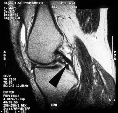
Question 23
A 48-year-old man has recurrent right knee pain. Figure 52a shows the sagittal proton density T2-weighted MRI scan, and Figure 52b shows the sagittal T2-weighted MRI scan at the same level. The arrow is pointing to a
Explanation
Question 24
Figure 53 shows a thoracolumbar specimen as viewed from posterior to anterior following removal of all posterior elements. Which of the following structures does the red string pass under?
Explanation
Question 25
A 5-year-old girl sustained a comminuted Salter-Harris type IV fracture of the left distal tibia 2 years ago. The AP radiograph shown in Figure 54a reveals a growth arrest and a 1.4-cm limb-length discrepancy. The ankle is in approximately 20 degrees of varus. Figure 54b shows a coronal reconstruction image of the distal tibial physis, and Figure 54c shows a sagittal reconstruction image of the same area. On the sagittal reconstruction image, the bar extends from the 9-mm mark to the 24-mm mark in 3-mm increments. On the coronal image, the bar extends from the 9-mm mark to the 24-mm mark, also in 3-mm increments. A map of the physeal bar based on these measurements is shown in Figure 54d. Initial treatment should consist of
Explanation
Question 26
During the ilioinguinal approach to the acetabulum, severe hemorrhage is encountered while dissecting near the superior pubic ramus. Which of the following anatomical structures is most likely injured?
Explanation
Question 27
When placing screws in the acetabulum during total hip arthroplasty, drilling into the posterior-inferior quadrant places which of the following structures at highest risk?
Explanation
Question 28
During a direct lateral (Hardinge) approach to the hip, the gluteus medius is split longitudinally. To prevent denervation of the anterior portion of the gluteus medius and minimus, the proximal split should not extend beyond what distance from the tip of the greater trochanter?
Explanation
Question 29
A 24-year-old male undergoes open reduction and internal fixation of a posterior wall acetabular fracture via a Kocher-Langenbeck approach. Retraction of the sciatic nerve is required. Which portion of the sciatic nerve is at greatest risk of iatrogenic injury, and what clinical deficit would be observed?
Explanation
Question 30
A patient undergoes an ilioinguinal approach for an anterior column acetabular fracture. The surgeon is working in the middle window of this approach. Which of the following structures dictates the medial boundary of this specific window?
Explanation
Question 31
Which of the following best describes the precise anatomic origin of the reflected head of the rectus femoris?
Explanation
Question 32
An orthopaedic surgeon is using the anterior (Smith-Petersen) approach to the hip. What is the true internervous plane utilized in the superficial dissection of this approach?
Explanation
Question 33
When inserting screws into the acetabulum during a total hip arthroplasty, the "safe zone" for screw placement to avoid major neurovascular injury is which quadrant?
Explanation
Question 34
A 14-year-old male athlete presents with sudden onset of severe groin pain after forcefully kicking a soccer ball. Radiographs reveal an avulsion fracture of the lesser trochanter. Which of the following muscles is responsible for this injury?
Explanation
Question 35
During an anterior intrapelvic (Stoppa) approach to the acetabulum, brisk arterial bleeding is encountered while dissecting over the superior pubic ramus near the symphysis. This is most likely due to an injury to an anastomosis between which two vascular systems (Corona Mortis)?
Explanation
Question 36
What is the primary blood supply to the adult femoral head?
Explanation
Question 37
A 35-year-old female experiences numbness and tingling in the anterolateral aspect of her thigh after prolonged wearing of a tight utility belt. Compression of which nerve is the most likely cause, and where does it typically exit the pelvis?
Explanation
Question 38
During hip arthroscopy, prolonged traction against a perineal post can lead to a specific neuropathy. Which of the following clinical findings is most characteristic of this complication?
Explanation
Question 39
When repairing a complete proximal hamstring avulsion, the surgeon must be careful to identify and protect the sciatic nerve. At the level of the ischial tuberosity, what is the anatomical relationship of the sciatic nerve to the hamstring origin?
Explanation
Question 40
In a patient with a posterior pelvic ring disruption, which ligament is considered the primary and strongest stabilizer against vertical shear forces?
Explanation
Question 41
The medial circumflex femoral artery (MCFA) is at risk during a posterior approach to the hip if the dissection extends too far inferiorly. The MCFA typically passes between which two muscles before entering the hip capsule?
Explanation
Question 42
A patient sustains a pelvic fracture involving the greater sciatic notch. Which of the following structures exits the pelvis through the greater sciatic foramen but superior to the piriformis muscle?
Explanation
Question 43
During an open reduction of a developmental dysplasia of the hip (DDH) via an anterior approach, the surgeon must tenotomize the iliopsoas. What nerve is most at risk of injury just medial to the iliopsoas muscle belly at the level of the pelvic brim?
Explanation
Question 44
In evaluating a patient with a pelvic ring injury, the presence of an open book pelvis (APC II or III) implies failure of the symphysis pubis and the anterior sacroiliac ligaments. Which pelvic ligament, if intact, prevents pure vertical displacement and distinguishes an APC II from an APC III injury?
Explanation
Question 45
The obturator nerve provides motor innervation to the adductor compartment of the thigh. If the nerve is completely transected within the obturator canal, which muscle in the medial compartment will retain partial innervation?
Explanation
Question 46
Which of the following landmarks serves as the primary anatomic reference for the inferior limit of the superficial interval in the Watson-Jones (anterolateral) approach to the hip?
Explanation
Question 47
A patient undergoes a pelvic osteotomy and develops persistent weakness in hip external rotation. The surgeon suspects injury to the nerve to the quadratus femoris. This nerve typically leaves the pelvis through the greater sciatic foramen and runs deep to which of the following structures?
Explanation
Question 48
During a posterior approach to the hip, the surgeon identifies the medial femoral circumflex artery (MFCA) to protect the blood supply to the femoral head. What is the correct anatomic course of the main branch of the MFCA?
Explanation
Question 49
When placing screws into the acetabulum during a total hip arthroplasty, which quadrant is considered the 'safe zone' to avoid major neurovascular injury?
Explanation
Question 50
A patient undergoes an anterior pelvic ring fixation via an ilioinguinal approach. During dissection along the superior pubic ramus, brisk bleeding occurs. This is most likely due to injury of an anastomosis between which two vascular systems?
Explanation
Question 51
During a direct anterior approach to the hip (Smith-Petersen), the superficial internervous plane is utilized. Which nerves supply the muscles defining this superficial plane?
Explanation
Question 52
In approximately 15-20% of the population, the sciatic nerve has an anatomical variation in its relationship with the piriformis muscle. What is the most common variant?
Explanation
Question 53
A 45-year-old female presents with persistent lateral hip pain and a Trendelenburg gait after a fall. MRI reveals an isolated avulsion of the gluteus medius tendon. Which aspect of the greater trochanter is the primary footprint for the gluteus medius?
Explanation
Question 54
An adolescent water skier sustains a sudden, forceful hip flexion with knee extension, resulting in an ischial tuberosity avulsion fracture. Which of the following muscles shares a conjoint tendon origin at this site?
Explanation
Question 55
The sacrospinous ligament is a critical anatomical landmark during pelvic surgery. Which two spaces does this ligament separate?
Explanation
Question 56
A patient complains of perineal numbness and fecal incontinence after a difficult vaginal delivery. The nerve responsible for these symptoms exits the pelvis through the greater sciatic foramen and re-enters through the lesser sciatic foramen. What is this nerve?
Explanation
Question 57
During a direct lateral (Hardinge) approach to the hip, proximal splitting of the gluteus medius must be limited to avoid denervating the anterior portion of the muscle. What is the generally accepted safe distance from the tip of the greater trochanter?
Explanation
Question 58
A 24-year-old athlete presents with snapping hip syndrome. Ultrasound evaluation demonstrates snapping of a tendon over the iliopectineal eminence. Which structure inserts onto the lesser trochanter and is responsible for this internal snapping?
Explanation
Question 59
What ligament of the hip is considered the strongest in the human body, acting primarily to prevent hyperextension of the hip joint?
Explanation
Question 60
In evaluating an acetabular fracture on an anteroposterior pelvic radiograph, the ilioischial line is disrupted. This radiographic landmark represents which anatomic structure of the acetabulum?
Explanation
Question 61
During an open reduction and internal fixation of a symphysis pubis diastasis, the surgeon must be aware of the boundaries of the femoral canal to avoid incarcerating a hernia or injuring vascular structures. What forms the medial boundary of the femoral ring?
Explanation
Question 62
The ligamentum teres of the hip contains a small artery that supplies a minor portion of the femoral head in adults. This artery is a branch of which of the following vessels?
Explanation
Question 63
A patient undergoes pelvic lymph node dissection and subsequently presents with weakness in hip adduction and paresthesias over the medial aspect of the thigh. Which nerve was most likely injured as it courses through the pelvis?
Explanation
Question 64
During an anterolateral approach to the hip (Watson-Jones), the internervous plane is between the tensor fasciae latae and the gluteus medius. What is the nerve supply to these two muscles?
Explanation
Question 65
The hip joint capsule is reinforced by several ligaments. Which capsular ligament is located posteriorly and is primarily responsible for limiting internal rotation of the hip in extension?
Explanation
Question 66
A 30-year-old male sustains a posterior hip dislocation. Post-reduction, he exhibits a foot drop and weakness in great toe extension, but plantar flexion is preserved. Which portion of the sciatic nerve is most vulnerable in this injury?
Explanation
Question 67
In the setting of a hip arthroscopy, the surgeon must be cautious when establishing the anterolateral portal to avoid injury to a major nerve. The lateral femoral cutaneous nerve (LFCN) is at risk. What is the typical anatomical course of the LFCN as it exits the pelvis?
Explanation
Question 68
During a modified Stoppa approach for a pelvic ring fracture, the surgeon encounters massive arterial hemorrhage just posterior to the superior pubic ramus. This bleeding is most likely originating from an anastomosis between which of the following vessels?
Explanation
Question 69
When placing acetabular screws during a total hip arthroplasty, the acetabulum is divided into four quadrants using a line from the anterior superior iliac spine through the center of the acetabulum and a second perpendicular line. A misdirected screw in the anterosuperior quadrant places which of the following structures at greatest risk?
Explanation
Question 70
A surgeon uses the direct lateral (Hardinge) approach to the hip, which involves splitting the gluteus medius. To avoid denervating the anterior portion of the gluteus medius and tensor fasciae latae, the split should not extend proximally from the tip of the greater trochanter more than:
Explanation
Question 71
An anterior (Smith-Petersen) approach to the hip is selected for an open reduction of a developmental dysplasia of the hip (DDH). What represents the superficial internervous plane for this approach?
Explanation
Question 72
In a 65-year-old patient sustaining a displaced intracapsular femoral neck fracture, the primary blood supply to the femoral head is typically disrupted. Which of the following arteries provides the majority of the blood supply to the adult femoral head?
Explanation
Question 73
Following a total hip arthroplasty via an anterior approach, a patient complains of burning pain and numbness over the anterolateral aspect of the operative thigh. The nerve responsible for this complication typically exits the pelvis in which location?
Explanation
Question 74
A patient with a complex pelvic ring injury presents with profound weakness in hip adduction and an area of decreased sensation over the distal medial thigh. An injury to the obturator nerve is suspected. Which of the following adductor muscles will likely retain partial function due to dual innervation?
Explanation
Question 75
During a piriformis-sparing posterior approach to the hip, the surgeon visualizes an anatomic variant of the sciatic nerve. In approximately 10% of the population, a portion of the sciatic nerve pierces or passes superior to the piriformis. Which neural element is most commonly involved in this variant?
Explanation
Question 76
The ilioinguinal approach provides excellent exposure of the anterior column of the acetabulum. The exposure is traditionally divided into three distinct anatomical 'windows'. Which structures are primarily located and mobilized within the middle window?
Explanation
Question 77
A 24-year-old male sustains a vertical shear pelvic fracture following a fall from height. Which ligamentous complex provides the most significant resistance to vertical displacement of the hemipelvis?
Explanation
Question 78
A 16-year-old elite track athlete presents with acute, severe pain over the anterior pelvis after forcefully kicking a ball. Radiographs demonstrate an avulsion fracture of the anterior inferior iliac spine (AIIS). Which muscle is responsible for this avulsion?
Explanation
Question 79
Following a technically challenging hip arthroscopy performed on a traction table, the patient reports severe perineal numbness and erectile dysfunction. This complication is most likely caused by direct compression of which nerve against the perineal post?
Explanation
Question 80
During a posterior (Kocher-Langenbeck) approach to the hip for a posterior wall acetabular fracture, the short external rotators are sharply detached. The superior border of which muscle must be preserved intact to protect the deep branch of the medial circumflex femoral artery?
Explanation
Question 81
Which ligament is considered the strongest ligament in the human body and acts as the primary restraint to hyperextension of the hip joint?
Explanation
Question 82
To minimize tension on the sciatic nerve while placing retractors during a Kocher-Langenbeck approach for acetabular fracture fixation, the ipsilateral lower extremity should be placed in which of the following positions?
Explanation
Question 83
During a surgical approach to the hip, understanding the vascular anatomy is critical to prevent avascular necrosis of the femoral head. Which of the following branches provides the predominant blood supply to the adult femoral head?
Explanation
Question 84
During a posterior approach to the hip, the short external rotators must be identified and tagged. Which of the following structures exits the pelvis through the lesser sciatic foramen?
Explanation
Question 85
The ilioinguinal approach to the acetabulum provides access to the anterior column. The 'middle window' of this approach is bounded by which of the following structures?
Explanation
Question 86
During an anterior intrapelvic (modified Stoppa) approach, the surgeon must be cautious of the 'corona mortis'. This vascular structure represents an anastomosis between which two systems?
Explanation
Question 87
When placing screws into the acetabulum during a total hip arthroplasty, the quadrant system is used to identify safe zones. Placement of a screw into the anterosuperior quadrant places which structure at highest risk of injury?
Explanation
Question 88
A direct anterior (Smith-Petersen) approach is used for a hip arthroplasty. During the superficial dissection, which nerve is most at risk of iatrogenic injury?
Explanation
Question 89
To avoid denervation of the hip abductors during a direct lateral (Hardinge) approach to the hip, the proximal split of the gluteus medius should be limited to what maximum distance from the tip of the greater trochanter?
Explanation
Question 90
A patient exhibits a positive Trendelenburg sign after sustaining a penetrating injury to the posterior pelvis. The injured nerve is responsible for innervating which of the following muscle groups?
Explanation
Question 91
A patient with severe right hip osteoarthritis is advised to use a cane for ambulation. To most effectively reduce the joint reactive forces across the right hip, how should the cane be used?
Explanation
Question 92
The artery of the ligamentum teres provides a small and variable blood supply to the femoral head. It is typically a terminal branch of which of the following arteries?
Explanation
Question 93
When performing a posterior (Kocher-Langenbeck) approach to the acetabulum, what is the true internervous plane utilized?
Explanation
Question 94
A 16-year-old sprinter presents with acute buttock pain after feeling a 'pop' while running. Radiographs reveal an avulsion fracture of the ischial tuberosity. Which of the following muscles originates at this anatomical site?
Explanation
Question 95
During surgical exposure of the posterior pelvic ring, the pudendal nerve must be protected. What is the anatomic path of the pudendal nerve relative to the sacrospinous and sacrotuberous ligaments?
Explanation
Question 96
During fracture fixation via the modified Stoppa approach, mobilization of the obturator neurovascular bundle is required to visualize the quadrilateral plate. Through which structure does this bundle exit the true pelvis?
Explanation
Question 97
An adolescent soccer player sustains a pelvic avulsion fracture resulting from a forceful kick. Radiographs confirm an avulsion of the anterior superior iliac spine (ASIS). Which muscle is primarily responsible for this injury?
Explanation
Question 98
A 15-year-old athlete experiences sudden anterior groin pain while sprinting. Imaging demonstrates an avulsion fracture of the anterior inferior iliac spine (AIIS). This injury is caused by the sudden contraction of which muscle?
Explanation
Question 99
During an anterolateral (Watson-Jones) approach to the hip, which vascular structure crosses the surgical interval and typically requires ligation to achieve adequate deep exposure?
Explanation
Question 100
The hip joint capsule is reinforced by several strong ligaments. Which ligament is the strongest in the body and acts primarily to prevent hyperextension of the hip joint?
Explanation
None
