العربية
Part of the Master Guide

Orthopedic Board Review Set 675: 100 MCQs for ABOS, OITE, FRCS – Hip Focus

Orthopedic Hip & Knee 2026 MCQs: Board Review Questions & Answers (Part 4)

27 Apr 2026 51 min read 88 Views
Figure for Hpkn 2007 MCQs - Part 4 - Question 76

Key Takeaway

Your ultimate guide to Orthopedic Hip & Knee 2026 MCQs: Board Review Questions & Answers (Part 4) starts here. Top-rated Orthopedic Hip & Knee 2026 MCQs bank. Practice with clinical case questions, orthopedic surgery board review, and evidence-based answers updated for 2026.

Orthopedic Hip & Knee 2026 MCQs: Board Review Questions & Answers (Part 4)

Comprehensive 100-Question Exam


00:00

Start Quiz

Question 1

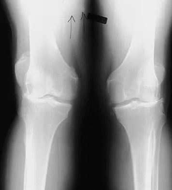
A 37-year-old man who works in a factory has isolated, lateral unicompartmental pain about his knee with activities. Nonsurgical management has failed to provide relief. The radiograph shown in Figure 45 reveals a tibiofemoral angle of approximately 15 degrees which is clinically correctable to neutral. What is the best surgical option in this patient?


Explanation

Patients with a valgus alignment about the knee can have lateral compartment arthritis. Similar to a high tibial osteotomy, a supracondylar femoral osteotomy is indicated in younger patients who have a more active lifestyle and isolated unicompartmental disease. In this young patient who works in a factory and has a valgus knee, a medial closing wedge supracondylar femoral osteotomy is the treatment of choice. The role of arthroplasty is limited in younger patients. Mathews J, Cobb AG, Richardson S, et al: Distal femoral osteotomy for lateral compartment osteoarthritis of the knee. Orthopedics 1998;21:437-440.

Question 2

Figure 46 shows the AP radiograph of an active 80-year-old patient with an acetabular fracture. The fracture was initially managed nonsurgically; however, the patient is now scheduled to undergo total hip arthroplasty. What is the treatment of choice for the contained acetabular bone defect?


Explanation

Acetabular fractures can result in a relative or actual acetabular bone defect. The medial blow-out fracture of the acetabulum has united well in this patient. It is likely that a medial shell of bone will be present during hip arthroplasty. The femoral head may be used as morcellized or structural bone to augment the medial defect and is preferred to structural allograft. Bipolar hip arthroplasty is notorious for medial migration in patients without a medial bone defect; therefore, it will not be a good choice in this patient. Filling the defect with methylmethacrylate cement, though an option, is not the best option in this active patient with an extensive medial defect. A double-bubble acetabular cup is used for patients with deficiency of the bone in the dome region. Mears DC: Surgical treatment of acetabular fractures in elderly patients with osteoporotic bone. J Am Acad Orthop Surg 1999;7:128-141.

Question 3

A 28-year-old woman who is an avid runner reports pain about the left hip with activities. Nonsurgical management has failed to provide relief. An MRI arthrogram is shown in Figure 47. What is the most likely diagnosis?


Explanation

The MRI arthrogram reveals dye extravasation into the labrum, consistent with a labral tear. The MRI findings are not typical of osteonecrosis, stress fracture, or transient osteoporosis. There is no increase in bone marrow edema in the neck or femoral head. Guanche CA, Sikka RS: Acetabular labral tears with underlying chondralmalacia: A possible association with high-level running. Arthroscopy 2005;21:580-585.

Question 4

Figure 48a shows the full-leg standing radiograph of a patient with a prior femoral fracture. Figure 48b shows the lateral view of the same joint. The patient is scheduled to undergo total knee arthroplasty. Because the mechanical axis of the lower extremity in patients with a prior femoral fracture may be disrupted, which of the following should be used during surgery to restore the mechanical axis of the lower extremity in this patient?


Explanation

48b The radiograph shows hardware that was used for fixation of a prior femoral fracture. The mechanical axis of the lower extremity in this patient is nearly normal (3 degrees valgus), and the deformity at the healed fracture site (14 degrees) does not appear to affect the joint alignment and is acceptable. Use of a routine knee prosthesis will be possible in this patient. To avoid hardware removal, extramedullary jigs and/or computerized navigation may be used to measure and restore the long axis of the femur. The use of a hinged prosthesis does not influence the mechanical axis directly. Extra-articular osteotomy is occasionally needed to reverse severe deformities. Papadopoulos EC, Parvizi J, Lai CH, et al: Total knee arthroplasty following distal femoral fractures. Knee 2002;9:267-274.

Question 5

Figure 49 shows a histologic section of the lung in a patient who died during total hip arthroplasty. What unexpected finding is seen in the pulmonary capillaries?


Explanation

Sudden death during total hip arthroplasty has been reported. In a report from the Mayo Clinic, intraoperative death occurred during cemented total hip arthroplasty in 23 patients. Fat and marrow embolization during preparation of the femur or cementing of the femoral component was believed to be responsible for the cardiopulmonary collapse that occurred during arthroplasty. Although fat and marrow emboli were found in the pulmonary capillaries of most of the patients on autopsy, this histologic section shows two particles of cement in the pulmonary capillaries. Parvizi J, Holiday AD, Ereth MH, et al: The Frank Stinchfield Award. Sudden death during primary hip arthroplasty. Clin Orthop 1999;369:39-48.

Question 6

After trial placement of components in a primary total knee arthroplasty, the knee is unable to come to full extension, but the flexion gap is appropriately balanced. After adequate soft-tissue releases have been performed, what is the next most appropriate action to balance the reconstruction?


Explanation

The reconstruction requires additional resection of the distal femur to allow increased extension while maintaining the current flexion gap tension. Resecting more proximal tibia or decreasing the tibial polyethylene thickness will decrease flexion tension as well as extension tension. Adding posterior femoral augments and using a larger femoral component will increase flexion tension. Ayers DC, Dennis DA, Johanson NA, et al: Common complications of total knee arthroplasty. J Bone Joint Surg Am 1997;79:278-311.

Question 7

Figure 50 shows the cross table lateral radiograph of a 31-year-old paratrooper who has recalcitrant groin pain. The pain is worse after activities such as standing or sitting (driving). Examination reveals that pain can be reproduced by internal rotation of the leg with the hip and knee in 90 degrees of flexion. Extensive nonsurgical managment has failed to provide relief. What is the treatment of choice?


Explanation

The radiograph reveals the classic "bump" that is seen in patients with femoroacetabular impingement (FAI). Ganz and associates described two types of FAI. This patient has cam impingement, which describes a nonspherical femoral head being forced into the acetabulum during hip motion and resulting in labral and chondral injury. Hip arthroscopy and labral debridement is unlikely to control the symptoms because the underlying anatomic abnormality is often difficult to address with arthroscopy. The treatment involves surgical dislocation of the hip with preservation of the blood supply to the femoral head, removal of the asphericity on the femoral side (femoral osteoplasty), and removal of the acetabular rim (acetabular osteoplasty) if the latter is found to contribute to impingement. Ganz R, Gill TJ, Gautier E, et al: Surgical dislocation of the adult hip a technique with full access to the femoral head and acetabulum without the risk of avascular necrosis. J Bone Joint Surg Br 2001;83:1119-1124. Ganz R, Parvizi J, Beck M, et al: Femoroacetabular impingement: A cause for early osteoarthritis of the hip. Clin Orthop 2003;417:112-120.

Question 8

During total knee arthroplasty, the patella is noted to subluxate laterally despite a lateral retinacular release. Which of the following methods is most likely to improve patellar stability?


Explanation

Slight external rotation of the tibial component will cause a net medialization of the tibial tubercle when the knee is articulated. This will help centralize the extensor mechanism over the trochlear groove and minimize the tendency for lateral subluxation. Internal rotation of the femoral component increases the risk of patellar instability. Anterior translation of the tibial component moves the patellar tendon insertion posteriorly, and may increase force on the patella but should not substantially alter patellar tracking. Clinical studies have shown no patellofemoral benefits to the use of fixed- or mobile-bearing designs. Thicker patellar components will not improve tracking, and may compound the problem. Pellicci PM, Tria AJ Jr, Garvin KL (eds): Orthopaedic Knowledge Update: Hip and Knee Reconstruction 2. Rosemont, IL, American Academy of Orthopaedic Surgeons, 2000, pp 207, 323-337 Pagnano MW, Trousdale RT, Stuart MJ, et al: Rotating platform knees did not improve patellar tracking: A prospective, randomized study of 240 primary total knee arthroplasties. Clin Orthop 2004;428:221-227.

Question 9

A 73-year-old man has stiffness after undergoing primary posterior cruciate ligament-retaining total knee arthroplasty 18 months ago. Extensive physiotherapy, dynamic splinting, and manipulations under anesthesia have failed to result in improvement. Examination reveals range of motion from 30 degrees to 60 degrees of flexion. The components are well fixed, and the evaluation for infection is negative. In discussing the possibility of revision arthroplasty, the patient should be advised that


Explanation

Stiffness following primary total knee arthroplasty remains a vexing problem. Treatment options have included extensive physical therapy, dynamic splinting, manipulation under anesthesia, arthroscopic arthrolysis, open arthrolysis with polyethylene exchange, and ultimately revision arthroplasty. Results are not as gratifying as would be expected. Babis and associates performed an open arthrolysis and polyethylene exchange on seven patients who were followed for a mean of 4.2 months. The results were poor. The mean improvement in arc of motion was only 20 degrees. Nicholls and Dorr treated 13 patients for stiffness. Only 40% of those patients obtained good to excellent results. Four patients (30%) required manipulation because of recurrent stiffness postoperatively. They noted they could not predictably improve the arc of motion with a revision operation. Haidukewych and associates reported on 15 patients who underwent revision of well-fixed components after total knee arthroplasty for stiffness. Of the 15 patients, 10 (66%) were satisfied with the outcome revision. Interestingly, they noted that in patients for whom the total arc of motion did not improve but who regained near full extension, there was a greater amount of satisfaction with the procedure than for those who did not regain full extension. Babis GC, Trousdale RT, Pagnano MW, et al: Poor outcomes of isolated tibial insert exchange and arthrolysis for the management of stiffness following total knee arthroplasty. J Bone Joint Surg Am 2001;83:1534-1536. Nicholls DW, Dorr LD: Revision surgery for stiff total knee arthroplasty. J Arthroplasty 1990;5:S73-S77.

Question 10

A 62-year-old patient is seen for routine follow-up after undergoing cementless total hip arthroplasty 2 years ago. The patient reports limited range of motion that severely affects daily activities. A radiograph is shown in Figure 51. Management should now consist of


Explanation

The patient has symptomatic postoperative heterotopic ossification after total hip arthroplasty. Postoperative prophylactic treatments include nonsteroidal anti-inflammatory drugs (usually indomethacin) or low-dose irradiation. The heterotopic ossification shown here is quite mature; therefore, nonsurgical management will not be successful. Surgical excision of grade III or IV heterotopic ossification should be followed with postoperative irradiation to minimize the chances of recurrence. Ayers DC, Evarts CM, Parkinson JR: The prevention of heterotopic ossification in high-risk patients by low-dose radiation therapy after total hip arthroplasty. J Bone Joint Surg Am 1986;68:1423-1430.

Question 11

What bilateral surgical intervention is considered inappropriate based on the findings shown in the radiograph in Figure 52?


Explanation

The radiograph reveals osteonecrosis of both femoral heads with reasonably maintained joint surfaces. There may be some slight flattening of the femoral heads. Hip arthrodesis is difficult to perform because of the necrotic bone. Its use in patients with osteonecrotic hips is limited because of the 80% bilaterality; therefore, it is not an acceptable alternative. All the other options are acceptable interventions. Mont MA, Jones LC, Sotereanos DG, et al: Understanding and treating osteonecrosis of the femoral head. Instr Course Lect 2000;49:169-185.

Question 12

Figure 53a shows the AP radiograph of a 70-year-old patient who is scheduled to undergo unicompartmental knee arthroplasty. Figure 53b shows the immediate postoperative radiograph, and the radiograph shown in Figure 53c, obtained 6 months after surgery, shows a medial tibial plateau fracture. The etiology of the fracture is best related to


Explanation

53b 53c While all of the above may contribute to the etiology of a tibial plateau fracture following unicompartmental knee arthroplasty, the recent literature has clearly noted that pin placement for fixation of tibial resection guides is the most critical factor associated with a tibial plateau fracture following unicompartmental knee arthroplasty. Vince and Cyran suggest that fractures associated with unicompartmental knee arthroplasty might be avoidable by limiting the number and paying attention to the location of the pin holes that are created to secure the tibial resection guides. Brumby and associates suggest avoiding multiple guide pin holes in the proximal tibia for unicompartmental knee arthroplasty. They currently recommend the use of one centrally placed pin and an ankle clamp to stabilize the resection guide. Yang and associates note that a medial tibial plateau fracture in association with minimally invasive unicompartmental knee arthroplasty can be eliminated by avoiding fixation pins close to the medial tibial cortex. Brumby SA, Carrington R, Zayontz S, et al: Tibial plateau stress fracture: A complication of unicompartmental knee arthroplasty using 4 guide pinholes. J Arthroplasty 2003;18:809-812. Yang KY, Yeo SJ, Lo NN: Stress fracture of the medial tibial plateau after minimally invasive unicompartmental knee arthroplasty: A report of 2 cases. J Arthroplasty 2003;18:801-803.

Question 13

During impaction of a cementless acetabular component, the posterior column was fractured and found to be displaced. Which of the following is considered the most appropriate surgical option?


Explanation

Acetabular bone loss presents a challenge during reconstruction. A cementless hemispherical cup can be used in most patients provided that the acetabular rim, particularly the posterior column, is intact. When the posterior column is disrupted, fixation with a reconstruction plate and/or the use of an antiprotrusio cage is recommended. The latter is particularly important when the posterior column is fractured and displaced, such as in this patient. Under these circumstances, reduction of the fracture and application of an antiprotrusio cage is recommended. In this particular type of case, some surgeons may elect to retain the hemispherical cup and apply an antiprotrusio cage over the cup ("cage over cup" technique). Berry DJ: Antiprotrusio cages for acetabular revision. Clin Orthop 2004;420:106-112.

Question 14

Which of the following factors increases the risk of sciatic nerve injury in primary total hip arthroplasty (THA)?


Explanation

Injury to the sciatic nerve is a relatively rare but serious complication of THA. Dissection of the sciatic nerve is not typically done during primary THA, although the nerve can be identified during the surgical approach. An anterolateral approach to THA would not necessarily be associated with any greater incidence of sciatic nerve injury than other approaches. Screw fixation for the acetabular component is often a matter of surgeon preference. Provided that the anatomic safe zones for screw fixation (posterior inferior and posterior superior) are recognized, injury to the sciatic nerve from acetabular screws can be minimized. Restoration of anatomic length is important in primary THA. Overlengthening can result in sciatic nerve palsy. Developmental dysplasia of the hip can lead to a congenitally shortened extremity with concomitant congenital shortening of the associated neurovascular structures. Overlengthening of the extremity during THA for developmental dysplasia of the hip can lead to sciatic palsy. Osteonecrosis is not an associated risk factor for sciatic nerve palsy. DeHart MM, Riley LH Jr: Nerve injuries in total hip arthroplasty. J Am Acad Orthop Surg 1999;7:101-111.

Question 15

A 68-year-old woman who underwent a right total hip arthroplasty 1 year ago has dislocated her hip five times since surgery. Radiographs show a retroverted acetabular component. What is the best treatment for this patient?


Explanation

The most common cause of recurrent dislocation following total hip arthroplasty continues to be component malposition. Component malposition should be addressed prior to any other treatment options, such as increasing soft-tissue tension with increased femoral offset or greater trochanteric advancement. A larger femoral head size may help, but correcting the component malposition should give more predictable results. A retroverted acetabular component should be revised to 15 degrees to 20 degrees of anteversion, matching the patient's anatomy with an abduction angle close to 45 degrees. Daly PJ, Morrey BF: Operative correction of an unstable total hip arthroplasty. J Bone Joint Surg Am 1992;74:1334-1343. Jolles BM, Zangger P, Leyvraz PF: Factors predisposing to dislocation after primary total hip arthroplasty: A multivariate analysis. J Arthroplasty 2002;17-282-288.

Question 16

Figure 54 shows the preoperative radiograph of a 45-year-old woman who is considering total hip arthroplasty with her orthopaedic surgeon. What femoral characteristic is a typical concern in this patient?


Explanation

Developmental dysplasia of the hip (DDH) leads to early arthritis of the hip as seen in this patient. Although DDH is believed to mostly affect the acetabulum, most patients with DDH also have anatomic aberrations of the femur. Using three-dimensional computer models generated by reconstruction of CT scans, dysplastic femurs were shown to have shorter necks and smaller, straighter canals than the controls. The shape of the canal became more abnormal with increasing subluxation. The studies also have shown that the primary deformity of the dysplastic femur is rotational, with an increase in anteversion of 5 degrees to 16 degrees, depending on the degree of subluxation of the hip. The rotational deformity of the dysplastic femur arises within the diaphysis between the lesser trochanter and the isthmus and is not attributable to a torsional deformity of the metaphysis. Osteopenia is not a concern in a patient with an excellent cortical index (thick cortices and narrow canal). Femoral varus or bowing of the femur is not a typical finding in patients with DDH. Noble PC, Kamaric E, Sugano N, et al: Three-dimensional shape of the dysplastic femur: Implications for THR. Clin Orthop 2003;417:27-40.

Question 17

A 68-year-old man with no significant medical history underwent a total knee arthroplasty 4 years ago. A radiograph is shown in Figure 55. He reports that he had no problems with the knee until 6 weeks ago when he noted the gradual onset of pain following a colonoscopy. Examination reveals a painful, swollen knee. Knee aspiration reveals a WBC count of 40,000/mm3. Management should consist of


Explanation

The treatment of choice for a late hematogenous infection is two-stage resection arthroplasty and reimplantation, with parenteral antibiotics prior to reimplantation. This is particularly true when septic loosening has occurred as in this patient. Open irrigation and debridement with polyethylene exchange has been used successfully when the duration of symptoms is 3 weeks or less. Long-term suppressive antibiotics are most commonly used when the patient's medical condition precludes further surgery. Delayed reimplantation has been shown to be superior to immediate reimplantation in multiple studies. Little data support the use of arthroscopic irrigation and debridement. Swanson KC, Windsor RE: Diagnosis of infection after total knee arthroplasty, in Callaghan JJ, Rosenberg AG, Rubash HE, et al (eds): The Adult Knee. Philadelphia, PA, JB Lippincott, 2003, vol 2, pp 1485-1491.

Question 18

Which of the following substances makes up the majority by weight of the extracellular matrix for articular cartilage?


Explanation

The extracellular matrix consists of water, proteoglycans, and collagen. Water makes up the majority (approximately 65% to 80%) of wet weight; 95% of the collage is type II with much smaller amounts of other collagens, including types IV, VI, IX, X, and XI. The exact functions of these other collagens are unknown, but they are believed to be important in matrix attachment and stabilization of the diameter of collagen fibrils. Buckwalter JA, Einhorn TA, Simon SR (eds): Orthopaedic Basic Science: Biology and Biomechanics of the Musculoskeletal System, ed 2. Rosemont, IL, American Academy of Orthopaedic Surgeons, 2000, pp 444-445.

Question 19

A 58-year-old woman is seen in the emergency department after falling at home. History reveals that she underwent right total knee arthroplasty 2 years ago. Radiographs are shown in Figures 56a and 56b. What is the most appropriate treatment?


Explanation

56b The radiographs show an oblique periprosthetic distal femoral fracture. Of the options listed, open reduction and internal fixation is the most appropriate surgical option because a well-fixed, posterior stabilized closed box femoral component is present. Nonsurgical methods are not favored because of the highly displaced, unstable fracture pattern and prolonged immobility. Revision with a stemmed component is an option but would sacrifice more bone stock in this younger patient. Moran MC, Brick GW, Sledge CB, et al: Supracondylar femoral fracture following total knee arthroplasty. Clin Orthop 1996;324:196-209. Raab GE, Davis CM III: Early healing with locked condylar plating of periprosthetic fractures around the knee. J Arthroplasty 2005;20:984-989.

Question 20

A patient with a valgus knee and lateral compartment bone loss undergoes a total knee arthroplasty using posterior condylar referencing instrumentation. Six months after surgery, the patient reports significant anterior knee pain, and radiographs reveal severe lateral patellar tilt. Management should consist of


Explanation

Severe valgus deformity is frequently accompanied by hypoplasia of the lateral femoral condyle. Posterior referencing instrumentation can substantially internally rotate the femoral component with respect to the transepicondylar axis and Whiteside's line. The femoral component malrotation must be corrected to properly address this problem. Berger RA, Della Valle CJ, Rubash HE: Patellofemoral problems in total knee arthroplasty, in Callaghan JJ, Rosenberg AG, Rubash HE, et al (eds): The Adult Knee. Philadelphia, PA, JB Lippincott, 2003, vol 2, pp 1245-1258.

Question 21

Figures 57a through 57c show the radiographs of a patient who has pain, discomfort, and a popping sensation localized to the posterior aspect of the knee after undergoing primary left total knee arthroplasty 6 months ago. Examination reveals that the patient is able to ambulate without a limp. There is no significant swelling, erythema, or effusion. Range of motion is 0 degrees to 115 degrees, and a palpable crepitation or snapping is detected at the posterior lateral joint line. What is the most likely diagnosis?


Explanation

57b 57c Popliteal snapping syndrome represents the most likely diagnosis. Barnes and Scott noted that the popliteus tendon can be a potential source of internal derangement after total knee arthroplasty. They noted that it can be subluxated anteriorly and posteriorly over a retained lateral femoral condyle osteophyte. Allardyce and associates described the condition as a popliteus condition, snapping as it rolls over a retained lateral femoral condylar osteophyte. Patellar clunk syndrome is a distinct syndrome associated with the patella and has been reported in posterior stabilized knees. In addition to crepitation with range of motion, the patella literally snaps or jumps as the knee is taken from flexion to extension. Beight JL, Yao B, Hozack WJ, et al: The patellar "clunk" syndrome after posterior stabilized total knee arthroplasty. Clin Orthop 1994;299:139-142. Barnes CL, Scott RD: Popliteus tendon dysfunction following total knee arthroplasty. J Arthroplasty 1995;10:543-545.

Question 22

Which of the following is the primary mechanism of polyethylene wear in the hip?


Explanation

Although previous theories on acetabular wear implicated fatigue cracking and delamination as primary wear mechanisms, these have actually manifested as major modes of polyethylene wear in knees. The primary mechanism of wear in polyethylene acetabular components appears to be adhesion and abrasion. In an analysis of 128 components retrieved at autopsy or revision surgery, wear appeared to occur mostly at the surface of the components and was the result of large strain plastic deformation and orientation of the surface layers into fibrils that subsequently ruptured during multidirectional motion. It was also shown conclusively that 32-mm heads displayed significantly more wear (volumetric wear) than either 22-mm or 26-/28-mm heads (1-mm increase in size increased volumetric wear by 10%). The wear at the articulating surface was characterized by highly worn polished areas superiorly and less worn areas inferiorly separated by a ridge. Abrasion was very common, occurring after adhesion and plastic deformation of polyethylene fibrils, and abrasion secondary to third-body wear. Wear rates decreased with longer survival of components, indicating a "bedding in" phenomenon, arguing against oxidative and fatigue wear. Crevice corrosion occurs in fatigue cracks with low oxygen tension (under screw heads, etc). Oscillatory fretting consists of cyclical abrading of the outer surface from small movements. Fatigue and delamination is predominant in total knee arthroplasty where stresses are maximum just below the surface of the polyethylene component, causing fatigue over time with subsequent delamination. In contrast, hip wear occurs primarily at the surface of the polyethylene component. Jasty M, Goetz DD, Bragdon CR, et al: Wear of polyethylene acetabular components in total hip arthroplasty: An analysis of one hundred and twenty-eight components retrieved at autopsy or revision operations. J Bone Joint Surg Am 1997;79:349-358. Beaty JH (ed): Orthopaedic Knowledge Update 6. Rosemont, IL, American Academy of Orthopaedic Surgeons, 1999, pp 47-53. Bell CJ, Walker PS, Abeysundera MR, et al: Effect of oxidation on delamination of ultrahigh-molecular-weight polyethylene tibial components. J Arthroplasty 1998;13:280-290.

Question 23

Which of the following complications may occur subsequent to resurfacing hip arthroplasty for osteonecrosis of the hip but not after total hip arthroplasty?


Explanation

Advocates of resurfacing hip arthroplasty cite preservation of the proximal femoral bone stock as the main advantage of this procedure over total hip arthroplasty. Fracture of the retained femoral neck has been reported following resurfacing arthroplasty. The exact etiology of the latter is unknown. Technical errors, such as notching of the femoral neck or possibly disruption of the blood supply to the femoral head during extensive soft-tissue exposure, may result in femoral neck fracture. Gabriel JL, Trousdale RT: Stem fracture after hemiresurfacing for femoral head osteonecrosis. J Arthroplasty 2003;18:96-99.

Question 24

Which of the following statements best describes results that have been reported with roentgen stereophotogrammetric analysis (RSA)?


Explanation

Migration of total hip femoral components has been measured by RSA, a technique that affords accuracy of 2 degrees and 0.5 mm. Several published studies on total hip arthroplasty femoral components have established the importance of this technique. Both cemented and cementless components migrate, with the rate of migration suggesting the adequacy of fixation of a component. Migration of 1 mm to 2 mm (occurring in either the varus-coronal plane and retroversion-transverse plane, or both) has been associated with a higher risk of loosening of the component.

Question 25

Osteonecrosis of the large joints may develop in patients with which of the following conditions?


Explanation

Osteonecrosis of major joints can occur in patients exposed to corticosteroids, alcohol, and antiseizure medications, as well as patients with hemaglobulinopathy, such as sickle cell anemia. In addition, patients with primary APS who had not taken corticosteroids were also found to be at high risk for osteonecrosis of the hip. In one study of 30 patients with primary APS, asymptomatic osteonecrosis was evident in 20%. A recent article has also found a high association between idiopathic osteonecrosis of the hip and collagen II mutation. None of the other conditions has been shown to be associated with a higher risk of osteonecrosis. Tektonidou MG, Malagari K, Vlachoyiannopoulos PG, et al: Asymptomatic avascular necrosis in patients with primary antiphospholipid syndrome in the absence of corticosteroid use: A prospective study by magnetic resonance imaging. Arthritis Rheum 2003;48:732-736.

Question 26

During a posterior-stabilized total knee arthroplasty, trial reduction is performed. The knee is noted to be symmetric and balanced in flexion, but tight in extension. What is the most appropriate next step to balance the knee?


Explanation

Resecting more distal femur increases the extension gap without affecting the flexion gap. This is the appropriate management for a knee that is balanced in flexion but tight in extension.

Question 27

A 65-year-old woman undergoes total knee arthroplasty. Intraoperatively, the surgeon notes that the patella tracks laterally and has a tendency to subluxate during flexion. Which of the following component adjustments would most effectively improve patellar tracking?


Explanation

External rotation of the femoral component lateralizes the anterior femoral sulcus, thereby decreasing the Q angle and improving central patellar tracking. Conversely, internal rotation of either the femoral or tibial components increases the Q angle and exacerbates lateral maltracking.

Question 28

A 55-year-old man receives a THA using a highly cross-linked polyethylene liner. What is the primary biochemical purpose of subjecting the polyethylene to a heating process (melting or annealing) immediately following gamma irradiation?


Explanation

Heating highly cross-linked polyethylene after gamma irradiation eliminates residual free radicals trapped in the polymer chains. This critical step prevents long-term in vivo oxidation, which is a primary cause of polyethylene degradation and late wear.

Question 29

A 24-year-old hockey player presents with persistent anterior groin pain exacerbated by hip flexion.

An AP pelvis radiograph reveals a prominent 'crossover sign'. What is the primary pathomorphology associated with this radiographic finding?


Explanation

The crossover sign on an AP pelvis radiograph indicates acetabular retroversion, where the anterior wall projects more laterally than the posterior wall in the cranial aspect of the joint. This finding is a hallmark of pincer-type femoroacetabular impingement (FAI).

Question 30

During a posterior-stabilized total knee arthroplasty using an anterior referencing system, the surgeon evaluates the gaps and notes that the joint is tight in flexion but symmetric and balanced in extension. Which of the following is the most appropriate next step?


Explanation

A tight flexion gap with a balanced extension gap can be addressed by downsizing the femoral component in an anterior referencing system, which translates the posterior condyles anteriorly and opens the flexion space. Increasing (not decreasing) the posterior tibial slope is an alternative option.

Question 31

A patient undergoes a primary THA via a direct anterior approach. Postoperatively, they report a burning sensation and numbness over the anterolateral aspect of the operative thigh. Which nerve is most likely affected, and where is it most vulnerable during this surgical exposure?


Explanation

The lateral femoral cutaneous nerve (LFCN) is at high risk during the superficial dissection of the direct anterior approach, which exploits the interval between the tensor fasciae latae and the sartorius. Injury to the LFCN results in anterolateral thigh paresthesia and dysesthesia.

Question 32

A 72-year-old man presents with knee pain 2 years following a TKA. Synovial fluid analysis reveals a WBC count of 4,500 cells/µL with 85% PMNs. Which of the following additional findings would definitively confirm a periprosthetic joint infection (PJI) according to the major criteria of the 2018 International Consensus Meeting?


Explanation

According to the 2018 ICM criteria, the presence of a sinus tract communicating with the joint or two positive cultures with phenotypically identical organisms are major criteria that definitively diagnose PJI. The other options are minor criteria that require a scoring system tally.

Question 33

A 55-year-old man presents with medial-sided knee pain. Radiographs reveal medial compartment osteoarthritis. He is being considered for a medial unicompartmental knee arthroplasty (UKA). Which of the following is considered an absolute contraindication to UKA?


Explanation

Inflammatory arthritis is an absolute contraindication to unicompartmental knee arthroplasty due to the global nature of the disease, which will predictably progress to involve the preserved compartments.

Question 34

A 68-year-old woman undergoes a primary total hip arthroplasty via a posterior approach. Six weeks postoperatively, she experiences recurrent posterior dislocations. Radiographic evaluation shows the cup position.

The acetabular component is measured in 25 degrees of abduction and 5 degrees of retroversion. What is the most appropriate management?


Explanation

The acetabular component is malpositioned in retroversion and under-abducted (normal target is 40-45 degrees abduction and 15-20 degrees anteversion). Revision to correct the version and abduction is the most appropriate management for recurrent instability.

Question 35

A 62-year-old woman complains of anterior knee pain and a clunking sensation 1 year after a posterior-stabilized total knee arthroplasty. Examination reveals patellar maltracking with a lateral tilt. Radiographs and a CT scan demonstrate internal rotation of both the tibial and femoral components. Which of the following is the most likely consequence of this combined malrotation?


Explanation

Internal rotation of either the femoral or tibial components increases the Q-angle, leading to lateral patellar tracking, tilt, and subluxation. Combined internal rotation significantly exacerbates this lateralizing effect.

Question 36

A 65-year-old man with ankylosing spondylitis and a completely fused lumbosacral spine is scheduled for a primary total hip arthroplasty (THA). How does this patient's spinopelvic stiffness alter the targeted functional safe zone for acetabular cup positioning compared to a patient with normal spinal mobility?


Explanation

A stiff spine prevents the normal posterior pelvic tilt that occurs during sitting, which naturally increases functional acetabular anteversion. To prevent anterior impingement and posterior dislocation when sitting, the cup may require increased anteversion.

Question 37

Intraoperative assessment during a primary posterior-stabilized total knee arthroplasty (TKA) reveals a flexion gap that is excessively tight, while the extension gap is perfectly balanced. Which of the following modifications is the most appropriate step to achieve a balanced knee?


Explanation

A tight flexion gap with a balanced extension gap requires increasing the flexion space without altering the extension space. Downsizing the femoral component using an anterior referencing guide resects more posterior femoral bone, thereby selectively increasing the flexion gap.

Question 38

Which of the following is considered an absolute contraindication to a medial unicompartmental knee arthroplasty (UKA)?


Explanation

Inflammatory arthropathy, such as rheumatoid arthritis, is an absolute contraindication to UKA due to the systemic, pan-articular nature of the disease. Age and weight criteria have become relative over time, and minor flexion contractures are typically correctable.

Question 39

A 70-year-old man presents with a painful, swollen right knee 4 years after a primary TKA. Joint aspiration yields synovial fluid with a white blood cell count of 45,000 cells/mcL and 92% polymorphonuclear neutrophils. Which of the following is the most appropriate next step in management?


Explanation

The patient has a chronic periprosthetic joint infection based on modern cell count criteria (>3,000 cells/mcL and >80% PMNs for chronic TKA PJI). The gold standard surgical treatment for a chronic PJI in the United States remains a two-stage revision arthroplasty.

Question 40

A 65-year-old man is scheduled to undergo a total hip arthroplasty (THA). Preoperative radiographs reveal spontaneous fusion of the lumbar spine from L2 to S1 secondary to diffuse idiopathic skeletal hyperostosis. How does this spinal stiffness affect acetabular dynamics and dislocation risk during the transition from standing to sitting?


Explanation

In a patient with a stiff spine, the pelvis cannot appropriately tilt posteriorly when transitioning from standing to sitting. Consequently, acetabular anteversion changes minimally (fails to increase), leading to anterior impingement and a high risk of posterior dislocation.

Question 41

A 55-year-old man presents with worsening groin pain 7 years after receiving a metal-on-polyethylene total hip arthroplasty with a large-diameter cobalt-chromium femoral head. Radiographs show well-fixed components without osteolysis. A MARS MRI demonstrates a thick-walled cystic mass communicating with the joint. Joint aspiration yields sterile fluid with markedly elevated cobalt levels. What is the most likely diagnosis?


Explanation

Trunnionosis (mechanically assisted crevice corrosion) occurs at the head-neck junction and can present in metal-on-polyethylene THA. It often manifests as an adverse local tissue reaction (ALTR) with elevated serum or joint fluid cobalt levels and a sterile cystic mass on MRI.

Question 42

A 68-year-old woman complains of recurrent knee swelling and a sense of instability when descending stairs 2 years after a primary posterior-stabilized total knee arthroplasty (TKA). Physical examination reveals a stable knee in full extension to varus and valgus stress, but marked anteroposterior translation at 90 degrees of flexion. Which of the following intraoperative technical errors is the most likely cause of this presentation?


Explanation

Flexion instability with a stable extension gap is characterized by a loose flexion gap. This can be caused by excessive posterior slope of the tibial cut, undersizing the anteroposterior femoral component, or excessive femoral internal rotation.

Question 43

When evaluating histologic tissue samples from a patient undergoing revision total hip arthroplasty for an adverse local tissue reaction (ALTR) associated with a metal-on-metal articulation, which of the following findings is most characteristic of ALVAL (aseptic lymphocyte-dominated vasculitis-associated lesion)?


Explanation

ALVAL is a type IV hypersensitivity response to metal wear debris. It is histologically characterized by a predominant perivascular lymphocytic infiltrate, differentiating it from the macrophage-dominated response seen in traditional polyethylene wear.

Question 44

A 78-year-old woman sustains a fall 5 years after a primary cementless THA.

Radiographs demonstrate a fracture around the femoral stem. Intraoperative assessment confirms that the femoral stem is grossly loose, but there is adequate cortical bone distal to the fracture in the diaphysis. What is the most appropriate management for this Vancouver B2 periprosthetic fracture?


Explanation

A Vancouver B2 fracture is characterized by a fracture around a loose stem with adequate distal bone stock. The gold standard of treatment is revision arthroplasty using a long cementless stem (extensively porous-coated or fluted tapered) that achieves diaphyseal fixation bypassing the fracture by at least two cortical diameters.

Question 45

A 72-year-old female with a history of recurrent THA dislocations was revised to a dual mobility articulation 3 years ago. She now presents with a new-onset, painless "clunk" with hip motion. Radiographs demonstrate an asymmetric, eccentric position of the metallic femoral head within the acetabular shell. What is the most likely diagnosis?


Explanation

Intra-prosthetic dislocation is a complication unique to dual mobility constructs, occurring when the inner metallic head dissociates from the outer mobile polyethylene liner. Radiographically, it appears as an asymmetric or eccentric position of the inner femoral head relative to the outer shell.

Question 46

According to the 2018 International Consensus Meeting (ICM) criteria for diagnosing periprosthetic joint infection (PJI), which of the following synovial fluid biomarkers is considered a highly specific major criterion for confirming the diagnosis?


Explanation

According to the 2018 ICM criteria, synovial fluid alpha-defensin is an advanced biomarker with high specificity and sensitivity for PJI. It is incorporated as a major criterion or a heavily weighted minor criterion depending on the specific scoring system applied.

Question 47

A 64-year-old woman is 1 year status post a posterior-stabilized TKA. She reports a painful catching sensation and an audible "clunk" at approximately 30 to 45 degrees of extension from a flexed position.

What is the most appropriate definitive management for this condition if conservative measures fail?


Explanation

Patellar clunk syndrome occurs primarily in posterior-stabilized TKA designs when a fibrous nodule forms at the superior pole of the patella and catches in the intercondylar box of the femoral component during extension. Treatment is arthroscopic or open excision of the fibrous nodule.

Question 48

A 45-year-old female presents with persistent anterior groin pain 14 months after a primary THA. The pain is exacerbated when actively lifting her leg into a vehicle. Radiographs show the acetabular component in 15 degrees of anteversion with no signs of loosening, but a cross-table lateral view demonstrates the anterior edge of the cup is completely flush with the anterior acetabular rim. After 6 months of failed physical therapy and corticosteroid injections, what is the best surgical intervention?


Explanation

Iliopsoas impingement post-THA presents with pain on active hip flexion. Because the acetabular component is well-fixed, appropriately anteverted, and flush (not significantly overhanging >8mm), iliopsoas tenotomy is the treatment of choice over component revision.

Question 49

A 55-year-old male with severe tri-compartmental knee osteoarthritis is scheduled for TKA. He has a history of a healed midshaft femoral fracture with a residual 22-degree extra-articular coronal varus deformity. Attempting an intra-articular resection to correct this deformity would compromise the collateral ligament attachments. What is the most appropriate surgical management?


Explanation

Extra-articular deformities >20 degrees in the coronal plane typically cannot be compensated for with intra-articular resections alone without violating collateral ligament attachments. The appropriate management is a simultaneous or staged extra-articular corrective osteotomy and TKA.

Question 50

The direct anterior approach (DAA) to the hip is increasingly popular due to its internervous plane. This surgical approach exploits the interval between muscles supplied by which of the following nerve pairs?


Explanation

The direct anterior approach to the hip uses the internervous plane between the tensor fasciae latae (supplied by the superior gluteal nerve) and the sartorius (supplied by the femoral nerve). Deep to this, the plane is between the rectus femoris (femoral nerve) and gluteus medius (superior gluteal nerve).

Question 51

In the concept of true kinematic alignment for total knee arthroplasty, the primary goal is to co-align the axes of the prosthetic components with the three kinematic axes of the native knee. Which axis serves as the primary reference for positioning the femoral component?


Explanation

Kinematic alignment aims to restore the pre-arthritic joint lines. The primary reference is the cylindrical axis of the femoral condyles, which dictates the primary flexion-extension axis of the knee, rather than standard mechanical alignment axes.

Question 52

A 22-year-old elite hockey player presents with chronic, activity-limiting groin pain. An AP pelvis radiograph demonstrates a "crossover sign" and projection of the ischial spine into the pelvic basin. These radiographic findings are most indicative of which pathology?


Explanation

The crossover sign (anterior wall crossing lateral to the posterior wall) and the ischial spine sign (visibility of the ischial spine inside the pelvic ring on an AP radiograph) are classic radiographic indicators of focal or global acetabular retroversion, causing pincer-type femoroacetabular impingement.

Question 53

A 65-year-old woman presents with a catching sensation and pain in her anterior knee 1 year after a primary posterior-stabilized total knee arthroplasty. Range of motion is 0 to 120 degrees, and the catch occurs as the knee extends from 90 degrees of flexion. What is the primary etiology of this complication?


Explanation

Patellar clunk syndrome occurs primarily in posterior-stabilized TKA due to a fibrous nodule forming at the superior pole of the patella. This nodule catches in the femoral intercondylar notch (cam-post mechanism) during active extension from a flexed position.

Question 54

A 72-year-old man has a painful total hip arthroplasty (THA) 4 years postoperatively. Serum ESR is 45 mm/hr and CRP is 22 mg/L. Joint aspiration yields 2,500 WBC/uL with 75% PMNs. According to the 2018 ICM criteria, what is the next best step to confirm a periprosthetic joint infection?


Explanation

The patient has indeterminate fluid results (WBC < 3000 but elevated inflammatory markers). Checking synovial alpha-defensin or synovial CRP provides high-yield minor criteria under the 2018 ICM guidelines to confirm or rule out PJI.

Question 55

A 55-year-old active male underwent a THA with a ceramic-on-ceramic bearing surface. Two years later, he reports an audible squeaking sound during hip flexion, but denies pain. What is the most significant risk factor for this phenomenon?


Explanation

Squeaking in ceramic-on-ceramic THA is strongly associated with component malposition, specifically edge loading from suboptimal acetabular cup inclination and anteversion. While usually painless, it can correlate with increased wear and stripe formation if edge-loading persists.

Question 56

A 78-year-old woman sustains a fall 8 years after a primary cementless THA. She is unable to bear weight. Radiographs show a displaced fracture around the distal third of the femoral stem, with evidence of prior stem subsidence and severe proximal osteolysis.

What is the most appropriate management?


Explanation

A periprosthetic fracture around a loose stem with adequate distal bone stock is classified as Vancouver B2. The standard of care is revision of the femoral component using a long cementless, diaphyseal-engaging stem to bypass the fracture and provide stability.

Question 57

A 45-year-old man has medial compartment knee osteoarthritis and a 10-degree varus deformity. He is being evaluated for a medial opening wedge high tibial osteotomy (HTO). Which of the following is an absolute contraindication to this procedure?


Explanation

A flexion contracture of greater than 15 degrees is generally considered an absolute contraindication for a high tibial osteotomy. Other strict contraindications include inflammatory arthritis, lateral compartment arthritis, and severe symptomatic patellofemoral arthritis.

Question 58

A 62-year-old woman with a metal-on-metal THA presents with new-onset groin pain and a palpable anterior mass. Serum cobalt and chromium levels are significantly elevated. MRI reveals a large cystic fluid collection with thick walls. What is the most appropriate management?


Explanation

This patient has an adverse local tissue reaction (ALTR/pseudotumor) secondary to metal wear debris. Management requires revision THA to a non-metal-on-metal bearing (e.g., ceramic-on-polyethylene) along with thorough debridement and pseudotumor excision.

Question 59

A 68-year-old man requires a TKA for severe osteoarthritis. He has a history of a femoral shaft fracture resulting in a 15-degree coronal plane extra-articular varus deformity.

How should this deformity ideally be managed during the TKA to ensure a balanced knee?


Explanation

Extra-articular deformities of the femur < 20 degrees in the coronal plane can typically be managed with compensatory intra-articular bone cuts, provided ligamentous balance is achievable. If the deformity is >20 degrees or compromises collateral balance, an extra-articular osteotomy may be required.

Question 60

A 70-year-old man is scheduled for a THA. He has a history of a rigid multilevel lumbar spinal fusion from L2 to the sacrum. How should acetabular cup placement be adjusted to minimize the risk of posterior dislocation during sitting?


Explanation

Patients with a lumbosacral fusion have a stiff spine (spinopelvic stiffness) and lack the normal increase in posterior pelvic tilt during sitting. To prevent anterior impingement and subsequent posterior dislocation during flexion, the acetabular cup should be placed with increased anteversion and inclination.

Question 61

Which of the following patients is the most appropriate candidate for a metal-on-metal hip resurfacing arthroplasty (HRA)?


Explanation

Ideal candidates for hip resurfacing are young, active males with primary osteoarthritis and large femoral heads. Contraindications include female sex (higher failure rates/metallosis), renal insufficiency (impaired metal ion excretion), large head cysts, and inflammatory arthritis.

Question 62

A 70-year-old man presents with knee pain 15 years after a primary cruciate-retaining TKA. Radiographs show eccentric polyethylene wear and a large uncontained osteolytic lesion in the medial tibial metaphysis. The components are radiographically stable. What is the most appropriate treatment?


Explanation

In the setting of significant osteolysis and extensive eccentric wear, the structural support of the tibial tray is compromised, even if radiographically fixed. Revision TKA with stemmed components is required to bypass the metaphyseal bone defect and provide rigid diaphyseal fixation.

Question 63

A 68-year-old woman sustains a complete patellar tendon rupture 2 years after a primary TKA. Primary repair is attempted but fails. She undergoes extensor mechanism reconstruction with a synthetic mesh. What is the optimal postoperative rehabilitation protocol?


Explanation

Following extensor mechanism reconstruction (using synthetic mesh or allograft) in the setting of TKA, prolonged immobilization in full extension for 6-8 weeks is critical. This protects the reconstruction from excessive tension while host tissue ingrowth and healing occur.

Question 64

A 55-year-old man undergoes a medial unicompartmental knee arthroplasty (UKA). Postoperatively, radiographs reveal an overcorrection of the mechanical axis into 3 degrees of valgus. Which of the following is the most likely late complication of this specific alignment error?


Explanation

Overcorrection of the mechanical axis into valgus during a medial UKA significantly increases contact pressures in the unresurfaced lateral compartment. This leads to accelerated lateral compartment arthritis, which is a primary mode of late failure in UKA.

Question 65

A 65-year-old man reports progressive, insidious left groin pain 6 years after a primary metal-on-polyethylene total hip arthroplasty.

His serum inflammatory markers are normal. Joint aspiration yields sterile, dark, cloudy fluid with a white blood cell count of 1,200 cells/uL and 60% neutrophils. An MRI with metal artifact reduction sequence (MARS) reveals a thick-walled cystic mass communicating with the joint space. What is the most likely cause of this presentation?


Explanation

Trunnionosis involves mechanically assisted crevice corrosion at the modular head-neck junction, which can cause an adverse local tissue reaction (ALTR) even in metal-on-polyethylene THAs. It classically presents with groin pain, a sterile dark metallic effusion, and cystic pseudotumors on MARS MRI.

Question 66

A 22-year-old woman presents with recurrent lateral patellar dislocations after failing 6 months of targeted physical therapy.

Advanced imaging demonstrates a tibial tubercle-trochlear groove (TT-TG) distance of 23 mm and a Caton-Deschamps index of 1.35. Which of the following is the most appropriate surgical treatment?


Explanation

This patient has significant patella alta (Caton-Deschamps index > 1.2) and an abnormally elevated TT-TG distance (> 20 mm) contributing to instability. Management requires MPFL reconstruction to restore the primary medial restraint, combined with a tibial tubercle osteotomy (distalization and medialization) to correct the anatomic risk factors.

None

Clinic OS
Medically Verified Content by
Prof. Clinic OS
Consultant Orthopedic & Spine Surgeon
Chapter Index