Operative Management of Macrodactyly: Debulking, Epiphysiodesis, and Digital Shortening
Key Takeaway
Macrodactyly requires a staged, meticulous surgical approach to halt disproportionate growth and restore digital function. This comprehensive guide details the Tsuge debulking technique, including neural and adipose resection, alongside phalangeal epiphysiodesis and Barsky digital shortening. Emphasizing precise soft-tissue handling and osteotomy execution, these procedures aim to optimize cosmetic appearance and biomechanical utility while minimizing neurovascular compromise in the hypertrophic digit.
Comprehensive Introduction and Patho-Epidemiology
Macrodactyly is an exceptionally rare, profoundly complex congenital anomaly characterized by the disproportionate, hamartomatous overgrowth of the soft tissue, bone, and neurovascular elements of one or more digits. Unlike simple hypertrophy, true macrodactyly involves all tissue layers—subcutaneous fat, peripheral nerves, phalangeal bones, and the overlying integument—creating a multidimensional surgical challenge. The condition is phenotypically heterogeneous, presenting either as an isolated anomaly or as a manifestation of a broader syndromic condition. True macrodactyly must be carefully differentiated from pseudomacrodactyly, which is secondary to underlying pathologies such as vascular malformations (e.g., Klippel-Trenaunay syndrome), neurofibromatosis type 1 (NF1), Proteus syndrome, or enchondromatosis (Ollier disease).
Recent advancements in molecular genetics have fundamentally altered our understanding of the patho-epidemiology of isolated macrodactyly. It is now widely recognized that the majority of these cases are driven by somatic, post-zygotic activating mutations in the PIK3CA gene. This places macrodactyly firmly within the PROS (PIK3CA-Related Overgrowth Spectrum) family of disorders. The mutation leads to constitutive activation of the phosphoinositide 3-kinase (PI3K)/AKT/mTOR signaling pathway, which is a critical regulator of cellular proliferation, survival, and metabolism. Because the mutation occurs post-zygotically, the distribution of the overgrowth follows a mosaic pattern, explaining why the anomaly is typically unilateral, localized to specific digits (most commonly in the distribution of the median nerve), and highly variable in its severity.
Clinically, macrodactyly is classified into two distinct growth patterns: static and progressive. In the static form, the enlarged digit grows commensurately with the rest of the child's body, maintaining a consistent, albeit abnormal, proportion. In contrast, the progressive form—which is far more common and surgically demanding—is characterized by aggressive, disproportionate growth that rapidly outpaces the adjacent normal digits. This progressive hypertrophy often leads to severe angular deformities, premature osteoarthritis, and profound functional impairment. The progressive form is frequently associated with lipofibromatous hamartoma (LFH) of the median or digital nerves, also known historically as macrodystrophia lipomatosa, wherein the epineurium and perineurium are massively infiltrated by mature adipose tissue.
The psychosocial and functional burdens placed upon the patient cannot be overstated. The hypertrophic digit significantly impairs fine motor skills, disrupts normal grip mechanics, and frequently causes mechanical impingement during activities of daily living. Furthermore, the grotesque appearance of the digit often leads to severe psychological distress, social ostracization, and anxiety for both the child and the parents. Consequently, the orthopedic surgeon must approach macrodactyly not merely as a mechanical deformity to be resected, but as a complex neuro-orthopedic condition requiring highly individualized, staged interventions aimed at optimizing long-term hand function and psychosocial well-being.
Detailed Surgical Anatomy and Biomechanics
A profound mastery of the altered surgical anatomy in macrodactyly is the absolute prerequisite for any operative intervention. The normal architectural planes of the digit are entirely obliterated by the hamartomatous infiltration of fibrofatty tissue. The most striking and surgically treacherous anatomical distortion occurs within the neurovascular bundles. In a macrodactylous digit, the proper digital nerves are typically grossly enlarged, highly tortuous, and heavily infiltrated with mature adipose tissue. This lipofibromatous hamartoma expands the epineurium and separates the neural fascicles, making standard neurolysis nearly impossible. The nerve may occupy up to fifty percent of the volar compartment's cross-sectional area, displacing the flexor tendons and digital arteries from their normal anatomical beds.
The vascular anatomy in these digits is equally aberrant and represents the primary source of catastrophic postoperative complications. The digital arteries are frequently elongated, tortuous, and encased in dense, fibrotic fat. More importantly, the microvascular subdermal plexus—which is responsible for the viability of the skin flaps during debulking procedures—is highly unpredictable. The venous drainage system is often dysplastic, lacking the robust dorsal venous network seen in normal digits. This reliance on a tenuous, anomalous microvasculature dictates the absolute necessity of staging surgical procedures. Circumferential dissection or simultaneous bilateral debulking invariably disrupts this fragile vascular network, precipitating acute venous congestion or arterial ischemia that culminates in digital necrosis.
Osseous anatomy in macrodactyly is characterized by both longitudinal overgrowth and circumferential periosteal hypertrophy. The phalanges are massively thickened, with dense cortical bone and expanded medullary canals. In skeletally immature patients, the physes (growth plates) are hyperactive. It is critical to recall that the physes of the proximal, middle, and distal phalanges are located at their proximal ends, whereas the metacarpal physes (excluding the thumb) are located distally. This asymmetric, accelerated physeal growth frequently leads to clinodactyly, as one side of the physis outpaces the other. Furthermore, the articular cartilage hypertrophies, leading to joint incongruity, restricted range of motion, and early-onset degenerative joint disease at the proximal interphalangeal (PIP) and distal interphalangeal (DIP) joints.
The soft tissue envelope and ligamentous structures are similarly distorted. Grayson’s and Cleland’s ligaments, which normally stabilize the digital skin during flexion and extension, are thickened and contracted, tethering the skin to the hypertrophic neurovascular bundles. The nail complex is often massively enlarged, with a hyperplastic germinal matrix and a thickened, dystrophic nail plate. Preservation of the nail complex during shortening procedures requires a meticulous understanding of the arterial supply derived from the terminal branches of the proper digital arteries, which form a crucial arcade proximal to the eponychial fold. Any disruption of this arcade during dorsal flap elevation will result in ischemic necrosis of the entire nail matrix.
Exhaustive Indications and Contraindications
The decision-making process in the operative management of macrodactyly is inherently complex, requiring a delicate balance between aggressive tissue reduction and the preservation of digital viability. The primary goal of surgery is never to create a "normal" digit—as this is biologically impossible—but rather to arrest progressive growth, reduce circumferential bulk, correct angular deformity, and maintain a sensate, functional digit that does not impede the overall utility of the hand. Surgical intervention is typically indicated when the hypertrophic digit begins to interfere with hand function, rapidly outpaces the growth of adjacent digits, or causes significant psychosocial distress.
Indications for specific procedures vary based on the patient's skeletal maturity and the morphological characteristics of the overgrowth. Soft tissue and bony debulking (the Tsuge technique) is indicated for progressive circumferential hypertrophy where the digit possesses excessive volar and dorsal bulk. Phalangeal epiphysiodesis is the procedure of choice for arresting longitudinal growth in a skeletally immature patient, ideally timed when the affected digit reaches the length of the corresponding digit of the same-sex parent. The Barsky digital shortening technique is reserved for severe longitudinal macrodactyly in skeletally mature patients, or in children where massive overgrowth renders simple epiphysiodesis insufficient to achieve a functional length.
Contraindications to reconstructive surgery must be strictly observed to prevent catastrophic outcomes. Absolute contraindications include the presence of active local or systemic infection, severe baseline vascular insufficiency (e.g., a dysvascular digit that exhibits delayed capillary refill prior to any intervention), and cases where the entire hand is so globally involved that isolated digital procedures would yield no functional benefit. A critical absolute contraindication in surgical planning is the attempt to perform single-stage circumferential debulking; this violates the fundamental principles of vascular preservation and invariably leads to necrosis.
Relative contraindications revolve largely around patient and parental expectations. If the family expects a cosmetically perfect, fully mobile digit postoperatively, surgery should be delayed until extensive counseling aligns their expectations with surgical reality. Furthermore, severely stiff, insensate, and massively enlarged digits that impede the function of adjacent normal fingers are often better served by primary ray amputation rather than multiple, high-risk reconstructive salvage attempts.
| Clinical Scenario / Parameter | Primary Indications | Absolute Contraindications | Relative Contraindications |
|---|---|---|---|
| Soft Tissue Debulking (Tsuge) | Progressive circumferential bulk; symptomatic LFH; functional impairment due to width. | Single-stage bilateral/circumferential debulking; severe baseline ischemia. | Stiff, completely non-functional digit; unrealistic cosmetic expectations. |
| Phalangeal Epiphysiodesis | Skeletally immature patient; digit length matches same-sex parent; progressive longitudinal growth. | Skeletally mature patient (closed physes); active phalangeal osteomyelitis. | Severe pre-existing joint contracture where shortening arthrodesis is preferred. |
| Digital Shortening (Barsky) | Massive longitudinal overgrowth; skeletally mature patient; failure of prior epiphysiodesis. | Compromised dorsal vascular supply; prior necrosis of the eponychial fold. | Poor patient compliance with prolonged K-wire fixation; severe osteoporosis. |
| Ray Resection / Amputation | Massively stiff, insensate digit; recurrent macrodactyly after multiple surgeries; mechanical block to hand function. | Functioning, sensate digit that can be salvaged; parent/patient refusal. | Borderline functional digit in a young child where growth potential remains. |
Pre-Operative Planning, Templating, and Patient Positioning
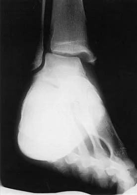
Thorough and meticulous preoperative planning is the cornerstone of successful macrodactyly reconstruction. The evaluation begins with high-quality, orthogonal plain radiographs of the affected hand and the contralateral normal hand. These radiographs are essential for assessing skeletal maturity, evaluating the congruency of the metacarpophalangeal (MCP), PIP, and DIP joints, and quantifying the extent of osseous hypertrophy. The surgeon must carefully template the proposed osteotomies, measuring the exact amount of bone to be resected to achieve a length commensurate with the adjacent normal digits. In pediatric patients, comparing the affected digit to the corresponding digit of the same-sex parent provides a highly reliable template for timing an epiphysiodesis.
Advanced imaging, specifically Magnetic Resonance Imaging (MRI) without contrast, is highly recommended to delineate the soft tissue architecture. MRI is the gold standard for evaluating the extent of fibrofatty infiltration within the peripheral nerves, classically demonstrating the pathognomonic "coaxial cable" sign associated with lipofibromatous hamartoma. The MRI allows the surgeon to map the trajectory of the displaced neurovascular bundles, assess the volume of the subcutaneous adipose tissue, and plan the safest surgical approach. Furthermore, in the modern era, genetic counseling and targeted tissue biopsy for PIK3CA somatic mutation analysis may be considered, particularly if the patient is a candidate for emerging targeted medical therapies (e.g., mTOR or PI3K inhibitors) as an adjunct to surgery.
Preparation for the operating room requires a highly specialized setup. General anesthesia or a comprehensive regional brachial plexus block is preferred, as these procedures are lengthy and require absolute patient immobility. The patient is positioned supine with the affected upper extremity extended on a radiolucent hand table, allowing for unhindered intraoperative fluoroscopy. A well-padded pneumatic arm tourniquet is applied to the proximal arm. Exsanguination is performed gently via elevation or the use of an Esmarch bandage, taking care not to aggressively compress the hypertrophic, potentially fragile vascular structures. The tourniquet is typically inflated to 250 mm Hg in adults, or approximately 100 mm Hg above the systolic blood pressure in pediatric patients.
Given the distorted and delicate nature of the neurovascular structures, optical magnification is non-negotiable. The surgeon must utilize surgical loupes with a minimum of 3.5x magnification, though many microsurgeons prefer the use of an operating microscope, particularly during the dissection of the lipofibromatous hamartoma or if epineural repair is anticipated. The surgical field is prepped with a standard chlorhexidine or povidone-iodine solution, and the hand is draped free to allow for intraoperative assessment of digital alignment, rotation, and cascade during osteotomy reduction and fixation.
Step-by-Step Surgical Approach and Fixation Technique
Soft Tissue and Bony Debulking (Tsuge Technique)
The Tsuge technique remains a foundational, comprehensive approach designed to concurrently address both the massive soft-tissue hypertrophy and the osseous overgrowth. Under tourniquet control, a midlateral incision is meticulously designed, extending the entire length of the involved digit. The midlateral line is defined by connecting the apices of the flexion creases when the finger is fully flexed. Placing the incision midlaterally is a critical biomechanical decision; it entirely avoids the creation of volar flexion contractures that frequently complicate Bruner or straight volar incisions in these patients. Furthermore, the midlateral approach provides direct, unhindered exposure to the neurovascular bundle, which is invariably displaced volarly and laterally by the hypertrophic adipose mass.
Once the skin flap is elevated, the surgeon faces the most perilous portion of the procedure: neurovascular dissection. The digital nerve and artery must be identified and painstakingly separated from the surrounding hamartomatous tissue. Extreme care must be taken to preserve the subdermal vascular plexus on the deep surface of the skin flap to prevent full-thickness necrosis. All excessive, lobulated adipose tissue is excised from the volar and dorsal compartments. If the digital nerve is massively enlarged by lipofibromatous hamartoma, management becomes highly nuanced. Tsuge originally recommended stripping and excising up to half of the neural fascicles to reduce bulk. However, modern hand surgeons recognize that aggressive fascicular stripping carries an unacceptably high risk of permanent sensory deficit and debilitating neuroma formation. Instead, contemporary practice favors a radical, microscope-assisted epineurotomy, where the epineurium is opened longitudinally and the interspersed fat is meticulously teased away from the intact fascicles. Kelikian’s alternative method—resecting a severely tortuous, redundant segment of the nerve entirely and performing a tension-free, end-to-end epineural repair—is reserved strictly for non-functional, massively redundant neural segments.
Following soft tissue debulking, the osseous overgrowth is addressed via a step-cut osteotomy to shorten the digit while preserving the DIP joint and nail complex. Using an oscillating microsaw, the surgeon resects matching sections of the volar half of the distal phalanx and the dorsal half of the middle phalanx. The remaining dorsal half of the distal phalanx is then reduced onto the remaining volar half of the middle phalanx. This interlocking step-cut provides excellent bony contact and inherent rotational stability. The osteotomy is rigidly fixed using two parallel 0.035-inch or 0.045-inch Kirschner wires (K-wires) driven retrogradely or antegrade. Finally, the redundant skin is draped over the newly contoured digit, overlapped, and excised. The closure must be completely tension-free, utilizing interrupted 5-0 nylon sutures, to accommodate the inevitable postoperative edema.
Phalangeal Epiphysiodesis
Epiphysiodesis is a highly effective, targeted, and minimally invasive procedure utilized to permanently halt the longitudinal growth of a macrodactylous digit in a skeletally immature patient. The precise timing of this procedure is critical; it is indicated when the growing child's affected digit reaches the exact length of the corresponding digit of the same-sex parent. Under tourniquet control, a midlateral incision is made along the length of the finger. The soft tissues are carefully elevated to expose the proximal, middle, and distal phalanges. The extensor mechanism is retracted dorsally, and the neurovascular bundle is protected and retracted volarly, providing clear visualization of the metaphyseal-epiphyseal junctions.
The surgeon must precisely identify the physes (growth plates) of the phalanges, which are anatomically located at the proximal end of each respective bone. Once identified, the periosteum is incised longitudinally and elevated to expose the cartilaginous physeal plate. Ablation of the physis must be absolute and comprehensive. Using a high-speed pediatric burr or a small surgical curet, the surgeon thoroughly eradicates the cartilaginous tissue, ensuring the burr reaches from the volar cortex entirely to the dorsal cortex, and from the radial to the ulnar margins.
Following mechanical debridement, the physeal gap is meticulously treated with electrocautery. This dual mechanical and thermal ablation ensures the complete destruction of the germinal matrix cells of the physis. Incomplete ablation is a severe technical error; leaving any active physeal cartilage will lead to asymmetric longitudinal growth, resulting in severe, progressive angular deformities (clinodactyly) as the child continues to grow. The wound is then thoroughly irrigated with sterile saline to remove all osteochondral debris, which could otherwise cause heterotopic ossification, and the incision is closed in a standard layered fashion.
Digital Shortening (Barsky Technique)
When a digit has grown massively out of proportion and the patient is either skeletally mature or possesses overgrowth so severe that epiphysiodesis alone is insufficient, the Barsky technique is employed. This powerful shortening procedure functions essentially as a shortening arthrodesis of the DIP joint while brilliantly preserving the aesthetic and functional integrity of the nail complex. Under tourniquet control, the surgeon designs a precise L-shaped incision. The longitudinal limb begins at the midlateral aspect of the PIP joint and extends distally to a level just proximal to the germinal matrix of the nail. The incision is then carried transversely across the dorsum of the finger, creating a robust, full-thickness dorsal flap that houses the entire nail complex.
Elevation of this dorsal flap is the most critical step of the Barsky procedure. The surgeon must work in the precise areolar plane above the extensor tendon insertion, meticulously preserving the terminal branches of the digital artery that supply the nail bed. Damage to the germinal matrix (located deep to the eponychial fold) during the transverse incision or the subsequent osteotomy will result in irreversible, severe nail dystrophy or complete loss of the nail plate. Once the flap is safely elevated and retracted distally, the DIP joint is fully exposed.
Using an oscillating saw, the surgeon performs the bony resection by removing the distal half of the middle phalanx and the proximal articular portion of the distal phalanx. The exact amount of bone resected is dictated by the preoperative templating and intraoperative comparison to the adjacent normal digits. The remaining distal phalanx (with the attached, viable nail complex) is then brought proximally to dock with the remaining middle phalanx. The surgeon must carefully assess the rotational alignment, ensuring the nail plate sits in perfect harmony with the adjacent digits. The osteotomy site is then rigidly fixed using crossed K-wires or a single longitudinal intraosseous wire. Redundant volar and lateral skin is excised to match the new length, and the wound is closed without tension.
Complications, Incidence Rates, and Salvage Management
The surgical management of macrodactyly is notoriously challenging and is associated with one of the highest complication profiles in congenital hand surgery. Patients and their parents must be extensively and transparently counseled regarding these risks prior to any intervention. The most devastating complication is vascular compromise, which can manifest as either venous congestion or arterial ischemia. Because the hypertrophic fat intimately intertwines with the dysplastic digital vessels, aggressive debulking can easily disrupt the tenuous microvascular supply. Venous congestion typically presents within the first 48 hours with a dark, swollen, and rapidly blistering digit, whereas arterial insufficiency presents with a pale, cold, and pulseless digit. If conservative measures (e.g., removing tight sutures, applying medicinal leeches for venous congestion) fail, the result is partial or complete digital necrosis.
Neurologic complications are also exceedingly common. Dissection, stripping, or resection of the digital nerve (as seen in the Tsuge or Kelikian techniques) will inevitably result in varying degrees of sensory loss. More concerning is the risk of painful neuroma formation. If a nerve is resected and the stump is not properly managed (e.g., by burying it deep into muscle or bone, or capping it), the resulting neuroma can cause intractable pain that severely limits overall hand function, rendering a technically successful debulking a functional failure. Joint stiffness is another ubiquitous complication. Extensive soft tissue dissection, combined with the prolonged postoperative splinting required for osteotomy healing, frequently leads to severe arthrofibrosis of the PIP and MCP joints.
Recurrence of the soft tissue hypertrophy is a frustrating reality, particularly regarding the fibrofatty infiltration associated with lipofibromatous hamartoma. The hamartomatous tissue has a high propensity for continued, albeit slower, growth even after aggressive resection. Patients must understand that a true "cure" is rarely achieved; the surgical goal is functional and cosmetic optimization, and multiple revision surgeries are often required throughout the patient's lifetime. When complications become insurmountable, or when a massively enlarged, stiff, and insensate digit physically blocks the function of the adjacent normal fingers, salvage management is required. In these severe cases, ray resection (amputation of the digit and its corresponding metacarpal) is the definitive salvage procedure, often dramatically improving the overall mechanics and aesthetics of the hand.
| Complication | Estimated Incidence | Prevention Strategy | Salvage / Management |
|---|---|---|---|
| Vascular Ischemia / Necrosis | 10% - 15% | Strict staging (3-month intervals); avoid circumferential debulking; preserve subdermal plexus. | Suture removal; medicinal leeches (venous); systemic vasodilators; eventual amputation if full necrosis occurs. |
| Neuroma / Sensory Loss | 20% - 30% | Avoid aggressive fascicular stripping; utilize microscopic epineural defatting; tension-free repairs. | Gabapentinoids; surgical excision of neuroma and burying stump into bone/muscle; targeted muscle reinnervation (TMR). |
| Joint Stiffness (Arthrofibrosis) | 40% - 50% | Meticulous hemostasis; early protected ROM protocols; avoid prolonged K-wire fixation across normal joints. | Aggressive hand therapy; dynamic splinting; surgical tenolysis or capsulotomy in severe, refractory cases. |
| Recurrence of Hypertrophy | 30% - 60% | Radical excision of all visible hamartomatous fat; complete physeal ablation during epiphysiodesis. | Repeat staged debulking; consideration of systemic mTOR inhibitors (Sirolimus) in PROS-related cases. |
| Angular Deformity (Clinodactyly) | 15% - 25% | Ensure complete, bi-cortical eradication of the physis during epiphysiodesis. | Corrective closing or opening wedge osteotomies of the affected phalanx. |
Phased Post-Operative Rehabilitation Protocols
The postoperative rehabilitation following macrodactyly reconstruction is as critical to the final outcome as the surgical execution itself. The protocol must be carefully phased to balance the absolute need for tissue healing and bony union against the high risk of severe joint arthrofibrosis. The rehabilitation timeline is highly dependent on whether an osteotomy or arthrodesis was performed, but generally follows a structured, three-phase approach overseen by a specialized certified hand therapist (CHT).
Phase 1: Immobilization and Tissue Healing (Weeks 0 to 3)
Immediately postoperatively, the hand is placed in a bulky, non-compressive soft dressing reinforced with a volar plaster splint. The hand is immobilized in the intrinsic-plus position (MCP joints flexed to 70-90 degrees, PIP and DIP joints in full extension) to maintain the collateral ligaments at their maximum length and prevent extension contractures. The primary goals during this phase are strict edema control, wound healing, and the prevention of hematoma formation. The extremity must be kept strictly elevated above heart level. Active and passive range of motion (ROM) of the uninvolved digits, wrist, elbow, and shoulder is initiated immediately to prevent proximal stiffness. The surgical dressings remain intact for the first 10 to 14 days, after which sutures are removed.
Phase 2: Early Protected Motion (Weeks 3 to 6)
If soft tissue debulking was performed without an osteotomy, early protected active ROM of the operated digit can begin at week 3. A custom thermoplastic splint is fabricated to protect the digit between exercise sessions. However, if a Tsuge step-cut osteotomy or a Barsky shortening was performed, the digit must remain protected until early radiographic union is observed, typically around 4 to 6 weeks. During this period, the K-wires remain in place, and motion at the fused or osteotomized joints is strictly contraindicated. The therapist focuses on aggressive scar management, utilizing silicone gel sheets, elastomer molds, and retrograde massage to soften the dense, fibrotic scar tissue that characteristically forms in these patients.
Phase 3: Strengthening and Functional Integration (Weeks 6 to 12+)
Once clinical and radiographic union of the osteotomy or arthrodesis site is confirmed, the K-wires are removed in the clinic. The patient is then transitioned to a rigorous protocol of active, active-assisted, and passive ROM exercises for all un-fused joints. Dynamic or static-progressive splinting may be introduced if PIP or MCP joint contractures are present. Strengthening exercises using therapeutic putty and hand grippers are gradually incorporated as tolerated. The ultimate goal of this phase is to integrate the newly contoured, shortened digit back into the patient's functional grasp and pinch patterns. The surgeon and therapist must also use this time to evaluate the digit for the next stage of reconstruction, adhering strictly to the minimum 3-month interval rule before attempting debulking on the contralateral side of the digit.
Summary of Landmark Literature and Clinical Guidelines
The operative management of macrodactyly has evolved significantly over the past century, guided by landmark anatomical studies, pioneering surgical techniques, and, more recently, groundbreaking genetic discoveries. A thorough understanding of this literature is essential for the academic orthopedic surgeon.
The foundational surgical principles were established in the mid-20th century. Tsuge's seminal paper in 1967 remains the bedrock for soft tissue and bony debulking. Tsuge was the first to systematically describe the combination of midlateral incisions, step-cut phalangeal osteotomies, and the controversial fascicular stripping of the digital nerve. While his nerve stripping technique has been largely superseded by modern microsurgical epineural defatting due to high neuroma rates, his approach to the osseous and soft tissue envelopes remains the gold standard. Similarly, Barsky’s 1967 publication detailed the elegant L-shaped dorsal flap technique, providing the first reliable method for massive digital shortening while preserving the critical anatomy of the nail complex. Kelikian’s contributions in 1974 further refined the management of the tortuous digital nerve, advocating for segmental resection and end-to-end repair in cases of severe lipofibromatous hamartoma.
In the modern era, the literature has shifted dramatically toward understanding the molecular etiology of the disease. The landmark discoveries by Keppler-N
