Operative Management of the Adducted Thumb and Intrinsic Hand Contractures
Key Takeaway
The adducted thumb represents a profound functional impairment, second only to complete thumb amputation. A supple first web space is mandatory for circumduction, opposition, and effective pinch kinematics. Severe web contractures force the thumb into adduction and external rotation, disrupting the delicate balance of intrinsic and extrinsic musculature. Surgical management requires meticulous soft tissue release, including intrinsic muscle modulation and capsular releases, to restore the saddle joint's complex biomechanical envelope and optimize hand function.
Comprehensive Introduction and Patho-Epidemiology
In the hierarchy of devastating hand pathologies, only the complete amputation or loss of the thumb causes more profound disability than a fixed, severe adduction contracture of the thumb, commonly referred to as a web contracture. The thumb is the absolute cornerstone of prehension, responsible for approximately forty to fifty percent of overall hand function. It is the singular digit endowed with the unique biomechanical capacity to bring its terminal sensory pad over the entire surface of any chosen finger, or to sweep across the distal palmar eminence. When this critical mobility is lost, the hand is effectively reduced to a rudimentary hook, severely compromising the patient's independence and ability to perform basic activities of daily living.
When the first web space becomes contracted, the thumb is tethered in a highly non-functional posture. This pathology rarely occurs in isolation; it is most frequently secondary to severe trauma, deep thermal or electrical burns, ischemic contractures (such as Volkmann’s ischemic contracture), spasticity stemming from upper motor neuron lesions (cerebral palsy, traumatic brain injury, stroke), or advanced rheumatologic diseases. A severe contracture inevitably forces the thumb into a position of adduction and external rotation, obliterating the ability to perform opposition, grasp, or pinch. The resulting functional deficit is profound, as the patient loses the ability to perform both precision pinch and power grasp maneuvers.
The patho-epidemiology of the adducted thumb is characterized by a progressive, multi-layered fibrotic cascade. Initially, the contracture may be limited to the cutaneous and subcutaneous tissues, particularly in the setting of thermal burns or superficial lacerations. However, as the chronicity of the condition increases, the pathology invariably extends into the deeper fascial layers, the intrinsic musculature, and ultimately the articular capsules of the trapeziometacarpal and metacarpophalangeal joints. In spastic conditions, the continuous, unopposed hypertonicity of the adductor pollicis and first dorsal interosseous muscles leads to myostatic contracture, where the muscle fibers physically shorten and are replaced by inelastic fibrous tissue.
Restoring the supple nature of the thumb web space and addressing the concurrent intrinsic contractures of the lesser digits remains a paramount, yet highly challenging, objective in reconstructive hand surgery. The orthopedic surgeon must approach these cases with a comprehensive understanding that the deformity is three-dimensional. It is not merely a loss of absolute distance between the first and second metacarpals, but a complex rotational and angular deformity that disrupts the entire kinematic chain of the hand. Successful operative management demands meticulous preoperative planning, a stepwise approach to soft tissue and articular release, and an unwavering commitment to prolonged postoperative rehabilitation.
Detailed Surgical Anatomy and Biomechanics
To fully appreciate the surgical management of the adducted thumb and intrinsic hand contractures, the orthopedic surgeon must possess an intimate, exhaustive understanding of the complex kinematics of the first ray and the intrinsic muscular architecture of the hand. The foundation of thumb mobility is the first carpometacarpal (CMC) joint, a highly specialized biconcave-biconvex saddle articulation between the trapezium and the base of the first metacarpal. This unique geometry permits a wide arc of circumductive movement, which is the absolute prerequisite for pinch and grasp. The joint allows for flexion, extension, abduction, adduction, and the critical rotational component of pronation required for true opposition. Stability of this joint is heavily reliant on the anterior oblique ligament (often referred to as the beak ligament), which prevents dorsal subluxation of the metacarpal base during powerful pinch maneuvers.
Effective thumb function relies on an exquisite, balanced interplay between the intrinsic and extrinsic musculature. The Abductor Pollicis Brevis (APB) acts as the primary positioning vector; it abducts and pronates the thumb metacarpal, stabilizing it in space to prepare for pinch. Conversely, the Adductor Pollicis, originating from the third metacarpal (transverse head) and the capitate and bases of the second and third metacarpals (oblique head), supplies the sheer power necessary for pinch and grasp by acting on the ulnar base of the proximal phalanx. The Flexor Pollicis Longus (FPL), as the sole extrinsic flexor, positions the distal phalanx in varying degrees of flexion, modulating the type of pinch utilized. The thumb web must remain entirely supple for these intricate movements to occur; even a mild fascial contracture will exponentially increase the workload on the APB, eventually leading to intrinsic fatigue, secondary joint subluxation, and limited opposition.
Beyond the thumb, the intrinsic musculature of the lesser digits plays a critical role in the pathoanatomy of the contracted hand. The lumbricals and interossei travel volar to the transverse metacarpal ligament and the axis of the metacarpophalangeal (MCP) joints, but dorsal to the axis of the proximal interphalangeal (PIP) joints. This unique anatomical routing allows them to flex the MCP joints and extend the PIP and distal interphalangeal (DIP) joints. In conditions such as rheumatoid arthritis, spastic hemiplegia, or compartment syndrome, these muscles become fibrotic and contracted, producing the classic "intrinsic-plus" deformity characterized by fixed MCP flexion and PIP extension.
Understanding the fascial architecture of the first web space is equally critical for successful surgical release. The web space is invested by the dorsal fascia, which is contiguous with the fascia over the first dorsal interosseous, and the volar fascia, which overlies the adductor pollicis. The distal edge of this fascial complex forms the natatory ligament of the first web space. In severe contractures, this fascial envelope becomes a dense, unyielding fibrotic sheet that binds the first and second metacarpals together. Surgical release must systematically dismantle these fascial tethers, often requiring complete excision of the fibrotic bands rather than mere incision, to restore the independent mobility of the first ray.
Exhaustive Indications and Contraindications
The decision to proceed with operative intervention for an adducted thumb and associated intrinsic contractures must be based on a rigorous clinical evaluation, weighing the potential functional gains against the substantial risks of complex hand reconstruction. The primary indication for surgery is a severe, fixed adduction contracture of the thumb that precludes functional opposition, grasp, or pinch, and which has proven entirely refractory to conservative measures such as serial casting, dynamic splinting, and targeted botulinum toxin injections. Furthermore, the presence of a severe intrinsic-plus deformity of the digits, characterized by fixed flexion contractures of the MCP joints and incomplete passive PIP flexion (a positive intrinsic tightness test), strongly mandates surgical release to restore the hand's capacity to participate in the newly reconstructed pinch mechanism.
However, the surgeon must be acutely aware of the absolute and relative contraindications to these extensive soft tissue and articular releases. An absolute contraindication to radical intrinsic release is the presence of an incompetent or ruptured extrinsic extensor mechanism (extensor digitorum communis). Releasing the intrinsic flexors of the MCP joint in a hand that lacks robust extrinsic extensor power will result in a catastrophic, irreversible loss of MCP extension, rendering the hand virtually useless. Additionally, severe, uncorrectable joint destruction or advanced arthropathy at the CMC or MCP joints may render soft tissue release futile; in such instances, primary arthrodesis is the more appropriate reconstructive pathway.
Relative contraindications include inadequate soft tissue coverage or profoundly compromised vascularity in the first web space. If a severe burn contracture has left thin, adherent, avascular scar tissue over the thumb web, a simple release will fail; the patient must be a candidate for complex flap coverage (such as a first dorsal metacarpal artery flap, a radial forearm flap, or a free tissue transfer) to import healthy, vascularized tissue into the defect. Furthermore, patients with severe cognitive impairment, unmanageable psychiatric conditions, or an inability to comply with the rigorous, months-long postoperative rehabilitation protocol are poor candidates, as postoperative non-compliance will inevitably lead to recurrent contracture and surgical failure.
| Clinical Parameter | Indications for Surgical Release | Relative Contraindications | Absolute Contraindications |
|---|---|---|---|
| Thumb Web Space | Fixed adduction contracture limiting ADLs; failure of splinting/Botox. | Poor local skin quality requiring complex free flap coverage. | Active, uncontrolled local soft tissue or bone infection. |
| Intrinsic Muscles | Positive Bunnell intrinsic tightness test; fixed MCP flexion. | Mild, flexible deformities correctable with therapy. | Incompetent extrinsic extensor mechanism (EDC rupture). |
| Articular Status | Supple joints or mild capsular contracture amenable to release. | Moderate osteoarthritis requiring concurrent procedures. | Severe, end-stage joint destruction (requires arthrodesis). |
| Patient Factors | High motivation, clear functional goals, strong support system. | Poor compliance history, severe uncontrolled spasticity. | Medically unstable for prolonged anesthesia; non-viable limb. |
Pre-Operative Planning, Templating, and Patient Positioning
Meticulous preoperative planning is the linchpin of successful outcomes in the surgical management of complex hand contractures. The clinical assessment must begin with a precise evaluation of the intrinsic tightness using the Bunnell test. The examiner holds the patient's MCP joints in passive extension and attempts to passively flex the PIP joints; resistance to PIP flexion indicates intrinsic tightness. The test is then repeated with the MCP joints in flexion; if PIP flexion improves, the diagnosis of intrinsic contracture is confirmed. The surgeon must also carefully differentiate between fixed articular contractures and musculotendinous tightness, as this will dictate the necessity for capsulotomies or collateral ligament releases during the procedure.
Imaging plays a vital role in the preoperative workup. Standard anteroposterior, lateral, and oblique radiographs of the hand and specific Robert's views of the thumb CMC joint are mandatory to assess for underlying osteoarthritis, joint subluxation, or osseous deformity that might preclude a simple soft tissue release. In cases of severe spasticity or prolonged contracture, the metacarpal heads may exhibit adaptive morphological changes or flattening. If extensive soft tissue scarring is present, particularly following crush injuries or electrical burns, advanced imaging modalities such as Magnetic Resonance Imaging (MRI) or high-resolution ultrasound may be employed to delineate the viability of the intrinsic musculature and the integrity of the neurovascular bundles.
Planning the cutaneous release and subsequent skin coverage of the first web space requires significant forethought. A simple Z-plasty is rarely sufficient for a severe adduction contracture, as it does not import new skin into the web space. The surgeon must template more complex local tissue rearrangements, such as a four-flap Z-plasty, a five-flap (jumping man) Z-plasty, or a V-Y advancement flap. For profound contractures where a large dead space will be created upon release of the first metacarpal, regional flaps must be marked preoperatively. The First Dorsal Metacarpal Artery (FDMA) flap, a neurovascular island flap harvested from the dorsum of the index finger, is an excellent choice and should be mapped out with a handheld Doppler prior to exsanguination.
Patient positioning and operating room setup must be optimized for microsurgical precision and ergonomic efficiency. The patient is positioned supine with the operative extremity extended on a radiolucent hand table. A well-padded pneumatic tourniquet is applied to the proximal arm. The surgeon and the assistant sit opposite each other, utilizing surgical loupe magnification (typically 3.5x to 4.5x) and a sterile field that allows for unencumbered manipulation of the limb. A mini C-arm fluoroscopy unit must be positioned perpendicular to the hand table, draped sterilely, and readily available for intraoperative assessment of articular congruity and K-wire trajectory during skeletal stabilization.
Step-by-Step Surgical Approach and Fixation Technique
Addressing the adducted thumb and concurrent intrinsic contractures requires a highly sequential, stepwise release of the tethering structures. The operation proceeds systematically from superficial to deep, continually reassessing the passive range of motion after each anatomical layer is dismantled. The ultimate goal is to achieve full passive abduction and pronation of the thumb, alongside supple extension of the digits, without placing undue tension on the neurovascular structures.
First Web Space Soft Tissue and Fascial Release
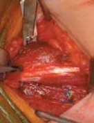
The procedure commences with the planned cutaneous incision over the first web space, typically utilizing the pre-marked complex Z-plasty or regional flap design. As the skin flaps are elevated, the surgeon encounters the dense, fibrotic dorsal and volar investing fascia of the first web space. This fascial layer, including the natatory ligament, must be radically excised rather than simply incised. Careful dissection is required to identify and protect the radial artery as it dives between the two heads of the first dorsal interosseous muscle, as well as the digital nerves to the thumb and the radial aspect of the index finger.
Once the fascia is cleared, the intrinsic muscles are evaluated. If the thumb remains adducted, the origin of the adductor pollicis must be addressed. For moderate contractures, a fractional lengthening of the muscle belly or a release of its origin from the third metacarpal shaft may suffice. In severe, unyielding cases, a complete release of the transverse and oblique heads of the adductor pollicis, and occasionally the mobilization of the first dorsal interosseous from the first metacarpal, is necessary. If, after complete musculofascial release, the first metacarpal remains stubbornly adducted, the pathology involves the trapeziometacarpal joint capsule. A dorsal capsulotomy of the first CMC joint is then performed, carefully preserving the volar beak ligament to prevent iatrogenic joint subluxation.
The Smith Technique for Intrinsic Contracture Release
When severe intrinsic tightness accompanies the adducted thumb, manifesting as fixed MCP joint flexion and PIP joint extension contractures, a radical intrinsic release is mandated. The technique described by Smith provides a comprehensive approach to dismantling the deforming intrinsic forces. A dorsal transverse incision is made just proximal to the metacarpophalangeal joints, extending from the index to the small finger. The surgeon must meticulously elevate the dorsal skin flaps, preserving the dorsal sensory branches of the radial and ulnar nerves, as well as the longitudinal venous drainage system, which is critical to preventing postoperative venous congestion and subsequent flap necrosis.
The extensor hood mechanism is identified over each digit. The lateral tendons of all the interossei and the abductor digiti quinti (ADQ) are isolated and resected precisely at the level of the metacarpophalangeal joints. This maneuver eliminates the primary deforming force driving MCP flexion and PIP extension. If the MCP joints remain flexed despite the intrinsic tendon resection, the sagittal bands are retracted distally to expose the underlying MCP joint capsule. The accessory collateral ligaments are identified and divided at their insertion into the volar plate. The true collateral ligaments are preserved if possible to maintain lateral stability, but in severe, long-standing contractures, a partial release of the true collateral ligaments may be unavoidable to achieve extension.
Volar Plate Arthrolysis and Skeletal Stabilization
The most perilous step of the intrinsic release involves addressing a contracted volar plate. The volar plate must be freed from its dense attachments to the base of the proximal phalanx. Utilizing a blunt probe, such as a Freer elevator, the surgeon gently sweeps proximally to separate any intra-articular adhesions between the volar plate and the metacarpal head. Aggressive sharp dissection in the volar compartment during this step risks catastrophic, irreversible injury to the digital neurovascular bundles; therefore, strict adherence to blunt dissection techniques in the deep volar space is absolutely mandatory.
Following extensive soft tissue and capsular release, the MCP joints are frequently highly unstable, and maintaining extension of the proximal phalanx proves difficult due to residual extrinsic flexor tension. Skeletal stabilization is achieved by inserting a Kirschner wire (typically 0.045-inch or 0.062-inch) obliquely across each affected metacarpophalangeal joint. The joint must be pinned in maximal safe extension. Crucially, before advancing the wire across the joint space, the surgeon must visually and fluoroscopically ensure that the base of the proximal phalanx articulates properly and concentrically with the metacarpal head. Pinning the joint in a dorsally subluxated position will lead to rapid, irreversible articular cartilage damage and devastating joint destruction.
Complications, Incidence Rates, and Salvage Management
The operative management of severe hand contractures is fraught with potential complications, demanding meticulous surgical technique and vigilant postoperative care. The most frequent complication is the recurrence of the contracture, which occurs in up to 20-30% of severe spastic or burn cases despite technically flawless surgery. Recurrence is almost universally linked to postoperative splinting non-compliance, inadequate initial soft tissue coverage, or the relentless progression of the underlying neurological or rheumatological disease. In cases of recurrence, revision surgery is exceedingly difficult due to altered anatomy and dense scar tissue, often necessitating more radical salvage procedures.
Neurovascular compromise is a devastating intraoperative complication. Injury to the digital nerves during the deep volar sweep for volar plate arthrolysis, or injury to the radial artery during the first web space release, can result in permanent sensory deficits or digital ischemia. Venous congestion of the dorsal skin flaps is another significant risk, particularly with the dorsal transverse incision used in the Smith technique. If the longitudinal dorsal veins are indiscriminately sacrificed, the resulting venous hypertension can lead to flap necrosis, wound dehiscence, and deep infection, exposing the underlying extensor tendons and joint capsules.
Salvage management for failed contracture releases or severe complications requires a pragmatic approach focused on achieving a stable, functional, albeit limited, hand. If the trapeziometacarpal joint becomes painfully arthritic or subluxated following a massive release, a primary CMC arthrodesis is the salvage procedure of choice, fusing the thumb in a functional position of palmar abduction and opposition. Similarly, if the MCP joints are destroyed by iatrogenic subluxation during K-wire pinning, MCP arthrodesis or silicone arthroplasty (in low-demand patients) may be required. When soft tissue coverage fails, the surgeon must immediately escalate to regional or free tissue transfer, such as a pedicled groin flap or an anterolateral thigh free flap, to salvage the limb.
| Complication | Estimated Incidence | Prevention Strategy | Salvage / Management |
|---|---|---|---|
| Recurrent Contracture | 20 - 30% | Aggressive prolonged splinting; adequate initial skin coverage. | Revision release with free tissue transfer; CMC/MCP arthrodesis. |
| Venous Flap Congestion | 5 - 10% | Preserve dorsal longitudinal veins; avoid excessive flap tension. | Leech therapy (Hirudo medicinalis); flap revision; skin grafting. |
| Pin Tract Infection | 10 - 15% | Meticulous pin care; avoid burying pins under tension. | Oral/IV antibiotics; early K-wire removal; localized debridement. |
| Digital Nerve Injury | < 2% | Use blunt instruments (Freer) for volar plate arthrolysis. | Immediate microsurgical primary epineurial repair or nerve grafting. |
| Iatrogenic Joint Subluxation | 3 - 5% | Fluoroscopic confirmation of concentric reduction prior to pinning. | Hardware removal, open reduction, revision pinning; late arthrodesis. |
Phased Post-Operative Rehabilitation Protocols
The success of an adducted thumb release and concurrent intrinsic contracture release relies as much on rigorous, highly structured postoperative rehabilitation as it does on meticulous surgical execution. The rehabilitation protocol must be phased, carefully balancing the need for tissue healing and graft/flap take with the absolute necessity of early motion to prevent recurrent fibrosis and tendon adhesions. This requires close, continuous collaboration between the orthopedic surgeon and a specialized certified hand therapist (CHT).
Immediate Postoperative Phase (Days 1-7)
In the immediate postoperative phase, the primary goals are the protection of the surgical reconstruction, the management of acute postoperative edema, and the initiation of safe, early motion of the unpinned joints. The hand is immobilized in a bulky, non-compressive soft dressing reinforced with a volar plaster splint. The thumb is meticulously positioned and held in maximal palmar abduction and pronation to maintain the newly created web space. Strict elevation of the extremity above the level of the heart is mandatory to combat the significant swelling associated with extensive dorsal transverse incisions and soft tissue releases.
Crucially, passive and active flexion exercises of the proximal interphalangeal (PIP) and distal interphalangeal (DIP) joints are initiated within one day of surgery. Because the MCP joints are pinned in extension, moving the PIP and DIP joints forces differential gliding of the flexor digitorum superficialis (FDS) and flexor digitorum profundus (FDP) tendons. This early motion is paramount to prevent adherence of the extrinsic flexor and extensor tendons to the surgical bed and maintains the glide of the newly released lateral bands, preventing the recurrence of the intrinsic-plus posture.
Intermediate Phase (Weeks 3-4)
The intermediate phase marks the transition from static stabilization to dynamic rehabilitation. The transarticular Kirschner wires stabilizing the MCP joints are typically removed at approximately three to four weeks postoperatively in the clinic setting, provided that adequate soft tissue healing has occurred. Immediately following pin removal, a custom thermoplastic dynamic extension splint is fabricated by the hand therapist. For the thumb, a rigid C-bar web spacer splint is utilized continuously, removed only for hygiene and structured therapy sessions, to physically block the recurrence of the adduction contracture.
Active range of motion (AROM) and active-assisted range of motion (AAROM) of the MCP joints are now commenced. The therapist focuses heavily on achieving full, independent flexion of the MCP joints while maintaining PIP extension, effectively reversing the preoperative intrinsic-plus pathology. Furthermore, targeted strengthening of the Abductor Pollicis Brevis (APB) and the extrinsic thumb musculature is initiated to provide dynamic muscular support to the newly acquired web space, ensuring the thumb remains functional in its abducted, pronated position.
Long-Term Management (Months 2-6)
The long-term management phase is focused on scar remodeling, functional integration, and the prevention of late recurrence. Night splinting of the thumb web space using the C-bar splint is continued for an absolute minimum of six months, as the biological forces of scar contracture peak during this prolonged remodeling phase. Discontinuing the splint prematurely is the most common cause of late surgical failure.
During this phase, therapy transitions heavily into functional occupational integration. Pinch strengthening exercises using varying resistances (e.g., therapy putty, dynamometers) are utilized. The patient is guided through complex, task-specific activities of daily living—such as picking up small coins, writing with a pen, and grasping large cylinders—to actively retrain the brain's motor cortex. This neuromuscular re-education is vital to ensure the patient actually utilizes the restored circumductive capacity of the trapeziometacarpal joint in their daily life, rather than reverting to compensatory, non-functional movement patterns.
Summary of Landmark Literature and Clinical Guidelines
The operative management of the adducted thumb and intrinsic contractures is built upon decades of foundational orthopedic research and evolving clinical guidelines. The seminal work by Brand and Milford, detailed extensively in Campbell’s Operative Orthopaedics, established the modern paradigm for addressing the cutaneous and fascial components of the first web space. Their description of the sliding flap and the absolute necessity of deepening the web space to restore the mechanical advantage of the APB remains a cornerstone of reconstructive hand surgery. They definitively proved that without a supple web space, tendon transfers for opposition are doomed to fail due to insurmountable mechanical resistance.
The surgical approach to the intrinsic-plus deformity was revolutionized by Smith, whose comprehensive technique for intrinsic contracture release remains the gold standard. Smith's detailed anatomical studies highlighted the specific deforming vectors of the lateral bands and the interossei, demonstrating that precise resection at the MCP level could reliably restore the balance between the intrinsic and extrinsic forces. His emphasis on the concurrent release of the volar plate and the critical need for temporary skeletal stabilization with K-wires drastically reduced the incidence of recurrent MCP flexion contractures.
Modern clinical guidelines, particularly those addressing spastic hand deformities, emphasize a multidisciplinary, step-wise approach. Current consensus statements dictate that surgical release should only be undertaken after a comprehensive trial of conservative modalities, including targeted botulinum toxin A (Botox) injections into the adductor pollicis and first dorsal interosseous muscles, coupled with aggressive serial casting. Surgery is reserved for fixed myostatic contractures that fail to yield to chemodenervation. Furthermore, contemporary literature strongly advocates for the use of regional flaps, such as the FDMA flap, over simple skin grafts in severe cases, as vascularized tissue provides vastly superior long-term pliability and resistance to secondary scar contracture.
📚 Medical References
- Brand PW, Milford LW: Web deepening with sliding fl ap for adducted thumb in the hand. In Crenshaw AH, ed: Campbell’s
- Smith RJ: Non-ischemic contractures of the intrinsic muscles of the hand. J Bone Joint Surg Am.
- Bunnell S: Ischaemic contracture, local, in the hand. J Bone Joint Surg Am.
