Ulnar Clubhand & Deficiencies: Surgical Management Guide
Key Takeaway
Ulnar clubhand, or paraxial ulnar hemimelia, is a rare congenital longitudinal deficiency of the postaxial upper extremity. Management requires a highly individualized, staged approach. Initial nonoperative serial casting corrects soft tissue contractures, while surgical interventions—such as ulnar anlage excision, syndactyly release, first web space deepening, and metacarpal osteotomies—address progressive radial bowing and optimize prehension. This guide details the biomechanical principles, classifications, and step-by-step operative techniques for managing ulnar deficiencies.
Comprehensive Introduction and Patho-Epidemiology
Ulnar deficiencies represent a profoundly complex and highly heterogeneous spectrum of congenital malformations, universally characterized by the longitudinal failure of formation along the postaxial (ulnar) border of the upper extremity. The most frequently encountered clinical presentation within this spectrum is a partial deficiency of the ulna combined with the absence of the ulnar two digits, a condition classically and colloquially referred to as ulnar clubhand. Synonymous nomenclature pervasive throughout the academic and historical literature includes ulnar dysmelia, paraxial ulnar hemimelia, and congenital absence of the ulna. Unlike its preaxial counterpart (radial clubhand), ulnar deficiency presents with a unique set of biomechanical and anatomical challenges that demand a highly nuanced, individualized surgical approach.
Embryologic and Genetic Foundations
The embryological insult responsible for ulnar longitudinal deficiency occurs between the fourth and seventh weeks of gestation, a critical window during which the upper limb bud undergoes rapid differentiation and elongation. Normal limb development is strictly governed by a complex interplay of signaling centers, most notably the apical ectodermal ridge (AER) for proximodistal growth and the zone of polarizing activity (ZPA) for anteroposterior (radioulnar) patterning. Ulnar deficiencies are fundamentally postaxial anomalies, suggesting a primary disruption in the ZPA, which is heavily regulated by the Sonic Hedgehog (SHH) signaling pathway. A localized failure in SHH expression or an altered response by the postaxial mesoderm leads directly to the hypoplasia or complete agenesis of the ulnar-sided structures, including the ulna, ulnar carpus, and the ulnar digits.
Despite our evolving understanding of limb embryogenesis, the exact etiology of this rare anomaly remains largely idiopathic, and its occurrence is almost exclusively sporadic. The singular historical report suggesting a familial inheritance pattern was documented by Roberts in 1886, who observed the deformity across three successive generations. However, exhaustive modern genetic analyses and consensus maintain that sporadic, de novo mutation or localized intrauterine vascular insults are the primary drivers. Unlike radial deficiencies, which have strong syndromic associations, genetic testing in isolated ulnar clubhand rarely yields a definitive heritable mutation, reinforcing the sporadic nature of the condition.
Epidemiologic Profile
Epidemiologically, ulnar deficiencies are exceedingly rare congenital hand anomalies, presenting a diagnostic and therapeutic challenge even to high-volume congenital hand surgeons. Their relative incidence is estimated to be one-tenth to one-third that of radial deficiencies, translating to an approximate occurrence of 1 in 100,000 to 1 in 150,000 live births. The condition exhibits a slight male predilection, and unlike radial clubhand, which is frequently bilateral, ulnar clubhand is unilateral in roughly 70% to 80% of documented cases. When bilateral involvement does occur, it is typically asymmetrical in its severity.
Associated Systemic and Musculoskeletal Anomalies
A critical clinical pearl for the evaluating orthopedic surgeon is understanding the pattern of associated anomalies. Anomalies associated with ulnar deficiencies, in stark contrast to radial deficiencies (which frequently present with VACTERL, TAR, or Holt-Oram syndromes), are almost exclusively limited to the musculoskeletal system. Extensive systemic workups—such as echocardiograms and renal ultrasounds—are significantly less likely to yield visceral anomalies. However, surgeons must meticulously screen for associated musculoskeletal conditions. Commonly associated anomalies include clubfoot (talipes equinovarus), fibular longitudinal deficiencies (paraxial fibular hemimelia), spina bifida, proximal focal femoral deficiency (PFFD), mandibular defects, and congenital absence of the patella. The presence of these concurrent musculoskeletal anomalies necessitates a multidisciplinary orthopedic approach, often requiring synchronized surgical staging between upper and lower extremity teams.
Detailed Surgical Anatomy and Biomechanics
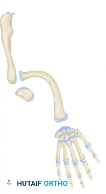
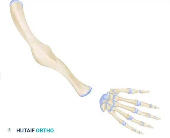
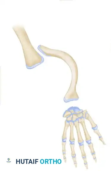
The pathoanatomy of ulnar clubhand is defined by varying degrees of osseous deficiency along the ulnar aspect of the hand and forearm, compounded by severe soft tissue contractures and predictable biomechanical derangements. The forearm is typically foreshortened and frequently exhibits pronounced bowing, creating a complex three-dimensional deformity.
Osteology and the Ulnar Anlage
The defining anatomical feature in many partial ulnar deficiencies is the presence of the ulnar anlage—a dense, unyielding fibrocartilaginous band representing the undeveloped distal ulna. Straub first elucidated the pathophysiology of this structure, noting that it typically spans the gap between the hypoplastic proximal ulna (or the distal humerus in cases of complete ulnar agenesis), the distal radius, and the ulnar carpus. This anlage lacks a functional physis and therefore possesses zero growth potential. As the radius continues its longitudinal growth, the anlage acts as an inflexible tether, functioning much like a bowstring. This tethering effect forces the radius and carpus into severe ulnar deviation, causes progressive bowing of the radial shaft (with radial convexity), and ultimately drives the proximal radius into subluxation or frank dislocation at the radiocapitellar joint.
Carpal bone deformities are ubiquitous in this patient population due to severe structural deficiency and altered mechanical loading during development. The pisiform and hamate are typically absent, reflecting the postaxial field defect. Furthermore, coalitions of the remaining carpal bones are frequently observed, most commonly involving the capitate, trapezoid, and trapezium. Digital malformation occurs in up to 89% of patients; the small and ring fingers are usually absent, and syndactyly of the remaining digits is a common finding. In approximately two-thirds of patients, the long and index fingers, as well as the thumb, may also be absent or severely hypoplastic, drastically altering prehensile capabilities.
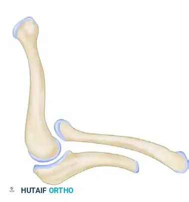
Radiocarpal and Elbow Joint Kinematics
The biomechanics of the ulnar-deficient limb are severely compromised. Ulnar deviation of the hand strongly correlates with the degree of radial bowing and an increased ulnar slope to the distal radius articular surface. This slope concurrently drives a supination deformity of the forearm, as the distal radius rotates around the tethering anlage. At the elbow, the joint is frequently restricted in its arc of motion. Radial head dislocation, a direct consequence of the distal tethering, creates a mechanical block to elbow flexion and extension. In severe cases, the proximal radius may fuse with the humerus, presenting as a frank humeroradial synostosis. Consequently, the patient relies heavily on compensatory shoulder motion to position the hand in space.
Soft Tissue and Neurovascular Aberrations
The soft tissue envelope and neurovascular anatomy in ulnar clubhand are highly aberrant and require meticulous preoperative consideration. The ulnar artery and ulnar nerve are frequently hypoplastic or entirely absent. In their stead, the limb relies on a dominant median nerve and a robust interosseous arterial system. Muscular anatomy is similarly unpredictable; the flexor carpi ulnaris (FCU) and extensor carpi ulnaris (ECU) are often absent or merge into the fibrocartilaginous anlage. The extrinsic flexors and extensors to the ulnar digits are missing, and those to the remaining radial digits may be fused or poorly differentiated. This profound lack of normal musculotendinous units severely limits the availability of local donors for tendon transfer procedures, forcing the surgeon to rely on the limited available flexor carpi radialis (FCR) or extensor carpi radialis longus/brevis (ECRL/ECRB).
Exhaustive Indications and Contraindications
Surgical intervention in ulnar deficiencies is highly individualized, and a algorithmic approach must be employed based on the patient's specific anatomical deficits and functional requirements. The primary goal of any intervention is to maximize prehension, optimize hand positioning in space, and correct progressive deformities that threaten long-term limb function.
Surgical Decision-Making Matrix
Primary indications for early surgical intervention include the presence of complex syndactyly, progressive radial bowing secondary to an ulnar anlage, dislocation of the radial head resulting in a severe limitation of elbow extension, and severe internal rotation deformity of the humerus. Syndactyly release is prioritized early (typically around 12 to 18 months of age) to allow for independent digital growth and function. If the radial head is dislocated and creates a rigid mechanical block to elbow extension, the creation of a one-bone forearm (radioulnar synostosis) is strongly indicated to restore a functional arc of motion and provide a stable platform for the hand.
Conversely, surgical restraint is often required. If the patient has a stable, functional elbow with an acceptable arc of motion despite radiographic abnormalities, aggressive skeletal reconstruction is contraindicated. The surgeon must recognize that a stiff, straight arm is functionally inferior to a deformed but mobile arm. Furthermore, if functional pronation and supination are preserved, the surgical creation of a one-bone forearm is an absolute contraindication, as the iatrogenic loss of forearm rotation will drastically decrease overall limb utility.
Absolute and Relative Contraindications
The decision to proceed with complex tendon transfers or skeletal realignments must be weighed against the patient's baseline function. Tendon transfers (such as the Flatt procedure) strictly require a good, stable, and passive range of motion in the border digits. Tendon transfers will predictably fail if placed across stiff, contracted, or arthrogrypotic joints.
| Indication / Contraindication Category | Specific Clinical Scenarios | Surgical Rationale |
|---|---|---|
| Absolute Indications | Progressive radial bowing >30 degrees; Tethering ulnar anlage; Complex syndactyly of border digits. | Excision of anlage prevents irreversible joint destruction and severe bowing. Syndactyly release prevents growth tethering. |
| Relative Indications | Mild to moderate first web space contracture; Absent thumb with functional index finger. | Web space deepening and rotational osteotomies can optimize prehension and pinch mechanics. |
| Absolute Contraindications | Creation of a one-bone forearm in a patient with functional pronation/supination. | Fusing the forearm destroys rotational kinematics, severely limiting the patient's ability to position the hand for ADLs. |
| Relative Contraindications | Tendon transfers in digits with severe, rigid joint contractures (passive ROM < 30 degrees). | Transferred muscle units lack the excursion and power to overcome rigid joint contractures, leading to functional failure. |
Pre-Operative Planning, Templating, and Patient Positioning
Thorough preoperative planning is the cornerstone of successful surgical outcomes in the management of ulnar clubhand. The surgeon must synthesize clinical examination findings with advanced imaging modalities to construct a precise, stage-based surgical blueprint.
Advanced Imaging and Radiographic Templating
Standard orthogonal radiographs of the entire upper extremity (including the shoulder, humerus, elbow, forearm, and hand) are mandatory. These usually demonstrate a pathognomonic pattern: an absent or hypoplastic distal ulna, a bowed radius with an increased ulnar slope along its distal articular surface, and varying degrees of carpal agenesis.
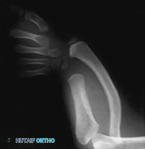
A critical diagnostic pitfall exists in the evaluation of neonates and infants: it is often exceedingly difficult to definitively determine the presence or absence of the proximal ulna on standard radiographs, as cartilaginous mineralization may not occur until the child reaches 12 to 18 months of age. Therefore, serial radiographs or advanced imaging modalities such as high-resolution Ultrasound or non-contrast Magnetic Resonance Imaging (MRI) are heavily relied upon. MRI is particularly invaluable for delineating the extent of the cartilaginous ulnar anlage, identifying occult carpal coalitions, and confirming the presence of a proximal ulnar segment before planning a complex one-bone forearm reconstruction.
Classification-Driven Surgical Strategy
Accurate classification dictates the surgical algorithm. The Swanson classification evaluates the longitudinal ulnar deficiency, while the Cole & Manske classification assesses the functional capacity of the thumb and first web space.
Swanson Classification of Ulnar Deficiency:
* Type 1: Hypoplasia or partial defect of the ulna. This is the most common variant.
* Type 2: Total defect of the ulna.
* Type 3: Total or partial defect of the ulna with humeroradial synostosis.
* Type 4: Total or partial defect of the ulna associated with congenital amputation at the wrist.
Cole & Manske Classification:
Recognizing that prehension is the ultimate functional goal, this system classifies hands based on thumb and first web involvement.
* Type A: Normal first web space and thumb.
* Type B: Mild first web and thumb deficiency.
* Type C: Moderate-to-severe first web and thumb deficiency; potential loss of opposition, malrotation of the thumb into the plane of the other digits, thumb-index syndactyly, and absent extrinsic tendon function.
* Type D: Absent thumb.
Anesthesia, Positioning, and Tourniquet Application
Surgical procedures are performed under general anesthesia, often supplemented with a regional brachial plexus block (supraclavicular or axillary) to provide superior postoperative analgesia and minimize vasospasm in the dysplastic limb. The patient is positioned supine with the operative extremity extended on a radiolucent hand table. A sterile pneumatic tourniquet is applied to the proximal arm; however, due to the foreshortened nature of the limb, a pediatric or infant-sized tourniquet is often required. Exsanguination is achieved via elevation or a sterile Esmarch bandage, taking extreme care not to traumatize the delicate soft tissues. Fluoroscopy must be readily available and positioned to allow for unimpeded orthogonal views of the entire limb throughout the procedure.
Step-by-Step Surgical Approach and Fixation Technique
The operative management of ulnar deficiencies encompasses a variety of procedures tailored to the patient's specific pathoanatomy. The following sections detail the technical execution of the most frequently utilized surgical interventions.
Excision of the Ulnar Anlage
Most authorities agree that the ulnar anlage must be excised to prevent irreversible radial bowing and shortening. Resection of the distal end of the fibrocartilaginous mass is recommended between the ages of 6 months and 2 years.
1. Incision: A longitudinal or lazy-S incision is made along the ulnar border of the distal forearm, extending from the mid-diaphysis to the ulnocarpal joint.
2. Dissection: The subcutaneous tissues are divided, and meticulous hemostasis is achieved. The surgeon must carefully identify and protect the dorsal sensory branch of the ulnar nerve (if present) and the ulnar artery.
3. Anlage Identification: The fibrocartilaginous anlage is identified as a dense, avascular band replacing the distal ulna. It is traced proximally to its junction with the osseous proximal ulna and distally to its insertion on the ulnar carpus and distal radius.
4. Resection: The anlage is sharply excised. A minimum of 1 to 2 centimeters of the fibrocartilage must be removed to prevent secondary tethering via scar formation. Extreme caution is exercised distally to avoid iatrogenic injury to the distal radial physis, which is often intimately associated with the anlage insertion.
5. Concomitant Osteotomy: If radial bowing exceeds 30 degrees at the time of presentation, a corrective closing-wedge osteotomy of the radius is performed concomitantly at the apex of the deformity, stabilized with smooth Kirschner wires (K-wires) or a pediatric dynamic compression plate.
Creation of a One-Bone Forearm (Radioulnar Synostosis)
If marked shortening and bowing of the radius are accompanied by severe forearm instability, radial head dislocation, and restriction of elbow motion, a one-bone forearm will significantly improve stability. This requires a viable proximal ulna.
1. Staging: The proximal radius (radial head) is typically excised several months prior to the creation of the one-bone forearm. Attempting simultaneous radial head excision and radioulnar synostosis is contraindicated due to the high risk of neurovascular compromise and compartment syndrome.
2. Approach: A dorsal longitudinal incision is utilized to expose both the distal aspect of the proximal ulna and the proximal aspect of the distal radius segment.
3. Preparation of Synostosis Site: The medullary canals of both the proximal ulna and the distal radius are opened and decorticated. The opposing bone ends are fashioned to maximize cortical contact, often utilizing a step-cut or oblique osteotomy technique.
4. Alignment and Fixation: The distal radius is aligned with the proximal ulna. The forearm is positioned in neutral rotation to 10 degrees of pronation, which is generally the most functional position for activities of daily living. Fixation is achieved using a stout, intramedullary K-wire passed antegrade through the olecranon into the distal radius, supplemented by a localized pediatric plate and screws if the bone caliber permits.
5. Bone Grafting: Cancellous autograft (typically harvested from the iliac crest) or allograft is packed meticulously around the osteotomy site to promote rapid osteogenesis.
First Web Space Deepening and Metacarpal Osteotomy
To optimize prehension in patients with Cole & Manske Type B or C deficiencies, deepening of the first web space and rotational correction of the metacarpals are executed in two distinct stages to protect the tenuous vascular supply.
Stage 1: Web Space Deepening
1. Incision and Flap Design: The first web space is deepened utilizing a broad four-flap Z-plasty or a jumping-man flap, depending on the severity of the adduction contracture.
2. Soft Tissue Release: Meticulous dissection is carried down to the underlying musculature. The fascia of the adductor pollicis and the origin of the first dorsal interosseous muscle are released from the metacarpal shaft to eliminate the dynamic deforming forces.
3. Closure: The flaps are transposed and inset without tension. A sterile, non-adherent dressing and a thumb spica splint are applied.
Stage 2: Metacarpal Osteotomy (Performed 3-6 months later)
1. Approach: A dorsal longitudinal incision is made over the targeted metacarpal.
2. Osteotomy: A rotational and/or closing-wedge osteotomy is performed at the metacarpal base utilizing an oscillating microsaw. The goal is to pronate and abduct the border digit to face the opposing digits, creating an effective, oppositional pinch.
3. Fixation: The osteotomy is secured with two crossed 0.035-inch or 0.045-inch smooth K-wires. The wires are cut outside the skin and bent to facilitate later removal in the clinic.
Tendon Transfers for Type II Deformities (Flatt Technique)
In patients lacking active extrinsic tendon function but possessing supple joints, tendon transfers are indicated. The Flatt technique utilizes wrist flexors or extensors extended by a tendon graft.
1. Donor Identification: The FCR, ECRL, or ECU are identified through limited longitudinal incisions at the wrist. The chosen donor is detached distally and mobilized proximally to ensure adequate excursion.
2. Graft Harvesting: The palmaris longus tendon (if present) or a strip of fascia lata is harvested to serve as an interpositional graft.
3. Proximal Coaptation: The proximal end of the graft is secured to the donor tendon utilizing a Pulvertaft weave. This weave provides superior biomechanical strength by passing the graft through the donor tendon a minimum of three times, maximizing the surface area for tenocyte migration. The weave is secured with 4-0 non-absorbable mattress sutures.
4. Distal Fixation: The graft is routed subcutaneously to the border digits. The distal ends are secured into the terminal phalanges utilizing a pull-out wire technique over a dorsal button. Tensioning is set with the wrist in neutral, ensuring that wrist extension initiates synergistic digital flexion (tenodesis effect).
Complications, Incidence Rates, and Salvage Management
Surgical intervention in the dysplastic upper extremity carries an inherently high risk of complications. The altered anatomy, diminutive size of the structures, and tenuous vascularity demand meticulous surgical technique and heightened vigilance.
Intraoperative and Early Postoperative Complications
Vascular compromise is the most devastating early complication. Because these limbs often rely on a single dominant vessel (frequently the interosseous or median artery), aggressive soft tissue releases or excessive tensioning during osteotomy fixation can precipitate acute ischemia. If capillary refill is delayed or absent upon tourniquet deflation, all compressive dressings and internal fixation must be immediately released, and the limb allowed to perfuse. Iatrogenic nerve injury, particularly to the superficial sensory branches during anlage excision, can lead to painful neuroma formation. Infection rates are generally low (<2%) but require prompt aggressive debridement if they occur, particularly in the presence of K-wires or bone grafts.
Late Complications and Deformity Recurrence
Recurrence of radial bowing is a frequent late complication, occurring in up to 30% of patients following anlage excision. This is often due to incomplete resection of the fibrocartilage or secondary tethering from dense scar tissue formation. Nonunion or delayed union is a significant risk following the creation of a one-bone forearm, given the poor osteogenic potential of the dysplastic bone and the limited soft tissue envelope. Pin tract infections from protruding K-wires are common and usually resolve with oral antibiotics and eventual pin removal.
Salvage Procedures and Revision Strategies
When primary procedures fail, salvage options become increasingly limited. In cases of recalcitrant nonunion of a one-bone forearm, revision internal fixation with rigid plate osteosynthesis and voluminous autologous iliac crest bone grafting is mandated. If recurrent radial bowing becomes severe and symptomatic, repeat osteotomies with application of a multi-planar external fixator (such as a Taylor Spatial Frame) may be required to achieve gradual correction and lengthen the foreshortened limb.
| Complication | Estimated Incidence | Prevention Strategy | Salvage / Management |
|---|---|---|---|
| Recurrent Radial Bowing | 20% - 30% | Resect minimum 1-2 cm of anlage; avoid distal physis injury. | Repeat corrective osteotomy; external fixation for gradual correction. |
| One-Bone Forearm Nonunion | 10% - 15% | Meticulous decortication; rigid intramedullary fixation; primary bone grafting. | Revision ORIF with rigid plating and autologous iliac crest bone graft. |
| Vascular Compromise / Ischemia | < 5% | Avoid excessive tension during deformity correction; release tourniquet prior to closure. | Immediate removal of fixation/dressings; vascular surgery consultation. |
| Pin Tract Infection | 15% - 20% | Frequent pin site care; avoid excessive skin tension around pins. | Oral antibiotics; early pin removal if clinical union is progressing. |
Phased Post-Operative Rehabilitation Protocols
The success of complex reconstructive procedures in ulnar clubhand is inextricably linked to a rigorous, specialized, and prolonged postoperative rehabilitation protocol. Therapy must be guided by a certified hand therapist (CHT) experienced in congenital anomalies.
Phase I: Immediate Postoperative Immobilization (Weeks 0-4)
Immediately following surgery, the limb is immobilized in a well-padded, long-arm bivalved cast or a bulky Robert Jones dressing reinforced with plaster splints. The primary goals during this phase are the protection of the osteotomies, tendon transfers, and soft tissue reconstructions, alongside the minimization of edema. The limb is kept strictly elevated. Skin sutures are typically removed at 14 days postoperatively under sedation in the pediatric population. For tendon transfers, the wrist is immobilized in a position that minimizes tension on the repair (e.g., mild flexion for flexor transfers).
Phase II: Early Mobilization and Splinting (Weeks 4-8)
At 4 to 6 weeks, pending radiographic evidence of early clinical union for osteotomies, the cast and protruding K-wires are removed in the clinic. The patient is transitioned to a custom-fabricated, removable thermoplastic splint.
* Osteotomies: Gentle active and active-assisted range of motion (ROM) exercises are initiated for the adjacent joints. Passive stretching is strictly avoided to prevent disruption of the healing callus.
* Tendon Transfers: The pull-out wires and dorsal buttons are removed at 3 to 4 weeks. A protected, active ROM protocol is initiated. The therapist focuses on teaching the child to activate the transferred muscle, utilizing biofeedback and play-based therapy to encourage synergistic movements (e.g., extending the wrist to close the fingers).
Phase III: Long-Term Functional Integration (Months 2-12+)
As bone healing consolidates and tendon transfers gain tensile strength, the splinting regimen is gradually weaned, initially during the day and eventually at night. Therapy shifts towards intensive functional integration, focusing on pinch mechanics, grasp strength, and bimanual activities of daily living. Occupational therapy plays a critical role in adapting tools and utensils to the child's specific prehensile capabilities. Long-term follow-up is mandatory until skeletal maturity to monitor for recurrence of deformity, progressive joint subluxation, and the potential need for secondary reconstructive procedures.
Summary of Landmark Literature and Clinical Guidelines
The evolution of surgical management for ulnar deficiencies is deeply rooted in a few seminal works that have established the modern standard of care. A comprehensive understanding of these foundational texts is essential for the academic orthopedic surgeon.
Foundational Classifications and Historical Context
The definitive morphological classification system was established by Swanson, Tada, and Yonenobu in their landmark 1984 paper. By delineating the four primary types of ulnar deficiency based on the presence and morphology of the ulna and radioulnar joints, they provided the first reliable framework for surgical prognostication. This was later refined by Havenhill et al. (2005), who introduced the "Type 0" designation to describe patients with an ulnar-deficient ray and carpus but a structurally normal ulna, highlighting the extreme variability of the postaxial defect.
The functional paradigm shift was driven by Cole and Manske (1997), who astutely recognized that the morphological appearance of the forearm was secondary to the functional capacity of the hand. Their classification system, based entirely on the thumb and first web space, revolutionized surgical planning by prioritizing the restoration of prehension over mere cosmetic alignment of the forearm.
Modern Outcomes and Evidence-Based Guidelines
The pathophysiology and imperative for early excision of the ulnar anlage were first comprehensively described by Straub in 1965. His assertion that the fibrocartilaginous band acts as an unyielding tether remains the cornerstone of early surgical intervention. Modern long-term outcome studies have validated Straub's theories, demonstrating that early excision (before age 2) significantly reduces the incidence of severe radial bowing and late radial head dislocation.
For tendon transfers in the dysplastic hand, the techniques popularized by Flatt remain the gold standard. Flatt's extensive work in congenital hand surgery emphasized the necessity of utilizing functional, expendable donors (
