Advanced Arthroplasty for Malignant Lesions: Reclaim Your Mobility

Key Takeaway
This topic focuses on Advanced Arthroplasty for Malignant Lesions: Reclaim Your Mobility, Arthroplasty for malignant lesions is a surgical procedure to reconstruct joints compromised by cancerous metastases. This intervention addresses severe bone weakening, pain, and pathologic fractures stemming from metastatic foci. It aims to restore skeletal stability, improve patient mobility, and enhance the quality of life for individuals experiencing advanced bone disease due to complex metastatic processes.
Comprehensive Introduction and Patho-Epidemiology
Metastatic bone disease (MBD) represents a profound clinical challenge that afflicts more than half of the 1.2 million patients newly diagnosed with cancer annually in the United States alone. As systemic therapies for primary carcinomas become increasingly efficacious, patient survival has paradoxically led to a rising incidence of skeletal metastases. Bony involvement is a major source of morbidity and mortality if not treated appropriately, precipitating intractable pain, hypercalcemia, spinal cord compression, and catastrophic pathologic fractures. The femur is the long bone most commonly affected by metastatic dissemination, with approximately 25% of all appendicular lesions localizing to the proximal third of the femur. Consequently, seventy-five percent of all surgical interventions for cancer that has metastasized to the appendicular skeleton are performed in the peritrochanteric and hip areas, necessitating advanced reconstructive arthroplasty techniques to restore function and mitigate pain.
The pathogenesis of skeletal metastasis is a complex, multistep cascade best understood through a modified "seed and soil" theorem. It is estimated that fewer than one in 10,000 neoplastic cells that escape into the systemic circulation from the primary tumor site possess the biologic machinery requisite to establish a viable metastatic focus in bone. To metastasize, a malignant cell must first detach from the primary mass, a process heavily dependent on the upregulation of degradative enzymes such as collagenases, hydrolases, cathepsin D, and various matrix metalloproteinases. Once the neoplastic cell intravasates into the vascular or lymphatic channel, it circulates throughout the body. It is theorized that these circulating tumor cells are protected from immune surveillance and hemodynamic shear stress by a fibrin-platelet clot; however, clinical trials utilizing heparin to disrupt this protective cloaking have not demonstrated a statistically significant alteration in metastatic outcomes. Local microenvironmental factors, notably integrins and specific chemokines (e.g., CXCL12), are instrumental in arresting the circulating metastatic cell at a remote osseous site. Once extravasated into the marrow cavity, the metastatic cell releases potent mediators, including tumor angiogenesis factor (TAF), which induces neovascularization and subsequently fuels the exponential growth of the metastatic focus.
Patients harboring advanced metastatic disease frequently manifest severe derangements in hematopoietic and calcium homeostasis. The infiltration of the marrow space often precipitates a normochromic, normocytic anemia accompanied by leukocytosis. In response to this profound anemia and marrow crowding, the reticuloendothelial system releases an increased number of immature hematopoietic precursors into the peripheral circulation—a phenomenon classically termed a leukoerythroblastic reaction, which is readily identifiable on a peripheral blood smear. Furthermore, hypercalcemia of malignancy is a life-threatening oncologic emergency seen in up to 30% of patients with extensive skeletal metastases. This is most frequently encountered in multiple myeloma, breast carcinoma, and non–small cell lung cancer, driven largely by tumor-mediated secretion of parathyroid hormone-related peptide (PTHrP) which aggressively stimulates osteoclastic bone resorption.
The radiographic and structural phenotype of the metastatic lesion—whether lytic, blastic, or mixed—is dictated by the tumor's interaction with native osteoblasts and osteoclasts. Blastic metastases, classically associated with prostate carcinoma, are often less painful and carry a lower incidence of pathologic fracture because the osteosclerotic response, albeit disorganized, does not weaken the bone as severely as osteolysis. However, not all prostate metastases are blastic; lytic variants exist, are exquisitely painful, and frequently lead to structural failure. Conversely, most tumors that metastasize from the breast demonstrate a mixed pattern of blastic and lytic areas within the same anatomic region. Bone destruction in purely lytic lesions (typical of renal cell carcinoma, thyroid carcinoma, and multiple myeloma) occurs not by direct tumor cell degradation of the mineralized matrix, but indirectly via the biologic hijacking of native osteoclasts through the RANK/RANKL pathway. These lytic lesions are frequently hypervascular; thus, before surgical intervention is undertaken for characteristic hemorrhagic tumors like thyroid or renal cell carcinoma, prophylactic transarterial embolization is strongly recommended to mitigate catastrophic perioperative exsanguination.
Detailed Surgical Anatomy and Biomechanics
Metastatic foci localizing to any part of the periacetabular or proximal femoral anatomy substantially compromise the mechanical integrity of the pelvic girdle and appendicular skeleton, placing the patient at an unacceptably high risk for pathologic fracture and subsequent nonunion. The hip joint is a highly constrained, multi-axial ball-and-socket articulation that must routinely withstand forces exceeding three to five times body weight during normal ambulation. The bony architecture of the acetabulum is classically described by Letournel and Judet as an inverted Y-shape, consisting of distinct anterior and posterior columns with their respective walls, which jut laterally to provide structural coverage for the femoral head. When neoplastic osteolysis disrupts these columns, the entire load-bearing axis of the hemipelvis is destabilized, predisposing the patient to central migration of the femoral head.

A rigorous understanding of these column mechanics is paramount for the orthopedic oncologist planning a complex reconstruction. The anterior column is defined as the osseous strut that extends from the anterior iliac crest, encompasses the anterior half of the acetabulum, and terminates at the pubic symphysis. The posterior column, which is significantly more massive and bears a greater proportion of the biomechanical load during the stance phase of gait, starts from the articulation of the superior gluteal notch with the sacrum, extends through the posterior half of the acetabulum, and descends through the ischium to the inferior pubic ramus. The acetabular dome, which serves as the superior weight-bearing region of the joint, represents the confluence of both the anterior and posterior columns and receives critical structural contributions from both the anterior and posterior walls.
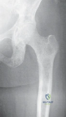
On the femoral side of the articulation, the proximal femur exhibits a highly evolved trabecular architecture designed to dissipate immense physiological loads. The femoral head itself is not truly spherical; rather, it is an ovoid structure that is congruent with the acetabulum only along its superior, weight-bearing portion. The internal architecture of the head, neck, and intertrochanteric area is defined by predictable patterns of principal and secondary bony trabeculations. The primary compressive group extends from the medial calcar vertically into the superior femoral head, while the primary tensile group arcs from the lateral cortex across the superior neck. Together, these form the head and neck arcade, enabling the proximal femur to withstand tremendous compressive, tensile, and torsional forces. The central area where these trabecular patterns intersect and leave a relative void is known as Ward's triangle, a natural zone of weakness that is frequently the epicenter for metastatic seeding and subsequent structural failure.

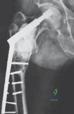
When metastatic disease infiltrates these critical anatomic zones, the biomechanical consequences are dire. Lytic lesions act as massive stress risers within the cortical and cancellous bone. According to engineering principles, a cortical defect whose diameter exceeds 50% of the bone's cross-sectional diameter reduces the bone's torsional strength by over 60%. In the proximal femur, the loss of the medial calcar buttress or the lateral tension band due to tumor osteolysis rapidly leads to varus collapse and catastrophic fracture. Because the native bone's healing potential is profoundly impaired by both the local tumor microenvironment and systemic adjuvant therapies (such as radiation and cytotoxic chemotherapy), the surgeon cannot rely on biological union. Therefore, arthroplasty constructs in this setting must be designed to bypass the affected anatomy entirely, transferring loads directly from the implant to healthy, distal cortical bone, typically requiring the extensive use of polymethylmethacrylate (PMMA) bone cement to achieve immediate, rigid, and durable fixation.
Exhaustive Indications and Contraindications
The decision to proceed with advanced arthroplasty for metastatic lesions of the hip and pelvis requires a nuanced synthesis of the patient's oncologic prognosis, systemic physiologic reserve, and local biomechanical stability. The overarching goals for surgical intervention in the patient with metastatic carcinoma to bone are the immediate relief of intractable pain, the prevention of impending pathologic fractures, the rigid stabilization of realized true fractures, the enhancement of mobility and overall quality of life, and, in highly selected solitary metastasis cases, improved overall survival. It is a generally accepted oncologic principle that a patient must have an estimated life expectancy of at least 6 weeks to warrant the physiologic insult of major operative intervention. For a patient who has sustained a pathologic fracture secondary to metastatic carcinoma, the average survival time is roughly 19 months, though this varies wildly by histology: prostate (29 months), breast (23 months), renal (12 months), and lung (4 months).
Prophylactic stabilization of impending lesions about the proximal femur and acetabulum should be aggressively pursued, as the morbidity and mortality associated with a completed pathologic fracture are exponentially higher than those of an elective, prophylactic reconstruction. This is particularly true for radioresistant tumors such as renal cell and thyroid carcinoma, where bony destruction is guaranteed to progress despite the best nonoperative modalities. Criteria for the performance of a prophylactic stabilization procedure include: 50% cortical lysis, a femoral lesion greater than 2.5 cm in diameter, an avulsion fracture of the lesser trochanter (which pathognomonically indicates deep infiltration of the intertrochanteric line), and persistent mechanical pain in the hip area 4 weeks following the completion of palliative radiation therapy.

To objectify the risk of impending fracture, the Mirels Scoring System is universally employed by orthopedic oncologists. The system assigns points (1, 2, or 3) based on four variables: Site (Upper limb, Lower limb, Peritrochanteric), Pain (Mild, Moderate, Functional), Lesion Type (Blastic, Mixed, Lytic), and Extent of cortical involvement (<1/3, 1/3 to 2/3, >2/3). A mean score of 7 or below indicates a low risk of fracture, suggesting that radiation therapy and bisphosphonates alone may be considered. A score of 8 suggests a borderline risk requiring close clinical judgment, while a score of 9 or above indicates a substantial risk of imminent mechanical failure, mandating prophylactic surgical intervention. Despite its utility, the Mirels score is not infallible; it fails to account for the specific histologic subtype, preexisting osteoporosis, patient body mass index, and functional demands, thus requiring the surgeon to synthesize the score with comprehensive clinical acumen.

Indications and Contraindications Matrix
| Parameter | Surgical Indications | Surgical Contraindications |
|---|---|---|
| Oncologic Status | Life expectancy > 6 weeks; solitary metastasis (curative intent possible); radioresistant tumor (renal/thyroid). | Life expectancy < 6 weeks; terminal multiorgan failure; rapidly progressive visceral crisis. |
| Biomechanical | Mirels Score $/ge$ 8; >50% cortical destruction; lesion >2.5cm; lesser trochanter avulsion; realized pathologic fracture. | Mirels Score $/le$ 7 with purely blastic lesion and minimal mechanical pain (manage with XRT). |
| Physiologic | Medically optimized; hemoglobin >8 g/dL; acceptable coagulation profile; able to participate in post-op rehab. | Profound, uncorrectable pancytopenia; severe coagulopathy; active systemic sepsis; inability to survive anesthesia. |
| Prior Treatment | Persistent mechanical pain 4 weeks post-XRT; failure of nonoperative hormonal/cytotoxic therapy to halt osteolysis. | Lesion currently responding well to systemic therapy with resolving pain and radiographic evidence of sclerosis. |
Pre-Operative Planning, Templating, and Patient Positioning
A methodical, multidisciplinary approach is absolutely mandatory in the workup of a patient presenting with presumed metastatic disease to the bone, particularly when the primary tumor is unknown. A thorough history and physical examination must be completed before exhaustive laboratory and radiographic analyses are initiated. Astonishingly, the primary carcinoma may be detected on physical examination (e.g., palpable breast mass, enlarged thyroid, digital rectal examination for prostate) in as many as 8% of patients. Initial laboratory analysis should be comprehensive, including a complete blood count to assess marrow reserve, erythrocyte sedimentation rate, comprehensive renal and hepatic panels, alkaline phosphatase (a marker of osteoblastic activity), and serum protein electrophoresis (SPEP) to rule out multiple myeloma.
Radiographic examination must follow a strict protocol. Routine radiographic screening in search of early metastatic disease is notoriously insensitive, as lytic changes become evident on plain radiographs only when trabecular and cortical destruction approaches 30% to 50%. However, for localized mechanical pain, an anteroposterior radiograph of the pelvis and full anteroposterior and lateral radiographs of the entire femur must be obtained to assess the entire bone segment. A plain chest radiograph is also mandatory, as approximately 45% of all primary tumors will be detected in the lung on initial screening. To assess the total skeletal burden, a whole-body technetium-99m staging bone scan is standard; however, the surgeon must remember that if the scan is entirely negative in the face of massive lytic destruction, multiple myeloma or a highly aggressive anaplastic carcinoma should be immediately suspected, as these do not provoke the osteoblastic response required for isotope uptake.
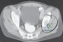
Advanced cross-sectional imaging is the cornerstone of preoperative templating. CT scans of the chest, abdomen, and pelvis should be performed routinely; CT of the lung can detect up to 15% of primary tumors missed on the plain radiograph. If a lesion is detected about the hip, and a detailed pelvic and hip CT has not been performed within the past 6 to 8 weeks, a dedicated fine-cut CT must be ordered. Intravenous contrast medium is generally not necessary for assessing bone stock, but is vital if assessing tumor proximity to major neurovascular bundles. A recent CT scan is particularly critical in the preoperative planning for an acetabular reconstruction to evaluate the integrity of the anterior and posterior columns. Furthermore, the use of PET/CT scanning is becoming the gold standard in the workup of patients with possible metastatic cancer, offering unparalleled sensitivity for identifying the primary source and mapping occult visceral and skeletal metastases.
If the lesion is unexpectedly found to be highly vascular or aneurysmal at the time of biopsy or initial imaging (classic for renal cell carcinoma and thyroid carcinoma), it is highly beneficial to perform a prophylactic transcatheter arterial embolization 24 to 48 hours prior to surgical intervention to drastically reduce perioperative bleeding. Patient positioning in the operating room must be meticulously planned based on the chosen surgical approach and the extent of the planned reconstruction. While the lateral decubitus position is standard for most posterior approaches to the hip, the surgeon must ensure that the entire femur is prepped and draped free in the event that a standard arthroplasty must be intraoperatively converted to a total femur replacement. Digital templating must account for bypassing the most distal extent of the lesion by a minimum of two cortical diameters to prevent stress risers at the tip of the cemented stem.
Step-by-Step Surgical Approach and Fixation Technique
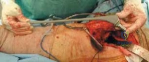
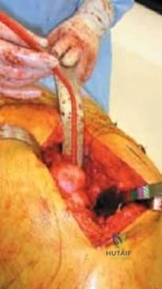
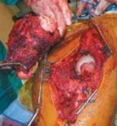
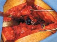
The surgical management of periacetabular and proximal femoral metastatic lesions demands a versatile approach capable of extensive exposure. The standard posterior approach to the hip is most frequently utilized, as it provides excellent visualization of the posterior column, the entire proximal femur, and allows for safe identification and protection of the sciatic nerve. In cases of massive anterior column destruction, an ilioinguinal or modified Stoppa approach may be required, though these are less common for purely palliative arthroplasty. Once the joint is accessed, the primary objective is rapid, aggressive intralesional debulking of the macroscopic tumor. Unlike primary bone sarcomas which require wide, en bloc resection with negative margins, MBD is generally treated with intralesional curettage using a high-speed burr, followed by adjuvant chemical or thermal ablation (e.g., argon beam coagulation, phenol, or hydrogen peroxide) to achieve local tumor control and prepare a clean osseous bed for cement interdigitation.
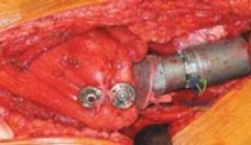
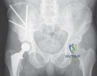
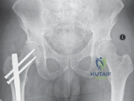
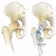
Acetabular reconstruction is dictated by the extent of bone loss, classically categorized by the Harrington Classification. Harrington Class I lesions represent minor bone loss with intact lateral cortices and a supportive medial wall; these can be managed with conventional cemented acetabular components, often utilizing a flanged cup to pressurize the PMMA. Class II lesions involve deficient medial walls but intact superior and lateral rims; these require the use of protrusio rings or titanium meshes combined with copious PMMA to recreate the medial buttress. Class III lesions are the most severe, characterized by massive destruction of the acetabular dome, medial wall, and columns, resulting in pelvic discontinuity. Reconstruction of Class III defects requires complex techniques such as the use of a Burch-Schneider anti-protrusio cage or saddle prosthesis. The cage is flanged into the intact ilium superiorly and slotted into the ischium inferiorly, bridging the massive defect. The void is then packed with PMMA, into which a standard polyethylene cup is cemented.
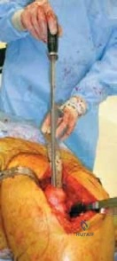
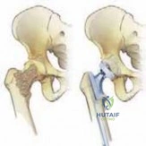
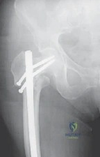
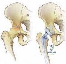
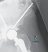
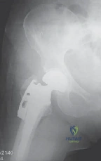
On the femoral side, cemented arthroplasty components are the definitive surgical option for impending and realized fracture management of the head, neck, and intertrochanteric areas. Because cancer patients, regardless of their age, have severely compromised bone healing potential secondary to systemic debilitation, radiation, and chemotherapy, biologic fixation (uncemented porous-coated stems) is generally contraindicated. Rigid fixation with PMMA augmentation is mandatory. The femoral canal must be aggressively reamed and brushed to remove all intramedullary tumor. Third-generation cementing techniques should be employed, utilizing a distal cement restrictor and retrograde injection of low-viscosity cement to achieve maximal interdigitation into the remaining healthy cancellous bone.
When the proximal femoral bone loss is extensive, standard primary stems are insufficient. In cases where the calcar is completely destroyed but the greater trochanter remains intact, a calcar-replacing endoprosthesis is utilized. If the entire proximal femur, including the abductor insertion, is obliterated by tumor, a proximal femoral replacement (megaprosthesis) is required. This modular system allows the surgeon to resect the proximal femur en bloc and reconstruct the defect with a titanium body and cemented distal stem. Soft tissue reconstruction is critical in these massive
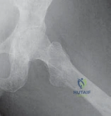
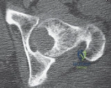
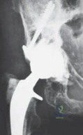
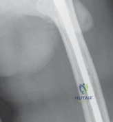
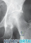
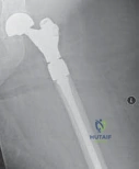
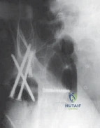
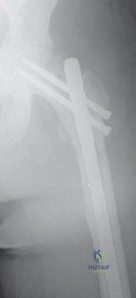
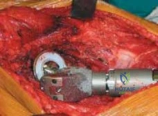
Clinical & Radiographic Imaging Archive