Elbow Cases Biceps: Your Guide to Tendonitis Diagnosis & Relief

Key Takeaway
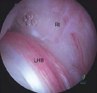
In this comprehensive guide, we discuss everything you need to know about Elbow Cases Biceps: Your Guide to Tendonitis Diagnosis & Relief. The Long head of the biceps tendonitis is characterized by anterior shoulder pain radiating to the biceps, worsened by overhead movements. Often observed in young athletes or those with repetitive use, diagnosis relies on specific physical findings. While this focuses on shoulder pain, a comprehensive understanding of biceps pathology is vital for assessing various musculoskeletal conditions, including **elbow cases biceps** pain and other associated issues.
Patient Presentation and History
A 44-year-old right-hand-dominant male presents to the orthopedic trauma clinic with acute right anterior elbow pain, swelling, and significant weakness following a lifting injury that occurred forty-eight hours prior to evaluation. The patient is a heavy manual laborer and an avid recreational weightlifter. He reports a six-month preceding history of insidious onset, aching anterior elbow pain that was previously diagnosed as distal biceps tendinopathy by his primary care physician. He had been managing this chronic tendinopathy with intermittent non-steroidal anti-inflammatory drugs, activity modification, and localized icing, with minimal sustained relief.
The acute event occurred while the patient was performing heavy eccentric biceps curls. He describes attempting to arrest a rapidly descending heavy barbell, applying a massive eccentric extension load to a flexed and supinated right forearm. He experienced an immediate, audible "pop" in the antecubital fossa, followed by sharp, tearing pain, rapid swelling, and an immediate subjective loss of strength in both elbow flexion and forearm supination.
His past medical history is unremarkable for systemic inflammatory arthropathies, diabetes mellitus, or renal osteodystrophy. However, he admits to a history of localized corticosteroid injections administered by an outside provider for his chronic tendinopathy three months prior to the acute rupture. He denies any recent fluoroquinolone antibiotic use. He is a current everyday smoker, consuming approximately one pack per day for the last twenty years, which represents a significant risk factor for microvascular compromise and subsequent tendinopathic degeneration at the hypovascular zone of the distal biceps tendon.
Clinical Examination Findings
Visual Inspection and Palpation
Upon initial inspection of the right upper extremity, there is a pronounced loss of the normal anterior contour of the distal arm. A classic "reverse Popeye" deformity is evident, characterized by proximal retraction of the biceps muscle belly into the mid-arm. Extensive ecchymosis is present across the antecubital fossa, extending distally along the volar aspect of the medial forearm, indicative of hematoma tracking along the fascial planes following the rupture of the bicipital aponeurosis (lacertus fibrosus).
Palpation reveals exquisite tenderness over the anterior elbow, specifically localized to the expected anatomical course of the distal biceps tendon and the radial tuberosity. The normal taut cord of the distal biceps tendon is completely absent within the antecubital space. The Biceps Crease Interval is significantly widened. In a normal anatomical state, the distance between the distal biceps muscle belly and the antecubital crease is typically less than six centimeters. In this patient, the Biceps Crease Interval measures approximately eight centimeters, strongly suggesting a complete rupture with significant proximal retraction.
Provocative Testing and Strength Assessment
The Hook Test, as described by O'Driscoll, is unequivocally positive. With the patient actively supinating the flexed elbow, the examiner's index finger cannot be hooked around the lateral edge of the distal biceps tendon, confirming discontinuity. The Passive Forearm Pronation Test also indicates a complete rupture; passive pronation of the forearm fails to produce the normal visible and palpable proximal translation of the biceps muscle belly.
Formal manual muscle testing demonstrates preserved but significantly weakened elbow flexion (grade 4/5), primarily driven by the intact brachialis and brachioradialis muscles. However, forearm supination strength is profoundly diminished (grade 3/5) compared to the contralateral extremity. This clinical discrepancy aligns with biomechanical studies demonstrating that while the brachialis is the primary elbow flexor, the distal biceps is the primary supinator of the forearm, and its loss results in a disproportionate functional deficit in supination torque and endurance.
Neurological and Vascular Evaluation
A meticulous neurovascular examination is paramount, given the proximity of critical structures to the zone of injury and the anticipated surgical field. The radial pulse is bounding and symmetric to the contralateral side. Capillary refill is brisk in all digits.
Neurological assessment reveals intact motor and sensory function of the median, ulnar, and radial nerves. Specific attention is directed to the Lateral Antebrachial Cutaneous nerve, which provides sensation to the lateral aspect of the forearm, and the Posterior Interosseous Nerve, assessed via active extension of the thumb and fingers at the metacarpophalangeal joints. Both nerves are clinically intact, establishing a critical baseline prior to any surgical intervention.
Imaging and Diagnostics
Standard Radiographic Evaluation
Standard anteroposterior and lateral radiographs of the right elbow are obtained to rule out associated osseous pathology. The radiographs demonstrate no evidence of acute fracture, radial head subluxation, or elbow joint effusion. Specifically, there is no evidence of a cortical avulsion fracture from the radial tuberosity, which, while rare, can occur in lieu of a mid-substance or insertional tendon failure. The radial tuberosity appears slightly hypertrophic with mild cystic changes, consistent with chronic traction tendinopathy and the patient's preceding history of anterior elbow pain. No heterotopic ossification or radioulnar synostosis is present.
Advanced Magnetic Resonance Imaging
To definitively characterize the extent of the tendon retraction, the integrity of the lacertus fibrosus, and the degree of underlying tendinosis, a non-contrast Magnetic Resonance Imaging scan of the right elbow is acquired. The imaging is performed utilizing the Flexed Abducted Supinated view, which optimally aligns the distal biceps tendon parallel to the longitudinal axis of the scanner, mitigating magic angle artifact and allowing for precise visualization of the tendon footprint on the radial tuberosity.

The Magnetic Resonance Imaging confirms a complete, full-thickness avulsion of the distal biceps tendon from its insertion on the radial tuberosity. The tendon edge is irregular and thickened, exhibiting intermediate signal intensity on T1-weighted images and hyperintensity on T2-weighted fluid-sensitive sequences, consistent with severe pre-existing angiofibroblastic tendinosis. The tendon stump is retracted approximately five centimeters proximal to the radial tuberosity. The lacertus fibrosus is identified as completely discontinuous, which explains the significant degree of proximal muscle retraction observed clinically. A large, complex fluid collection consistent with an acute hematoma is visualized filling the empty tendon sheath and extending into the antecubital fossa.
Ultrasonography in Tendon Pathology
While Magnetic Resonance Imaging serves as the gold standard for preoperative templating in complex or chronic cases, dynamic high-resolution ultrasonography was also utilized in the clinic for immediate confirmation. The ultrasound demonstrated an empty tendon sheath distally with a hypoechoic hematoma. Dynamic scanning during passive pronation and supination confirmed the lack of continuity between the muscle belly and the radial tuberosity, providing rapid, cost-effective, and highly specific point-of-care diagnostic validation.
Differential Diagnosis
The clinical presentation of acute anterior elbow pain and weakness following an eccentric load requires a systematic differentiation of several distinct pathologies. The table below outlines the primary differential diagnoses considered in this clinical scenario.
| Pathology | Mechanism of Injury | Key Clinical Findings | Imaging Characteristics |
|---|---|---|---|
| Complete Distal Biceps Rupture | Sudden eccentric extension load on a flexed, supinated forearm. | Positive Hook Test, Reverse Popeye deformity, severe loss of supination strength (>40%), widened Biceps Crease Interval. | MRI shows empty radial tuberosity footprint, retracted tendon stump, and fluid in the tendon sheath. |
| Distal Biceps Tendinopathy | Chronic overuse, repetitive microtrauma, heavy lifting without acute tearing. | Anterior elbow pain exacerbated by resisted supination. Hook test intact. No visible deformity. Normal strength but limited by pain. | MRI shows tendon thickening, intrasubstance hyperintense signal on T2, intact insertion, no retraction. |
| Brachialis Muscle Rupture | Direct trauma or violent elbow hyperextension. | Pain localized deep to the biceps. Weakness isolated to elbow flexion. Supination strength remains entirely normal. | MRI shows disruption of the brachialis muscle belly or myotendinous junction, intact distal biceps tendon. |
| Pronator Teres Syndrome | Insidious onset, repetitive pronation and gripping activities. | Aching pain in the proximal volar forearm. Paresthesias in the median nerve distribution. Negative Hook Test. | MRI typically normal. Electromyography may show focal slowing or denervation in median nerve distribution. |
| Avulsion Fracture of Radial Tuberosity | Similar to tendon rupture; eccentric loading. | Identical clinical presentation to tendon rupture. Crepitus may be palpable over the proximal radius. | Radiographs demonstrate a displaced bony fragment avulsed from the proximal medial radius. |
Surgical Decision Making and Classification
Indications for Operative Intervention
The decision to proceed with operative management is based on a comprehensive assessment of the patient's physiological age, functional demands, chronicity of the injury, and the specific biomechanical deficits associated with non-operative management.
In this 44-year-old manual laborer and weightlifter, non-operative management would yield unacceptable functional outcomes. Biomechanical and clinical outcome studies consistently demonstrate that non-operative treatment of complete distal biceps ruptures results in a permanent 40% to 50% deficit in sustained supination strength, a 30% deficit in peak elbow flexion strength, and a profound decrease in overall upper extremity endurance. Given the patient's occupational requirement for repetitive heavy lifting and torque generation, anatomic surgical reattachment is strongly indicated to restore native biomechanics and maximize functional recovery. Non-operative management is typically reserved for low-demand, sedentary, or elderly patients with significant medical comorbidities that preclude safe anesthesia.
Classification of Distal Biceps Pathology
Distal biceps pathology exists on a continuum, and precise classification dictates the surgical approach. This case represents an acute-on-chronic complete rupture.
The pathology can be classified based on chronicity:
1. Acute: Less than 3 to 4 weeks from injury. The tendon is typically mobile, and anatomic footprint restoration is straightforward.
2. Chronic: Greater than 4 weeks from injury. Characterized by severe tendon retraction, muscle atrophy, and scarring of the tendon stump to the underlying brachialis fascia, often requiring extensive mobilization, V-Y muscle lengthening, or allograft reconstruction (e.g., Achilles or semitendinosus autograft/allograft).
Furthermore, the anatomical insertion of the distal biceps consists of two distinct functional bundles. The short head inserts more distally and anteriorly on the radial tuberosity, functioning primarily as an elbow flexor. The long head inserts more proximally and posteriorly, functioning as the primary supinator. In this complete rupture, both functional footprints must be restored to optimize the biomechanical advantage of the reconstructed tendon.
Biomechanical Comparison of Fixation Constructs
The evolution of distal biceps repair has yielded multiple fixation techniques, including transosseous sutures, suture anchors, interference screws, and suspensory cortical buttons.
Biomechanical cadaveric studies have definitively established that the suspensory cortical button provides the highest ultimate load to failure (exceeding 400 Newtons) and the greatest resistance to cyclic displacement. The addition of an interference screw (the "tension-slide" technique) further increases construct stiffness and creates an aperture-level fixation that compresses the tendon directly against the bleeding cancellous bone of the radial tuberosity, optimizing the biological environment for tendon-to-bone healing. Given the patient's high functional demands and the presence of underlying tendinopathic tissue that may compromise suture-only purchase, a combined cortical button and interference screw construct is selected to provide immediate, rigid biomechanical stability and allow for early aggressive rehabilitation.
Surgical Technique and Intervention
Patient Positioning and Anesthesia
The patient is taken to the operating room and placed in the supine position on the operating table. General endotracheal anesthesia is induced to ensure complete muscle relaxation, which is critical for mobilizing the retracted tendon. Alternatively, a regional supraclavicular brachial plexus block can be utilized, provided it does not interfere with immediate postoperative neurological assessment. A non-sterile tourniquet is applied high on the proximal arm. The right upper extremity is prepped and draped in a standard sterile orthopedic fashion. The arm is positioned on a radiolucent hand table to allow for unencumbered fluoroscopic imaging and full range of motion of the elbow and forearm. The tourniquet is inflated to 250 mmHg following exsanguination with an Esmarch bandage.
Surgical Approach and Neurovascular Protection
A single-incision anterior approach is utilized. A transverse incision, approximately four centimeters in length, is made within the antecubital crease, minimizing the cosmetic footprint and reducing the risk of hypertrophic scarring.
Subcutaneous dissection is performed meticulously with tenotomy scissors. The Lateral Antebrachial Cutaneous nerve is immediately identified in the subcutaneous tissue overlying the brachioradialis. It is carefully mobilized, protected with a vessel loop, and retracted laterally. Iatrogenic injury to this nerve is the most common complication of the anterior approach, leading to painful neuromas and lateral forearm numbness.
The deep fascia is incised, and the internervous plane between the brachioradialis (innervated by the radial nerve) and the pronator teres (innervated by the median nerve) is developed. The recurrent radial artery and its venae comitantes (the "leash of Henry") are identified crossing the operative field. To ensure adequate exposure of the radial tuberosity and prevent catastrophic bleeding during retraction, these vessels are isolated, ligated with silk sutures, and divided.
Tendon Preparation and Anatomic Footprint Exposure
Blunt finger dissection is utilized to sweep proximally into the arm to locate the retracted distal biceps tendon stump. The tendon is delivered into the wound. The degenerative, tendinopathic distal aspect of the tendon is debrided back to healthy, organized collagenous tissue to ensure robust healing. A number two high-tensile strength non-absorbable suture is woven through the distal two centimeters of the tendon utilizing a modified Krackow locking stitch configuration. The suture limbs are then passed through the designated holes of a titanium suspensory cortical button.
Attention is then directed to the radial tuberosity. The forearm is maximally supinated to deliver the tuberosity anteriorly into the surgical field. This maneuver is absolutely critical; maximal supination wraps the Posterior Interosseous Nerve safely around the posterior aspect of the radial neck, moving it away from the trajectory of the drill.
The anatomic footprint on the ulnar aspect of the radial tuberosity is identified and cleared of residual soft tissue using a periosteal elevator and a curette. A guide pin is placed centrally within the footprint and directed obliquely across the radius, aiming from anterior-ulnar to posterior-radial, exiting the dorsal cortex.
Fixation Construct and Implant Deployment
A cannulated reamer, matched to the measured diameter of the prepared tendon stump (typically 7.0 to 8.0 millimeters), is advanced over the guide pin to drill the unicortical socket. The reaming is stopped precisely at the far cortex to preserve the posterior cortical bridge. A smaller 4.5-millimeter drill is then utilized to breach the posterior cortex, creating the channel for the cortical button.
Copious saline irrigation is utilized throughout the drilling process to mitigate thermal necrosis of the bone and to flush out osteogenic bone debris, which is a primary catalyst for postoperative heterotopic ossification and radioulnar synostosis.
The cortical button is passed through the transosseous tunnel utilizing the passing sutures. Once the button clears the posterior dorsal cortex, it is flipped via tension on the deployment suture. Fluoroscopy is utilized to confirm that the button is resting flush against the posterior cortex of the radius, free of soft tissue interposition.
The tension-slide technique is then executed. The tensioning sutures are pulled, docking the tendon stump deeply into the prepared unicortical socket. While maintaining tension on the sutures and holding the elbow in 30 degrees of flexion and neutral rotation, a bioabsorbable tenodesis interference screw is advanced into the socket anterior to the tendon. This provides rigid aperture fixation and compresses the tendon against the cortical wall. The remaining suture limbs are tied over the interference screw to complete the construct.
The wound is thoroughly irrigated. The fascia is left open to prevent compartment syndrome. The subcutaneous tissue is closed with interrupted absorbable sutures, and the skin is approximated with a running subcuticular suture. A sterile compressive dressing and a posterior plaster splint are applied with the elbow immobilized in 90 degrees of flexion and the forearm in neutral rotation.
Post Operative Protocol and Rehabilitation
Immediate Postoperative Immobilization
The immediate postoperative phase (Days 1 to 10) focuses on wound healing, edema control, and protection of the surgical repair. The patient remains in the posterior plaster splint applied in the operating room. Elevation and localized cryotherapy are highly recommended. Finger, wrist, and shoulder active range of motion exercises are initiated immediately to prevent distal and proximal joint stiffness and to promote lymphatic drainage. The patient is instructed on strict non-weight-bearing precautions for the operative extremity.
Early Range of Motion Phase
At the first postoperative visit (typically 10 to 14 days), the splint and sutures are removed. The patient is transitioned into a custom-molded hinged elbow brace. The brace is initially locked from 30 degrees of extension to 110 degrees of flexion.
During weeks 2 through 6, supervised physical therapy is initiated. The focus is on restoring passive and active-assisted range of motion. Passive forearm pronation and supination are permitted with the elbow flexed to 90 degrees to minimize tension on the repair. Active elbow extension is allowed, but active elbow flexion and active forearm supination are strictly prohibited to prevent eccentric loading of the healing tendon-to-bone interface. The extension block on the hinged brace is gradually advanced by 10 degrees per week, aiming for full extension by postoperative week six.
Progressive Strengthening and Return to Activity
At six weeks postoperatively, the hinged brace is discontinued. The tendon is expected to have achieved primary biological incorporation into the radial tuberosity. Active range of motion in all planes is initiated. Gentle isometric strengthening begins, focusing on the brachialis and triceps.
Between weeks 8 and 12, progressive isotonic strengthening is introduced. Light resistance exercises for elbow flexion and forearm supination are incorporated, starting with one-pound weights and advancing based on patient tolerance and the absence of pain.
From months 3 to 6, the patient enters the advanced strengthening and work-hardening phase. Heavy eccentric loading, plyometrics, and sport-specific training are gradually reintroduced. For this specific patient, a structured return to heavy manual labor and weightlifting is anticipated between 5 and 6 months postoperatively, contingent upon achieving symmetric upper extremity strength and passing functional capacity evaluations. Premature return to heavy eccentric lifting before the 5-month mark carries a significant risk of construct failure or re-rupture.
Clinical Pearls and Pitfalls
Neurovascular Protection Strategies
The surgical anatomy of the anterior elbow is unforgiving. The Lateral Antebrachial Cutaneous nerve is highly variable in its arborization over the anterior elbow. It must be actively sought out and protected during the initial subcutaneous dissection. A traction injury to this nerve can result in a debilitating dysesthetic pain syndrome that often overshadows the success of the tendon repair.
Protection of the Posterior Interosseous Nerve during the drilling phase is non-negotiable. The drill trajectory must be carefully planned. By maintaining the forearm in maximal supination, the radial tuberosity is rotated anteriorly, and the Posterior Interosseous Nerve is translated posteriorly and laterally, safely out of the path of the bicortical drill bit. Plunging the drill past the posterior cortex must be strictly avoided.
Mitigation of Heterotopic Ossification
Heterotopic ossification and the devastating complication of proximal radioulnar synostosis are primarily associated with the two-incision (Boyd-Anderson) approach due to the necessary subperiosteal dissection of the ulna to expose the radial tuberosity. However, it remains a risk in the single-incision approach if bone debris is not meticulously managed.
To mitigate this risk, the surgeon must employ copious, high-pressure saline irrigation during all reaming and drilling steps to flush out osteoprogenitor cells. Additionally, the use of a bone wax plug in the medullary canal or ensuring the interference screw completely seals the unicortical socket can prevent the extravasation of marrow contents into the interosseous space. Some surgeons advocate for a brief postoperative course of Indomethacin as prophylaxis in high-risk patients, though this must be weighed against the potential inhibitory effects of non-steroidal anti-inflammatory drugs on early tendon-to-bone healing.
Management of the Chronically Retracted Tendon
While this case presented as an acute-on-chronic rupture allowing for primary repair, surgeons must be prepared for the chronically retracted tendon. If the tendon cannot be mobilized to the radial tuberosity with the elbow flexed at 60 degrees, primary repair is under excessive tension and will likely fail.
In such scenarios, extensive proximal release of the biceps muscle belly from the underlying brachialis fascia is required. If excursion remains inadequate, a V-Y fractional lengthening of the biceps aponeurosis or the utilization of an interpositional allograft (such as an Achilles tendon with a bone block or a folded semitendinosus graft) becomes necessary to bridge the defect. Recognizing the transition from an acute, mobilizable tendon to a chronic, scarred retraction is critical for appropriate preoperative planning and patient counseling regarding the potential need for allograft reconstruction.