Comprehensive Master Guide · Medically Reviewed

Orthopedic Foot And Ank Review | Dr Hutaif Foot & Ankle -...

ORTHOPEDIC MCQS ONLINE 015FOOT and Ankle CLINICAL SITUATION FOR QUESTIONS 1 THROUGH 4 A B Figures 1a and 1b are the radiographs of a 17-year-old boy who plante…

37 Detailed Chapters
47 min read
Updated: Apr 2026
Dr. Mohammed Hutaif
Medically Reviewed by
Prof. Dr. Mohammed Hutaif
Verified Content Expert Reviewed

Quick Medical Answer

Looking for accurate information on ORTHOPEDIC MCQS ONLINE 015 FOOT AND ANKLE e? Lisfranc injuries are midfoot ligament disruptions, often missed initially. The Lisfranc ligament extends from the second metatarsal to the medial cuneiform; a "fleck sign" is an avulsion fracture at the second metatarsal base. Treatment for displaced injuries is typically open reduction and internal fixation (ORIF) with rigid screw/plate fixation. To review pubmed pmid view detailed evidence, consult relevant orthopedic literature.

Foot & Ankle Ortho MCQs: Review, View PubMed PMID Insights

Orthopedic Foot And Ank Review | Dr Hutaif Foot & Ankle -...

Comprehensive 100-Question Exam


00:00

Start Quiz

Question 1


A 32-year-old male sustains a Hawkins Type II fracture of the talar neck following a motor vehicle collision. Which of the following sources of blood supply is most likely to remain intact, often providing the sole remaining perfusion to the talar body?


Explanation

In a Hawkins Type II talar neck fracture (displaced neck fracture with subtalar subluxation/dislocation), the blood supply from the artery of the tarsal canal (the main supply to the body) and the artery of the tarsal sinus are typically disrupted. The deltoid branch of the posterior tibial artery, which enters the medial aspect of the talar body, is often the last remaining blood supply unless a Hawkins III or IV fracture occurs.

Question 2


A 24-year-old rugby player sustains an axial load to a plantarflexed foot. Radiographs reveal a 'fleck sign' adjacent to the base of the second metatarsal. The primary injured ligament connects which two osseous structures?


Explanation

The Lisfranc ligament is an interosseous ligament that connects the lateral aspect of the medial cuneiform to the medial base of the second metatarsal. It is the largest and strongest of the ligaments stabilizing the Lisfranc joint complex. A 'fleck sign' represents an avulsion of this ligament from the base of the second metatarsal.

Question 3

A 45-year-old female presents with severe bunion pain. Weight-bearing radiographs demonstrate a hallux valgus angle (HVA) of 42 degrees, an intermetatarsal angle (IMA) of 18 degrees, and clinical hypermobility of the first tarsometatarsal (TMT) joint. Which of the following procedures is most appropriate to effectively correct the deformity and minimize recurrence?


Explanation

A Lapidus procedure (first TMT arthrodesis) is indicated for severe hallux valgus deformities (IMA > 15-20 degrees, HVA > 40 degrees), particularly when there is hypermobility or instability at the first TMT joint. It corrects the intermetatarsal angle at the apex of the deformity and stabilizes the medial column.

Question 4


A 38-year-old recreational athlete sustains an acute, closed Achilles tendon rupture. He opts for nonoperative management. According to recent high-level evidence, which rehabilitation protocol yields a re-rupture rate most comparable to operative treatment?


Explanation

Recent high-level randomized controlled trials have demonstrated that functional rehabilitation protocols (early protected weight-bearing and range of motion in a functional orthosis) for nonoperative management of acute Achilles tendon ruptures result in re-rupture rates comparable to surgical repair, while avoiding surgical complications like wound breakdown or nerve injury.

Question 5

A 60-year-old female presents with a progressive, flexible flatfoot deformity and inability to perform a single-leg heel raise. Examination shows severe hindfoot valgus and >40% uncovering of the talonavicular joint. She reports lateral ankle impingement pain. According to the Johnson and Strom classification modified by Myerson, what is the most appropriate surgical management if conservative treatment fails?


Explanation

This patient has a flexible Stage IIb Adult Acquired Flatfoot Deformity (Posterior Tibial Tendon Dysfunction). Stage IIb is characterized by significant forefoot abduction (>30-40% talonavicular uncovering). Appropriate treatment includes correcting the deformity with a lateral column lengthening (e.g., Evans osteotomy) in addition to an FDL transfer and medial displacement calcaneal osteotomy (MDCO) to address both the valgus and the abduction.

Question 6


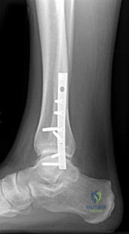
When performing an extensile lateral approach for open reduction and internal fixation of a displaced intra-articular calcaneus fracture, the surgical flap must be carefully elevated as a full-thickness layer to the periosteum to prevent necrosis. Which artery provides the primary blood supply to the apex of this lateral flap?


Explanation

The lateral calcaneal artery, a branch of the peroneal artery, provides the primary blood supply to the lateral flap used in the extensile lateral approach to the calcaneus. The flap must be full-thickness and elevated subperiosteally to preserve this vascular supply and minimize wound healing complications.

Question 7

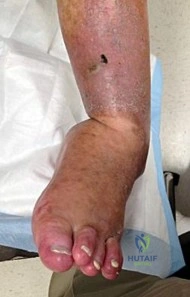
A 55-year-old patient with poorly controlled diabetes mellitus presents with a red, hot, and swollen left foot. Radiographs show periarticular osteopenia, fragmentation of bone, and early subluxation at the midtarsal joints. Infection has been definitively ruled out. According to the Eichenholtz classification, what stage is this, and what is the standard initial management?


Explanation

Eichenholtz Stage 1 (Development/Fragmentation) of Charcot arthropathy is characterized clinically by a red, hot, swollen foot and radiographically by bone fragmentation, joint subluxation, and debris. The gold standard initial treatment is immobilization in a total contact cast (TCC) and offloading to prevent further deformity until the acute inflammatory phase resolves (progressing to Stage 2 - Coalescence, and Stage 3 - Consolidation).

Question 8


In the setting of a complex ankle fracture, the anterior inferior tibiofibular ligament (AITFL) is typically attached to specific osseous landmarks. An avulsion fracture of the anterolateral distal tibia by the AITFL is eponymously known as which of the following?


Explanation

The Tillaux-Chaput fragment is an avulsion of the anterolateral distal tibia by the anterior inferior tibiofibular ligament (AITFL). A Wagstaffe-Le Fort fragment is an avulsion of the anteromedial fibula by the AITFL. The Volkmann fragment is an avulsion of the posterolateral tibia by the posterior inferior tibiofibular ligament (PITFL).

Question 9

A 26-year-old male sustains an inversion ankle sprain while his foot is maximally plantarflexed. He later develops persistent deep ankle pain. MRI reveals an osteochondral lesion of the talus. Based on the mechanism of injury, where is the lesion most likely located and what is its typical morphology?


Explanation

Osteochondral lesions of the talus follow the 'DIAL a PIMP' mnemonic: Dorsiflexion Inversion = Anterior Lateral (shallow, wafer-shaped, usually traumatic); Plantarflexion Inversion = Medial Posterior (deep, cup-shaped, more often morphologic or microtraumatic but can be acute). Given the plantarflexion and inversion mechanism, a posteromedial, deep cup-shaped lesion is expected.

Question 10


A 42-year-old runner complains of chronic medial heel pain radiating to the plantar aspect of the foot. Examination reveals maximal tenderness over the medial tuberosity of the calcaneus, exacerbated by palpation distal to the abductor hallucis. Entrapment of the first branch of the lateral plantar nerve (Baxter's nerve) is suspected. This nerve typically courses between which two muscle bellies?


Explanation

Baxter's nerve (the first branch of the lateral plantar nerve) is a common cause of recalcitrant heel pain. It typically becomes entrapped as it courses laterally between the deep fascia of the abductor hallucis muscle and the medial aspect of the quadratus plantae muscle.

Question 11


A 20-year-old male with Charcot-Marie-Tooth disease presents with a symptomatic bilateral cavovarus foot deformity. A Coleman block test is performed, which normalizes his hindfoot varus. Which of the following muscle imbalances is the primary driver initiating the plantarflexed first ray in this specific deformity?


Explanation

In Charcot-Marie-Tooth disease, the intrinsic muscles, tibialis anterior, and peroneus brevis weaken early. The peroneus longus and tibialis posterior remain strong. The strong peroneus longus overpowers the weak tibialis anterior, driving the first ray into plantarflexion, which leads to a forefoot-driven hindfoot varus deformity (corrected by the Coleman block test).

Question 12

A professional American football player forcibly hyperextends his first metatarsophalangeal (MTP) joint during a tackle. MRI confirms a complete rupture of the plantar plate (Grade III turf toe injury). Which of the following describes the most common anatomical site of failure in a complete plantar plate rupture requiring surgical repair?


Explanation

Turf toe represents a sprain or rupture of the first MTP joint plantar plate. The most common site of failure in a Grade III (complete tear) turf toe injury is at the distal insertion of the plantar plate onto the base of the proximal phalanx. Proximal tearing or sesamoid diastasis can occur but distal avulsion is classically targeted in surgical repair.

Question 13


A 28-year-old professional basketball player sustains a fracture at the base of the fifth metatarsal. Radiographs show a transverse fracture at the metaphyseal-diaphyseal junction extending into the 4th-5th intermetatarsal articulation (Zone 2). What is the recommended treatment to minimize nonunion and expedite return to play in this elite athlete?


Explanation

A Zone 2 fifth metatarsal fracture (Jones fracture) occurs at the metaphyseal-diaphyseal junction and involves the 4th-5th intermetatarsal articulation. Due to the watershed blood supply, it has a high rate of nonunion. In elite athletes, early intramedullary screw fixation is the gold standard to decrease nonunion rates and allow a faster return to play.

Question 14

A 30-year-old skier presents with lateral ankle pain and a snapping sensation behind the lateral malleolus after a fall. The injury occurred during forced ankle dorsiflexion and eversion. Radiographs show a thin cortical flake of bone avulsed from the posterolateral border of the fibula. What is the most likely diagnosis?


Explanation

The clinical presentation (snapping behind lateral malleolus, forced dorsiflexion/eversion) and radiographic finding ('fleck sign' from the posterolateral fibula) are pathognomonic for an injury to the superior peroneal retinaculum (SPR) and subsequent peroneal tendon subluxation or dislocation.

Question 15


A 58-year-old male presents with dorsal first MTP joint pain. Radiographs demonstrate advanced joint space narrowing, large dorsal osteophytes, and subchondral sclerosis. Clinically, he has severe pain at the extremes of motion AND during the mid-arc of motion (Coughlin and Shurnas Grade 4 hallux rigidus). Which surgical procedure is considered the most reliable gold standard for this patient?


Explanation

For advanced hallux rigidus (Coughlin and Shurnas Grade 3 and 4), particularly Grade 4 where pain is present in the mid-arc of motion, cheilectomy alone is insufficient because the joint cartilage is destroyed globally. First MTP joint arthrodesis is the gold standard, providing reliable pain relief and functional improvement.

Question 16

A 14-year-old boy presents with a history of recurrent ankle sprains and a painful, rigid flatfoot. Examination reveals restricted subtalar motion and peroneal spasticity. Lateral radiographs reveal a continuous bony contour forming a 'C-sign'. Which of the following describes the most likely anatomic location of the pathology?


Explanation

The 'C-sign' on a lateral radiograph is indicative of a talocalcaneal coalition, specifically bridging the medial talus to the sustentaculum tali (middle facet). A calcaneonavicular coalition is typically seen on an oblique radiograph as an 'anteater nose' sign.

Question 17


A 25-year-old male sustains a severe crush injury to his foot and develops compartment syndrome. The human foot is traditionally described as having nine anatomical compartments. Which of the following structures is exclusively located within the calcaneal compartment?


Explanation

The foot has 9 compartments: Medial, Lateral, Superficial (contains flexor digitorum brevis), Calcaneal (contains quadratus plantae), Adductor (contains adductor hallucis), and four interosseous compartments. The quadratus plantae resides in the calcaneal compartment, which communicates with the deep posterior compartment of the leg.

Question 18

Total Ankle Arthroplasty (TAA) has become an increasingly popular alternative to ankle arthrodesis for end-stage ankle osteoarthritis. According to current guidelines, which of the following is generally considered an ABSOLUTE contraindication for TAA?


Explanation

Absolute contraindications for Total Ankle Arthroplasty (TAA) include active infection, Charcot arthropathy, absent motor function/paralysis, severe uncorrectable malalignment, and avascular necrosis of the talus involving >50% of the talar body (as the implant requires sufficient viable bone for fixation).

Question 19


A 21-year-old track athlete presents with insidious onset of vague midfoot pain. Examination reveals point tenderness over the dorsal 'N-spot'. CT scan confirms a navicular stress fracture. This fracture typically originates in which specific region of the navicular due to a vascular watershed area and maximum shear stress?


Explanation

Navicular stress fractures typically occur in the central third of the bone, extending from the dorsal margin towards the plantar aspect. This area is a known relative avascular 'watershed' zone between the medial and lateral blood supplies and sustains high shear forces during the foot strike phase in athletes.

Question 20

During the surgical repair of an acute syndesmotic injury (high ankle sprain) without a medial malleolus fracture, the surgeon places a syndesmotic screw. Historically, teaching dictated fixing the syndesmosis with the ankle in maximal dorsiflexion to prevent overtightening. What anatomical characteristic of the talus was the basis for this traditional teaching, even though recent studies show anatomical reduction is the true determinant of postoperative motion?


Explanation

The traditional teaching of dorsiflexing the ankle during syndesmotic screw placement is based on the anatomy of the talar dome, which is trapezoidal and wider anteriorly. It was thought that fixing it in plantarflexion would over-constrain the mortise and prevent subsequent dorsiflexion, though modern literature shows that anatomical reduction of the fibula in the incisura is the critical factor.

Question 21

A 45-year-old male presents with chronic lateral foot and ankle pain 18 months after sustaining a closed, displaced intra-articular calcaneus fracture treated nonoperatively. He reports difficulty wearing standard closed shoes. Physical examination reveals limited subtalar motion and tenderness inferior to the lateral malleolus. What is the most likely pathophysiologic cause of his current symptoms?


Explanation

Nonoperative management of displaced calcaneus fractures frequently results in loss of calcaneal height, hindfoot varus, and lateral wall blow-out. This structural deformity leads to subfibular impingement and peroneal tendon entrapment, causing chronic lateral pain and difficulty with shoe wear.

Question 22

A 55-year-old female presents with a painful, progressive flatfoot deformity. Examination reveals an inability to perform a single-limb heel rise and significant flexible hindfoot valgus. Weight-bearing radiographs demonstrate a talonavicular coverage angle of 45 degrees and uncovering of the talar head. Which of the following surgical strategies is most appropriate for this stage of deformity?


Explanation

This patient has Stage IIb adult-acquired flatfoot deformity, characterized by a flexible deformity with significant forefoot abduction (>40% talonavicular uncovering). Management requires a soft tissue reconstruction (FDL transfer), hindfoot correction (MDCO), and a lateral column lengthening (e.g., Evans osteotomy) to correct the forefoot abduction.

Question 23

A surgeon is performing a minimally invasive percutaneous repair of an acute Achilles tendon rupture. To minimize the risk of iatrogenic nerve injury during suture passage, the surgeon must be acutely aware of the course of the sural nerve. At approximately what distance proximal to the calcaneal insertion does the sural nerve typically cross the lateral border of the Achilles tendon?


Explanation

The sural nerve courses distally and crosses the lateral border of the Achilles tendon approximately 9.8 to 12 cm proximal to its calcaneal insertion. Sutures placed blindly in the proximal stump at this level carry a high risk of sural nerve entrapment.

Question 24

A 62-year-old patient with poorly controlled type 2 diabetes presents with a globally swollen, erythematous, and warm right foot. Pedal pulses are palpable. Radiographs reveal fragmentation, periarticular debris, and subluxation at the tarsometatarsal joints. What is the most appropriate initial management?


Explanation

This patient is in the acute fragmentation phase (Eichenholtz Stage 1) of Charcot neuroarthropathy. The gold standard for initial management is immediate offloading with a total contact cast to arrest the progression of deformity until the inflammatory phase resolves.

Question 25

A 22-year-old female with Charcot-Marie-Tooth disease presents with bilateral cavovarus feet. A Coleman block test is performed, and her hindfoot varus completely corrects to a neutral alignment when the first metatarsal is allowed to drop off the block. What does this finding dictate regarding surgical planning?


Explanation

The Coleman block test evaluates hindfoot flexibility in a cavovarus foot. Correction of hindfoot varus indicates a flexible hindfoot driven by a rigid, plantarflexed first ray, meaning surgical correction must include a first metatarsal dorsiflexion osteotomy.

Question 26

A 21-year-old collegiate basketball player sustains a fracture of the fifth metatarsal. Radiographs show a transverse fracture at the metaphyseal-diaphyseal junction involving the fourth-fifth intermetatarsal articulation. He is treated with intramedullary screw fixation. To minimize the risk of nonunion or construct failure, which of the following is the most critical technical factor regarding the screw?


Explanation

In surgical fixation of Zone 2 (Jones) fractures in athletes, using the largest solid or cannulated screw that accommodates the medullary canal (usually 4.5-5.5 mm) provides superior biomechanical stability. Smaller screws have a high failure and nonunion rate.

Question 27

A 19-year-old cross-country runner presents with insidious onset dorsal midfoot pain. Plain radiographs are negative. A CT scan confirms an incomplete, non-displaced stress fracture of the dorsal cortex of the tarsal navicular. Why is this specific anatomic region highly susceptible to nonunion?


Explanation

Navicular stress fractures typically occur in the central third of the bone, which is an avascular watershed zone between the branches of the dorsalis pedis and medial plantar arteries. This tenuous blood supply significantly increases the risk of delayed union or nonunion.

Question 28

A 28-year-old male sustains an acute distal tibiofibular syndesmotic injury. During surgical stabilization, the surgeon meticulously evaluates the individual syndesmotic ligaments. Which of the following ligaments provides the greatest resistance to lateral displacement of the fibula and is mechanically the strongest?


Explanation

The posterior inferior tibiofibular ligament (PITFL) is the strongest component of the syndesmotic complex, contributing approximately 42% of the resistance to lateral fibular displacement. The AITFL contributes approximately 35%.

Question 29

A 58-year-old male presents with dorsal foot pain and stiffness of the great toe. Examination shows palpable dorsal osteophytes and pain exclusively at the extremes of motion. Radiographs demonstrate <50% joint space narrowing with preservation of the plantar joint space (Coughlin Grade 2 Hallux Rigidus). What is the most appropriate initial surgical intervention if conservative measures fail?


Explanation

For early-to-moderate hallux rigidus (Grade 1 or 2) with preserved plantar cartilage and pain primarily at extremes of dorsiflexion, a dorsal cheilectomy is the standard bone-preserving surgical option. Arthrodesis is reserved for end-stage (Grade 3 or 4) disease.

Question 30

A 22-year-old American football lineman sustains an acute hyperextension injury to his first MTP joint. MRI confirms a complete tear of the plantar plate. During surgical repair, where is the most common anatomic location of the plantar plate disruption?


Explanation

In severe 'turf toe' injuries requiring surgery, the plantar plate most commonly fails via avulsion from its distal insertion at the base of the proximal phalanx. Surgical repair involves reattaching the complex to the proximal phalanx base.

Question 31

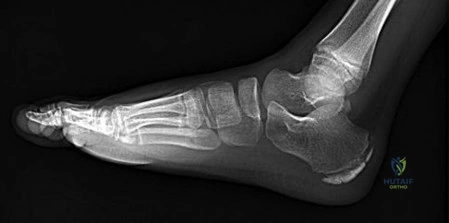
A 10-year-old boy presents with a history of recurrent lateral ankle sprains and a rigid flatfoot. Oblique radiographs of the foot demonstrate an elongated anterior process of the calcaneus, commonly known as the 'anteater nose' sign. Which of the following is the most likely diagnosis?


Explanation

The 'anteater nose' sign on an oblique radiograph of the foot is a classic radiographic finding for a calcaneonavicular coalition. This represents the elongated anterior process of the calcaneus approaching the navicular. Symptoms typically manifest between ages 8 and 12.

Question 32

Osteochondral lesions of the talus (OLTs) have distinct morphologic characteristics based on their location. Which of the following descriptions classically characterizes an anterolateral OLT?


Explanation

Anterolateral OLTs are classically described as shallow and wafer-shaped, and they are almost universally associated with a history of trauma (inversion and dorsiflexion). In contrast, posteromedial lesions are typically deep, cup-shaped, and may not have a clear traumatic etiology.

Question 33

A 45-year-old female presents with pain in the second webspace, radiating into the toes. Clinical examination reveals pain upon lateral compression of the forefoot accompanied by a palpable click (Mulder's sign). She undergoes surgical excision through a dorsal approach. Which of the following is a known risk specific to the dorsal approach compared to the plantar approach for this pathology?


Explanation

When excising a Morton's neuroma via a dorsal approach, the deep transverse metatarsal ligament must be divided to adequately expose and resect the neuroma. Failure to properly visualize and cut this ligament can lead to an incomplete resection or recurrent symptoms.

Question 34

A 26-year-old skier presents with snapping over the lateral malleolus after an acute eversion injury. Examination confirms subluxation of the peroneal tendons with resisted eversion. Surgical management involves repair of the superior peroneal retinaculum (SPR). To which structure must the SPR be anatomically reattached?


Explanation

Peroneal tendon subluxation is caused by an avulsion of the superior peroneal retinaculum (SPR) from its insertion on the posterolateral ridge of the fibula. Surgical repair involves reattaching the SPR to this posterolateral ridge, often alongside deepening of the fibular groove.

Question 35

A 68-year-old male with end-stage ankle osteoarthritis is being evaluated for a Total Ankle Arthroplasty (TAA). Which of the following represents an absolute contraindication to performing a TAA rather than an ankle arthrodesis in this patient?


Explanation

Extensive avascular necrosis of the talar body (>50%) is an absolute contraindication for Total Ankle Arthroplasty due to the high risk of catastrophic implant subsidence and failure. In such cases, arthrodesis (often tibiotalocalcaneal) is preferred.

Question 36

A 38-year-old male sustains a high-energy pilon fracture. CT imaging demonstrates significant comminution, but distinct fracture fragments are identifiable. The anterolateral distal tibial articular fragment, often avulsed during the injury, is attached to which ligament?


Explanation

The anterolateral distal tibial fragment in a pilon or syndesmotic injury is known as the Chaput fragment. It serves as the tibial attachment for the anterior inferior tibiofibular ligament (AITFL).

Question 37

A 50-year-old runner undergoes a complete surgical release of the plantar fascia for recalcitrant plantar fasciitis. Post-operatively, she reports relief of her heel pain but develops new-onset, severe pain along the lateral border of her midfoot. What is the most likely biomechanical cause of this new symptom?


Explanation

A complete release of the plantar fascia destroys the windlass mechanism, leading to arch depression, increased strain on the midfoot ligaments, and subsequent lateral column overload. This complication causes severe, recalcitrant lateral midfoot pain.

Question 38

A 25-year-old equestrian falls from a horse, sustaining a subtle Lisfranc injury with instability of the first, second, and third tarsometatarsal (TMT) joints. The surgeon opts for open reduction and internal fixation utilizing dorsal spanning plates rather than transarticular screws. What is the primary biomechanical and biologic advantage of using dorsal spanning plates in this scenario?


Explanation

Dorsal spanning plates 'bridge' the tarsometatarsal joints without violating the joint surfaces, thereby preventing iatrogenic damage to the articular cartilage. This is thought to lower the incidence of secondary post-traumatic osteoarthritis compared to transarticular screws.

Question 39

A 14-year-old female presents with insidious onset forefoot pain localized to the second metatarsophalangeal (MTP) joint. Radiographs demonstrate sclerosis, flattening, and early fragmentation of the second metatarsal head. What is the most likely diagnosis?


Explanation

Freiberg's infraction is an osteochondrosis or avascular necrosis of the metatarsal head, most commonly affecting the second metatarsal. It typically presents in adolescent females during their growth spurt.

Question 40

A 40-year-old roofer falls from a height and sustains a displaced intra-articular calcaneus fracture. He undergoes Open Reduction Internal Fixation (ORIF) via an extensile lateral approach. Postoperatively, he complains of numbness over the lateral aspect of his heel and foot. Which nerve was most likely injured during the surgical approach?


Explanation

The sural nerve is at high risk of injury during the extensile lateral approach to the calcaneus. It provides sensation to the lateral aspect of the heel and foot.

Question 41

A 55-year-old female presents with a progressive flatfoot deformity. Examination reveals a flexible hindfoot valgus and inability to perform a single-leg heel raise. Weight-bearing radiographs show more than 30% uncovering of the talonavicular joint on the AP view. Which of the following surgical interventions is most appropriate for this Stage IIb posterior tibial tendon dysfunction?


Explanation

Stage IIb adult acquired flatfoot deformity is characterized by a flexible deformity with significant forefoot abduction (>30% talonavicular uncovering). Adding a lateral column lengthening to an FDL transfer and MDCO is required to correct the severe forefoot abduction.

Question 42

A 22-year-old collegiate football lineman presents with severe pain at the base of the great toe after being tackled with his foot planted in extreme dorsiflexion. MRI confirms a complete tear of the plantar plate complex with proximal retraction of the sesamoids. What is the most appropriate management for this athlete?


Explanation

Grade 3 turf toe injuries involving a complete plantar plate tear with sesamoid retraction in elite athletes generally require surgical repair. This restores the push-off strength and normal biomechanics of the first MTP joint.

Question 43

A 20-year-old track athlete presents with insidious onset of vague dorsal midfoot pain. CT scan reveals an incomplete stress fracture of the tarsal navicular located in the central third of the bone. What anatomical feature primarily predisposes the navicular to stress fractures in this specific region?


Explanation

The central third of the tarsal navicular is a relative watershed area of avascularity. This limited blood supply predisposes the central portion of the bone to stress fractures and delayed healing.

Question 44

A 19-year-old professional basketball player sustains an acute, displaced fracture at the metaphyseal-diaphyseal junction of the fifth metatarsal. To minimize the risk of nonunion and expedite return to play, intramedullary screw fixation is planned. What is the optimal entry point for accurately placing the screw in the medullary canal?


Explanation

The optimal entry point for an intramedullary screw in a Jones fracture is 'high and inside' (dorsal and medial) on the proximal tuberosity of the fifth metatarsal. This trajectory best aligns with the straight medullary canal and avoids lateral cortex blowout.

Question 45

A 55-year-old female presents with progressive flattening of her left medial longitudinal arch. On examination, she is unable to perform a single-leg heel rise. Weight-bearing radiographs demonstrate >40% talonavicular uncoverage and an abnormally high talus-first metatarsal angle. Which of the following surgical procedures is most appropriate for this stage of posterior tibial tendon dysfunction (Stage IIb)?


Explanation

Stage IIb posterior tibial tendon dysfunction involves flexible pes planovalgus with significant forefoot abduction (>40% TN uncoverage). A lateral column lengthening is necessary in addition to FDL transfer and MDCO to adequately correct the forefoot abduction.

Question 46

During an extensile lateral approach for open reduction and internal fixation of a displaced intra-articular calcaneus fracture, protecting the primary blood supply to the apex of the corner flap is critical. Which of the following arteries is primarily responsible for perfusing this flap?


Explanation

The lateral calcaneal artery, a branch of the peroneal artery, provides the primary blood supply to the lateral cutaneous flap of the heel. An extensile lateral approach must maintain full-thickness subperiosteal dissection to avoid flap necrosis.

Question 47

A 22-year-old professional basketball player presents with lateral foot pain after landing awkwardly. Radiographs reveal an acute, non-displaced fracture at the metaphyseal-diaphyseal junction of the fifth metatarsal (Zone 2). What is the recommended treatment to minimize the risk of nonunion and allow the fastest return to play?


Explanation

Zone 2 base of 5th metatarsal fractures (Jones fractures) have a high rate of nonunion due to watershed vascularity. Intramedullary screw fixation is the gold standard for high-level athletes to promote reliable healing and expedite return to sports.

Question 48

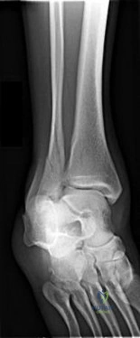
A 40-year-old male is undergoing open reduction and internal fixation of a bimalleolar equivalent ankle fracture. Intraoperatively, the Cotton test demonstrates syndesmotic instability. On an AP and Mortise radiograph, what is the normal limit for the tibiofibular clear space when evaluating reduction?


Explanation

The tibiofibular clear space is the most reliable radiographic parameter for syndesmotic integrity. It should measure < 6 mm on both AP and Mortise views, measured 1 cm proximal to the joint line.

Question 49

When performing a minimally invasive repair of an acute Achilles tendon rupture, care must be taken to avoid injury to the sural nerve. At what approximate distance proximal to the calcaneal insertion does the sural nerve typically cross the lateral border of the Achilles tendon?


Explanation

The sural nerve courses from medial to lateral and crosses the lateral border of the Achilles tendon approximately 9.8 cm proximal to its calcaneal insertion. Sutures placed proximal to this level carry a higher risk of nerve entrapment.

Question 50

A 60-year-old poorly controlled diabetic male presents with an acutely swollen, red, and warm right foot. He denies trauma, and his pedal pulses are bounding. Inflammatory markers are mildly elevated. Radiographs show early fragmentation and debris at the midfoot. What is the most appropriate immediate management?


Explanation

This is a classic presentation of acute Stage I (developmental/fragmentation) Charcot neuroarthropathy, characterized by bounding pulses and fragmentation. The gold standard for initial management is strict immobilization using a total contact cast to prevent further collapse.

Question 51

A 52-year-old avid runner presents with worsening great toe pain. Examination reveals less than 10 degrees of dorsiflexion and significant pain through the mid-arc of motion. Radiographs demonstrate severe joint space narrowing, subchondral sclerosis, and large circumferential osteophytes at the first metatarsophalangeal (MTP) joint. What is the most definitive and reliable surgical treatment?


Explanation

The patient has Coughlin/Shurnas Grade 4 hallux rigidus (pain in mid-arc of motion, severe radiographic changes). First MTP arthrodesis is the most reliable and durable procedure for advanced hallux rigidus, particularly in young, active patients.

Question 52

A 19-year-old track athlete experiences vague dorsal midfoot pain for three months. A CT scan confirms a non-displaced stress fracture of the tarsal navicular. In which anatomic zone of the navicular do these stress fractures most commonly occur due to an area of relative avascularity?


Explanation

Tarsal navicular stress fractures predominantly occur in the central third of the bone. This area represents a vascular watershed zone between the medial and lateral blood supplies, predisposing it to poor healing and nonunion.

Question 53

In patients with Charcot-Marie-Tooth (CMT) disease, a classic cavovarus foot deformity develops due to specific muscle imbalances. Which of the following combinations best describes the primary overpowering muscle forces driving the plantarflexed first ray and hindfoot varus?


Explanation

In CMT, the relatively strong peroneus longus overpowers the weak tibialis anterior, causing a plantarflexed first ray. Simultaneously, the strong tibialis posterior overpowers the weak peroneus brevis, driving hindfoot varus.

Question 54

A surgeon is performing a dorsal approach for the excision of a recurrent Morton's neuroma in the third web space. To gain adequate exposure and successfully identify and resect the neuroma, which of the following structures MUST be transected?


Explanation

The common plantar digital nerve (and the neuroma) lies plantar to the deep transverse metatarsal ligament. When using a dorsal surgical approach, this ligament must be transected to expose and resect the neuroma.

Question 55

A 28-year-old male develops severe foot pain and tense swelling following a severe crush injury. Compartment syndrome of the foot is suspected. Among the nine recognized fascial compartments of the foot, the calcaneal compartment contains which of the following vital structures?


Explanation

The calcaneal compartment is located in the hindfoot and contains the quadratus plantae muscle and the lateral plantar nerve. It communicates directly with the deep posterior compartment of the leg.

Question 56

A 13-year-old male sustains an external rotation injury to his ankle. Radiographs show a Salter-Harris III fracture of the anterolateral distal tibia (Tillaux fracture). This fracture pattern is primarily due to the avulsion pull of which of the following ligaments?


Explanation

A juvenile Tillaux fracture occurs due to the pull of the anterior inferior tibiofibular ligament (AITFL) on the anterolateral distal tibial epiphysis. This occurs uniquely in adolescents because the distal tibial physis closes central-to-medial, leaving the lateral aspect open and vulnerable to avulsion.

Question 57

A 60-year-old male is undergoing surgery for chronic insertional Achilles tendinopathy with a large Haglund's deformity. During debridement through a central tendon-splitting approach, it is determined that 65% of the Achilles tendon insertion must be detached and excised to adequately remove the degenerative tissue. What is the most appropriate next step in management?


Explanation

If more than 50% of the Achilles tendon insertion is debrided during surgery for insertional tendinopathy, the remaining tendon is biomechanically insufficient. Flexor hallucis longus (FHL) tendon transfer is the gold standard to augment the repair.

Question 58

A 68-year-old female with end-stage post-traumatic ankle osteoarthritis is being evaluated for a total ankle arthroplasty (TAA). Which of the following conditions represents an absolute contraindication to performing a TAA in this patient?


Explanation

Charcot neuroarthropathy, active infection, avascular necrosis of the talar body, and severe uncorrectable malalignment are absolute contraindications for Total Ankle Arthroplasty (TAA). Loss of protective sensation leads to rapid catastrophic implant failure.

Question 59

According to the Lauge-Hansen classification of ankle fractures, a Supination-External Rotation (SER) mechanism predictably injures structures in a sequential order. What represents Stage 3 of this injury pattern?


Explanation

The SER sequence is: Stage 1 (AITFL rupture), Stage 2 (spiral/oblique fibula fracture), Stage 3 (PITFL rupture or posterior malleolus fracture), and Stage 4 (deltoid rupture or medial malleolus fracture).

Question 60

A 24-year-old sprinter presents with acute plantar first MTP joint pain after forcefully pushing off the starting blocks. Radiographs reveal a radiolucency through the tibial sesamoid. Which of the following radiographic characteristics best distinguishes an acute sesamoid fracture from a congenital bipartite sesamoid?


Explanation

An acute sesamoid fracture typically presents with an irregular, sharp radiolucent line without sclerotic margins. In contrast, a bipartite sesamoid generally has smooth, corticated (sclerotic) edges and is often bilateral.

Question 61

A 52-year-old female presents with a progressive, painful flatfoot deformity. Clinical examination reveals an inability to perform a single-leg heel rise. Weight-bearing radiographs show a flexible hindfoot valgus and greater than 30% uncovering of the talonavicular joint with forefoot abduction. What is the most appropriate surgical management for this Stage IIb posterior tibial tendon dysfunction?


Explanation

Stage IIb adult-acquired flatfoot deformity involves a flexible hindfoot with significant forefoot abduction (>30% talonavicular uncovering). Surgical correction requires a medializing calcaneal osteotomy to correct hindfoot valgus, an FDL transfer to replace the dysfunctional PTT, and a lateral column lengthening to correct the forefoot abduction.

Question 62

A 40-year-old construction worker falls from a height and sustains a severely displaced, joint-depressed calcaneus fracture. The surgeon elects to perform an open reduction and internal fixation via an extensile lateral approach. Which of the following is the most common postoperative complication associated with this specific surgical approach?


Explanation

Wound edge necrosis and dehiscence at the apex of the flap is the most common complication of the extensile lateral approach to the calcaneus, occurring in up to 10-25% of cases. Careful soft tissue handling, subperiosteal dissection, and utilizing a 'no-touch' technique are critical to minimizing this risk.

Question 63

A 35-year-old recreational basketball player suffers an acute Achilles tendon rupture. Based on high-level level I evidence (e.g., the Willits trial) comparing operative repair versus non-operative management with early functional rehabilitation, what is the expected outcome profile?


Explanation

Recent level I evidence demonstrates that when early functional rehabilitation protocols are utilized, the re-rupture rates between operative and non-operative management of Achilles ruptures are not statistically different. However, operative management carries a higher risk of soft tissue complications, such as infection and sural nerve injury.

Question 64

A 58-year-old male with long-standing, poorly controlled type II diabetes presents with a red, hot, swollen left foot. He denies trauma. Pulses are bounding, and sensation to monofilament testing is absent. Radiographs demonstrate marked periarticular fragmentation, subchondral debris, and subluxation at the tarsometatarsal joints. According to the Eichenholtz classification, what stage of Charcot neuroarthropathy does this represent?


Explanation

Eichenholtz Stage I (Developmental/Fragmentation stage) is characterized by acute inflammation, osseous fragmentation, joint subluxation/dislocation, and debris formation. Stage II involves coalescence and absorption of fine debris, while Stage III shows consolidation and remodeling.

Question 65

A 21-year-old collegiate soccer player sustains a twisting injury to his foot and is diagnosed with an acute diaphyseal/metaphyseal junction fracture of the fifth metatarsal (Zone 2, Jones fracture). Given his desire to return to elite-level sports, what is the gold standard of treatment?


Explanation

Zone 2 fractures (Jones fractures) occur in a vascular watershed area and have a high risk of delayed union or nonunion. In competitive athletes, early percutaneous intramedullary screw fixation is the gold standard to expedite return to play and reduce nonunion risk.

Question 66

A 28-year-old female presents with chronic ankle pain following recurrent sprains. MRI reveals an osteochondral lesion of the talus (OCD). Which of the following best describes the characteristics and mechanism of a posteromedial talar dome lesion?


Explanation

Posteromedial talar dome lesions are typically deep, cup-shaped, and less likely to displace. They are classically caused by an inversion injury while the foot is plantarflexed, which compresses the medial talar dome against the tibial plafond.

Question 67

A patient with Charcot-Marie-Tooth disease presents with a bilateral cavovarus foot deformity. A Coleman block test is performed on the right foot, and the hindfoot varus corrects completely to a neutral alignment. What does this clinical finding indicate?


Explanation

The Coleman block test distinguishes between a flexible and rigid hindfoot varus in cavovarus feet. If placing the lateral foot on a block (allowing the plantarflexed first ray to drop) corrects the hindfoot varus to neutral, the hindfoot is flexible and the deformity is forefoot-driven.

Question 68

A 68-year-old male presents with severe pain in his first metatarsophalangeal (MTP) joint, particularly during toe-off. Examination reveals pain throughout the mid-range of motion. Radiographs demonstrate >50% joint space narrowing, a dorsal ring of osteophytes, and subchondral cysts (Coughlin/Shurnas Grade 4 Hallux Rigidus). What is the most reliable definitive surgical treatment?


Explanation

For advanced hallux rigidus (Grade 3 with pain at mid-range or Grade 4) with significant joint degeneration, first MTP arthrodesis is the gold standard. It provides predictable pain relief, corrects deformity, and allows for near-normal gait biomechanics.

Question 69

A professional American football lineman sustains a severe hyperextension injury to his great toe. Clinical exam shows marked swelling, ecchymosis, and a gross lack of resistance to passive MTP dorsiflexion. MRI confirms a complete rupture of the plantar plate with proximal retraction of the sesamoids. What is the most appropriate management?


Explanation

This is a Grade 3 'turf toe' injury characterized by a complete tear of the plantar plate complex and proximal migration of the sesamoids. Surgical repair is indicated in elite athletes to restore the push-off strength and prevent chronic instability.

Question 70

A 45-year-old female complains of burning and tingling in the plantar aspect of her foot, which worsens after standing. Examination reveals a positive Tinel's sign posterior to the medial malleolus. If an MRI is ordered and identifies a space-occupying lesion compressing the posterior tibial nerve, what is the most common etiology in this specific location?


Explanation

Tarsal tunnel syndrome is a compression neuropathy of the tibial nerve. When a specific space-occupying lesion is identified as the cause, venous varicosities are the most common finding, followed by ganglion cysts and lipomas.

Question 71

A 22-year-old skier experiences a sudden 'popping' sensation behind his lateral malleolus after forced dorsiflexion and eversion of his ankle. Examination reveals tenderness and visible swelling over the peroneal tendons, which subluxate anteriorly with active eversion. Which of the following is the most common anatomic variant of the superior peroneal retinaculum (SPR) injury in this condition?


Explanation

Acute peroneal tendon dislocation is typically caused by failure of the superior peroneal retinaculum (SPR). The most common mechanism of SPR failure is a periosteal avulsion from its attachment on the posterolateral distal fibula, sometimes creating a 'fleck sign' on radiographs.

Question 72

A 30-year-old male presents to the emergency department after a twisting injury to his ankle. Radiographs show a widened medial clear space and an isolated proximal third fibula fracture (Maisonneuve fracture). Given the location of the proximal fibula fracture, which nerve is at greatest risk of associated injury?


Explanation

A Maisonneuve fracture involves a pronation-external rotation injury that tears the syndesmosis and fractures the proximal fibula. The common peroneal nerve wraps around the fibular neck and is highly vulnerable to stretching or entrapment from injuries in this proximal region.

Question 73

A 12-year-old boy presents with a painful, rigid flatfoot and a history of recurrent ankle sprains. Peroneal spasticity is noted on examination. Oblique radiographs of the foot demonstrate an elongated anterior process of the calcaneus approaching the navicular. What is the classic radiographic name for this finding?


Explanation

The 'anteater nose sign' on an oblique radiograph of the foot is classic for a calcaneonavicular coalition. It represents the elongated tubular anterior process of the calcaneus extending toward the navicular. The 'C-sign' is typically seen on lateral radiographs in talocalcaneal coalitions.

Question 74

A 55-year-old runner has chronic insertional Achilles tendinopathy with a prominent Haglund deformity and extensive intratendinous calcification. Conservative management has failed. During surgery, aggressive debridement of the diseased tendon is necessary. At what threshold of Achilles tendon detachment/debridement is augmentation with a flexor hallucis longus (FHL) transfer generally recommended?


Explanation

During debridement of insertional Achilles tendinopathy, if more than 50% of the tendon footprint is compromised or resected to remove calcifications and degenerated tissue, augmentation with an FHL tendon transfer is widely recommended to restore plantarflexion power and prevent catastrophic rupture.

Question 75

According to the Lauge-Hansen classification system, what is the typical progression of osseous and ligamentous injury in a Supination-External Rotation (SER) ankle fracture?


Explanation

The Lauge-Hansen SER mechanism follows a sequential pattern: Stage 1 is the anterior inferior tibiofibular ligament (AITFL), Stage 2 is a spiral/oblique fracture of the lateral malleolus, Stage 3 is the posterior inferior tibiofibular ligament (PITFL) or posterior malleolus, and Stage 4 is the deltoid ligament or medial malleolus.

None

Detailed Chapters & Topics

Dive deeper into specialized chapters regarding orthopedic-mcqs-online-015-foot-and-ankle-e

37 Chapters
01
Chapter 1 51 min

Foot And Ankle Free Orthopedics Review | Dr Hutaif Foot -...

Test your knowledge with Dr. Hutaif's free Foot and Ankle Orthopedics review. Take this interactive MCQ quiz with a tim…

02
Chapter 2 74 min

Orthopedic Board Prep: Comprehensive Interactive Rheumatoid Foot Exam

Master the rheumatoid foot exam with our interactive orthopedic board prep. Use study and exam modes to test your skill…

03
Chapter 3 96 min

Hallux Rigidus Oral Questions: Your Expert Answer Guide

Foot and ankle structured oral questions7: Hallux rigidus EXAMINER : This 45-year-old male patient has presented with p…

04
Chapter 4 7 min

Unraveling Acquired Adult Flatfoot: Key Answers for Orthopedics

Foot and ankle structured oral questions5: Acquired adult flatfoot EXAMINER : I would like you to look at this clinical…

05
Chapter 5 81 min

Oral Questions Lateral: Ankle Instability Exam Prep

Foot and ankle structured oral questions1: Lateral ligament instability of the ankle EXAMINER . Tell me what this diagr…

06
Chapter 6 13 min

Hallux Valgus Exam Prep: Master Oral Questions Hallux Scenarios

Foot and ankle structured oral questions6: Hallux valgus EXAMINER : Please have a look at these clinical photographs an…

07
Chapter 7 12 min

Ankle Structured Oral: Expert Answers for Arthritis Cases

Foot and ankle structured oral questions2: Ankle arthritis EXAMINER : Describe the findings on this X-ray. ( Figure 4.2…

08
Chapter 8 74 min

Orthopedic Foot Review | Dr Hutaif Foot & Ankle Review - ...

ONLINE ORTHOPEDIC MCQS FOOT0 9 1 .        Figure 1 shows the radiograph of a 60-year-old woman who underwent a previous…

09
Chapter 9 60 min

Orthopedic With Answer Foot Review | Dr Hutaif Foot & A -...

Master your orthopedic foot review with Dr. Hutaif's interactive MCQ quiz. Test your knowledge, track your score, and c…

10
Chapter 10 43 min

Orthopedic Foot And Ank Review | Dr Hutaif Foot & Ankle -...

Question 1Figure 1 is the radiograph of a 48-year-old man. He is of normal height and weight, medically healthy,and in …

11
Chapter 11 18 min

Foot And Ankle Self Assessme Review | Dr Hutaif Foot & - ...

for more mcqs in the bank ORTHOPEDIC MCQS ONLINE BANK OITE 20 ORTHOPEDIC MCQS ONLINE BANK OITE 97 ORTHOPEDIC MCQS BANK …

12
Chapter 12 17 min

What to Expect: Ankle Surgery Postoperative Care & Instructions

Ankle arthrodesis ‌ ‌ 367 Surgery for Achilles tendinopathy 377 Ankle arthroplasty ‌ 371 Surgery for peroneal tendinopa…

13
Chapter 13 18 min

Mastering Foot & Ankle Interventions: Essential Ankle Surgery Insights

Ankle arthrodesis ‌ ‌ 367 Surgery for Achilles tendinopathy 377 Ankle arthroplasty ‌ 371 Surgery for peroneal tendinopa…

14
Chapter 14 10 min

Operative Correction of Tibiofibular Diastasis and Fibular Malunion: A Comprehensive Surgical Guide

Master operative correction of tibiofibular diastasis and fibular malunion. Learn expert surgical techniques to restore…

15
Chapter 15 16 min

Operative Management of Ingrown Toenails: A Comprehensive Surgical Guide

Master the operative management of ingrown toenails. This comprehensive surgical guide covers essential nail anatomy, b…

16
Chapter 16 11 min

Disorders of the Anterior Tibial Tendon: A Comprehensive Surgical Guide

Master the diagnosis and surgical management of anterior tibial tendon disorders, including tenosynovitis, acute ruptur…

17
Chapter 17 16 min

Operative Management of Talar Fractures: A Comprehensive Surgical Guide

Master the operative management of talar fractures with our expert surgical guide. Learn about vascular challenges, ana…

18
Chapter 18 19 min

Operative Management of Tarsal Malunions: A Comprehensive Surgical Guide

Master the operative management of lesser tarsal malunions. Discover reconstructive surgery techniques to restore a sta…

19
Chapter 19 10 min

Ankle Malunion Reconstruction: Surgical Techniques & Biomechanics

Explore the biomechanics and surgical techniques for ankle malunion reconstruction. Learn how fibular shortening drives…

20
Chapter 20 19 min

Partial Nail Fold and Nail Matrix Removal: Advanced Surgical Techniques

Master partial nail fold and matrix removal with our advanced surgical guide. Discover perionychial anatomy to prevent …

21
Chapter 21 11 min

Interdigital Neuroma Excision: The Dorsal Approach

Master the dorsal approach for Morton's neuroma excision. Explore surgical anatomy, pathophysiology, and techniques to …

22
Chapter 22 10 min

Anterior Tarsal Tunnel Syndrome: Operative Guide

Master the operative management of anterior tarsal tunnel syndrome. Explore deep peroneal nerve anatomy, diagnosis, and…

23
Chapter 23 20 min

Operative Management of Pes Planus and Tarsal Coalition: A Comprehensive Surgical Guide

Comprehensive orthopedic guide on the surgical management of pes planus and tarsal coalition. Covers biomechanics, surg…

24
Chapter 24 11 min

Operative Management of Recalcitrant Plantar Fasciitis and Heel Pain Syndrome

Discover the operative management of recalcitrant plantar fasciitis. Understand the pathoanatomy, key risk factors, and…

25
Chapter 25 16 min

Interdigital Neuroma (Morton's Toe): Comprehensive Surgical Management

Discover comprehensive surgical management for interdigital neuroma (Morton's toe). Explore its history, anatomical cau…

26
Chapter 26 10 min

Plantar Fasciotomy and Closing Wedge Metatarsal Osteotomy: The Gould Technique

Master the Gould technique for cavovarus foot reconstruction. Discover how plantar fasciotomy and metatarsal osteotomy …

27
Chapter 27 16 min

Comprehensive Surgical Management of Bunionette (Tailor’s Bunion)

Explore the pathoanatomy, causes, and comprehensive surgical management of a bunionette (tailor's bunion). Learn how to…

28
Chapter 28 19 min

Calcaneal Osteotomy: Dwyer & Crescentic Techniques

Master the biomechanics of calcaneal osteotomies. Learn how to correct complex hindfoot deformities using the Dwyer and…

29
Chapter 29 17 min

Operative Management of Anterior Tibial Tendon Pathology

Explore expert management of anterior tibial tendon pathology. Discover clinical evaluation, insertional tendinosis sym…

30
Chapter 30 15 min

Talocalcaneal Coalition: Comprehensive Diagnosis and Surgical Management

Comprehensive orthopedic guide on talocalcaneal coalition, detailing clinical presentation, CT imaging, resection techn…

31
Chapter 31 11 min

Subtalar Arthroereisis: Biomechanics, Surgical Techniques, and Clinical Outcomes

Master subtalar arthroereisis for flatfoot correction. Explore joint biomechanics, implant classifications, surgical te…

32
Chapter 32 11 min

Midfoot Fractures and Dislocations: A Comprehensive Surgical Guide

Discover expert surgical strategies for midfoot fractures and dislocations. Learn about biomechanics, navicular fractur…

33
Chapter 33 14 min

Metatarsal Stress Fractures: Comprehensive Diagnosis and Surgical Management

Comprehensive orthopaedic guide on metatarsal stress fractures, detailing biomechanics, conservative care, and step-by-…

34
Chapter 34 11 min

Operative Management of Sesamoid Pathology: Bone Grafting and Reconstruction

Master the surgical management of sesamoid nonunion, osteochondritis, and sesamoiditis. Evidence-based guide on bone gr…

35
Chapter 35 10 min

Surgical Management and Excision of the Tibial Sesamoid

Explore the biomechanics, pathoanatomy, and surgical management of the tibial sesamoid. Learn about excision techniques…

36
Chapter 36 11 min

Arthritis of the Foot: Inflammatory and Degenerative Management

A comprehensive orthopedic guide to inflammatory and degenerative arthritis of the foot, detailing biomechanics, nonope…

37
Chapter 37 11 min

Partial Nail Plate Removal: Comprehensive Surgical Technique and Clinical Guidelines

Master partial nail plate removal with our comprehensive surgical guide. Learn essential anatomy, operative steps, and …

Dr. Mohammed Hutaif
Medically Verified Content by
Prof. Dr. Mohammed Hutaif
Consultant Orthopedic & Spine Surgeon
Guide Overview