Full Question & Answer Text (for Search Engines)
Question 1:
A 24-year-old semi-professional soccer player presents with recurrent right ankle 'giving way' sensation after multiple inversion injuries over 18 months. He has failed a comprehensive 6-month physiotherapy program including bracing and proprioceptive training. On examination, he has tenderness over the anterior talofibular ligament (ATFL) and calcaneofibular ligament (CFL) regions.
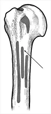
 Which of the following physical examination findings would most strongly indicate mechanical ankle instability and guide surgical consideration?
Options:
- Pain with palpation over the sinus tarsi
- A positive anterior drawer test with a 'clunk' and apprehension
- Generalized hypermobility on Beighton score
- Limited dorsiflexion range of motion
- Pain during a single-leg hop test
Correct Answer: A positive anterior drawer test with a 'clunk' and apprehension
Explanation:
A positive anterior drawer test with a 'clunk' and apprehension is a key indicator of mechanical instability, specifically ATFL insufficiency, which is the most common cause of recurrent lateral ankle instability. The 'clunk' suggests significant talar translation, and apprehension indicates the patient's recognition of the instability. While other findings like sinus tarsi pain, hypermobility, limited dorsiflexion, and pain on hop test are relevant to chronic ankle problems, they do not directly demonstrate the mechanical laxity of the lateral ankle ligaments as strongly as a positive anterior drawer test in this context. Generalized hypermobility might suggest a need for a reconstructive rather than just a repair procedure, but the mechanical instability is primarily shown by the drawer test.
Question 2:
Which of the following ligaments is the primary static restraint to anterior translation of the talus relative to the tibia, particularly in plantarflexion?
Options:
- Posterior talofibular ligament (PTFL)
- Calcaneofibular ligament (CFL)
- Anterior inferior tibiofibular ligament (AITFL)
- Anterior talofibular ligament (ATFL)
- Deltoid ligament
Correct Answer: Anterior talofibular ligament (ATFL)
Explanation:
The anterior talofibular ligament (ATFL) is the weakest and most commonly injured of the lateral ankle ligaments. It primarily resists anterior translation of the talus and internal rotation, especially when the ankle is in plantarflexion, which is the position of typical inversion injury. The CFL primarily resists inversion in dorsiflexion, the PTFL resists posterior talar translation, the AITFL (part of the syndesmosis) stabilizes the distal tibiofibular joint, and the deltoid ligament stabilizes the medial ankle.
Question 3:
A 30-year-old runner sustains an acute inversion injury to his right ankle. He presents to the emergency department unable to bear weight. Radiographs are ordered. According to the Ottawa Ankle Rules, which of the following findings would necessitate ankle radiographs?
Options:
- Pain in the malleolar zone and an inability to bear weight immediately after the injury and in the emergency department
- Ecchymosis and swelling over the lateral malleolus
- Tenderness over the medial malleolus only
- Limited range of motion in dorsiflexion
- Pain in the malleolar zone and tenderness over the base of the fifth metatarsal
Correct Answer: Pain in the malleolar zone and an inability to bear weight immediately after the injury and in the emergency department
Explanation:
The Ottawa Ankle Rules state that ankle radiographs are required if there is pain in the malleolar zone AND any of the following: inability to bear weight both immediately and in the emergency department (four steps), or bone tenderness at the posterior edge or tip of the lateral malleolus, or bone tenderness at the posterior edge or tip of the medial malleolus. Therefore, pain in the malleolar zone and an inability to bear weight immediately after the injury and in the emergency department is a clear indication for radiographs. Tenderness over the base of the fifth metatarsal would necessitate foot radiographs, not necessarily ankle radiographs, unless there is also malleolar zone pain. Ecchymosis, swelling, and limited range of motion are common symptoms but not standalone indications for radiographs according to Ottawa Ankle Rules.
Question 4:
In the context of chronic lateral ankle instability, what is the most common surgical procedure employed for primary repair?
Options:
- Chrisman-Snook reconstruction
- Watson-Jones tenodesis
- Modified Brostrom procedure (Brostrom-Gould)
- Anatomical reconstruction with allograft
- Peroneal tendon transfer
Correct Answer: Modified Brostrom procedure (Brostrom-Gould)
Explanation:
The Modified Brostrom procedure, often referred to as Brostrom-Gould, is the most common and gold standard surgical procedure for primary repair of chronic lateral ankle instability. It involves direct repair of the attenuated ATFL and CFL, often augmented with the inferior extensor retinaculum. Chrisman-Snook and Watson-Jones are older tenodesis procedures using peroneal tendons, which are non-anatomical reconstructions. Anatomical reconstruction with allograft is typically reserved for revision cases or patients with poor tissue quality/generalized laxity. Peroneal tendon transfer isn't a primary repair technique for instability.
Question 5:
Which of the following describes the anatomical relationship of the calcaneofibular ligament (CFL)?
Options:
- It originates from the distal anterior fibula and inserts onto the lateral talus.
- It runs anteriorly from the distal fibula to the calcaneus, deep to the peroneal tendons.
- It is an extracapsular ligament that runs posteriorly and inferiorly from the fibula to the calcaneus, superficial to the peroneal tendons.
- It is an extracapsular ligament that runs anteriorly and inferiorly from the fibula to the calcaneus, deep to the peroneal tendons.
- It originates from the posterior fibula and inserts onto the posterior talus.
Correct Answer: It is an extracapsular ligament that runs anteriorly and inferiorly from the fibula to the calcaneus, deep to the peroneal tendons.
Explanation:
The CFL is an extracapsular ligament that runs anteriorly and inferiorly from the distal fibula to the lateral aspect of the calcaneus. Crucially, it runs deep to the peroneal tendons. The ATFL originates from the distal anterior fibula and inserts onto the lateral talus. The PTFL originates from the posterior fibula and inserts onto the posterior talus. The CFL's deep relationship to the peroneal tendons is important for surgical approaches.
Question 6:
A patient presents with recurrent ankle sprains and complaints of the ankle 'giving way'. Clinically, you suspect chronic mechanical instability. Which of the following imaging modalities is most sensitive for evaluating the integrity of the ATFL and CFL?
Options:
- Plain radiographs (AP, lateral, mortise views)
- Stress radiographs (anterior drawer and talar tilt views)
- Computed Tomography (CT) scan
- Magnetic Resonance Imaging (MRI)
- Bone scintigraphy
Correct Answer: Magnetic Resonance Imaging (MRI)
Explanation:
Magnetic Resonance Imaging (MRI) is the most sensitive imaging modality for evaluating the integrity of soft tissue structures like the ATFL and CFL. It can show attenuation, scarring, or complete tears of these ligaments. While stress radiographs are excellent for demonstrating functional mechanical instability by measuring talar tilt and anterior translation, they don't directly visualize the ligaments' integrity as well as MRI. Plain radiographs are for bony anatomy, and CT scans are superior for bony detail but less so for soft tissues.
Question 7:
During an anterior drawer test of the ankle, which position of the ankle joint (in terms of plantarflexion/dorsiflexion) best isolates the ATFL?
Options:
- Neutral (0 degrees dorsiflexion/plantarflexion)
- Full dorsiflexion
- Slight dorsiflexion (10-15 degrees)
- Slight plantarflexion (10-20 degrees)
- Full plantarflexion
Correct Answer: Slight plantarflexion (10-20 degrees)
Explanation:
The anterior drawer test for the ankle is best performed with the ankle in slight plantarflexion (approximately 10-20 degrees). This position relaxes the CFL and PTFL, thereby isolating the ATFL, which is taut in plantarflexion and anterior translation. In dorsiflexion, the ATFL is relaxed, and the CFL becomes more taut, making assessment of the ATFL less specific.
Question 8:
What is the primary role of the calcaneofibular ligament (CFL) in ankle stability?
Options:
- Resisting internal rotation of the talus
- Preventing eversion of the hindfoot
- Resisting varus (inversion) stress, particularly in dorsiflexion
- Limiting anterior translation of the talus
- Stabilizing the syndesmosis
Correct Answer: Resisting varus (inversion) stress, particularly in dorsiflexion
Explanation:
The calcaneofibular ligament (CFL) is the primary static restraint to varus (inversion) stress of the ankle, particularly when the ankle is in a neutral or dorsiflexed position. While it also contributes to subtalar stability, its main role in ankle stability is resisting inversion. The ATFL limits anterior translation and internal rotation. The deltoid ligament prevents eversion, and syndesmotic ligaments stabilize the distal tibiofibular joint.
Question 9:
A 35-year-old male with chronic lateral ankle instability undergoes a modified Brostrom-Gould procedure. Which structure is commonly imbricated with the repaired ATFL and CFL to augment the repair?
Options:
- Peroneus brevis tendon
- Inferior extensor retinaculum
- Peroneus longus tendon
- Spring ligament
- Superior peroneal retinaculum
Correct Answer: Inferior extensor retinaculum
Explanation:
In the modified Brostrom-Gould procedure, the attenuated ATFL and CFL are directly repaired and imbricated. The repair is then augmented by incorporating a flap of the inferior extensor retinaculum over the repaired ligaments. This provides additional strength and stability to the lateral ankle complex. The peroneal tendons are used in tenodesis procedures (e.g., Chrisman-Snook), not typically in a Brostrom-Gould repair. The spring ligament is on the medial side, and the superior peroneal retinaculum stabilizes the peroneal tendons.
Question 10:
What is the most common concomitant injury found in patients with chronic lateral ankle instability?
Options:
- Achilles tendon rupture
- Posterior tibial tendon dysfunction
- Osteochondral lesion of the talus
- Syndesmotic injury
- Tarsal tunnel syndrome
Correct Answer: Osteochondral lesion of the talus
Explanation:
Osteochondral lesions (OCLs) of the talus are the most common concomitant injury found in patients with chronic lateral ankle instability, occurring in up to 50% of cases. The repeated episodes of 'giving way' and abnormal biomechanics can lead to impaction injuries of the talar dome. While other conditions listed can occur, OCLs are specifically and highly associated with chronic ankle instability. It is crucial to evaluate for these lesions pre-operatively, often requiring an MRI.
Question 11:
Which of the following is considered a dynamic stabilizer of the lateral ankle?
Options:
- Anterior talofibular ligament
- Calcaneofibular ligament
- Peroneus longus and brevis muscles
- Posterior talofibular ligament
- Interosseous membrane
Correct Answer: Peroneus longus and brevis muscles
Explanation:
Dynamic stabilizers are muscles and their tendons that cross a joint and contribute to its stability through active contraction. The peroneus longus and brevis muscles are the primary dynamic stabilizers of the lateral ankle, actively resisting inversion and providing eversion force. The ATFL, CFL, and PTFL are static stabilizers (ligaments). The interosseous membrane stabilizes the tibia and fibula proximally.
Question 12:
A 16-year-old female high school basketball player presents with persistent pain, swelling, and recurrent 'popping' sensation over the lateral ankle after an inversion injury 3 months ago. Examination reveals tenderness along the course of the peroneal tendons, and she is able to voluntarily sublux her peroneal tendons over the lateral malleolus. What is the most likely diagnosis?
Options:
- Chronic lateral ankle instability with ATFL tear
- Peroneal tendon subluxation/dislocation
- Ankle impingement syndrome
- Osteochondral lesion of the talus
- Sural nerve entrapment
Correct Answer: Peroneal tendon subluxation/dislocation
Explanation:
Recurrent 'popping' or 'snapping' over the lateral malleolus, combined with tenderness along the peroneal tendons and the ability to voluntarily sublux them, is highly suggestive of peroneal tendon subluxation or dislocation. This often occurs after an acute dorsiflexion-eversion injury, but can be mistaken for or coexist with lateral ankle sprains. While chronic instability, impingement, OCLs, and nerve entrapment can cause lateral ankle pain, the specific symptom of 'popping' and demonstrable subluxation points directly to peroneal tendon pathology.
Question 13:
What is the typical mechanism of injury for a lateral ankle sprain involving the ATFL?
Options:
- Dorsiflexion and eversion
- Plantarflexion and inversion
- Isolated dorsiflexion
- Isolated inversion
- Plantarflexion and eversion
Correct Answer: Plantarflexion and inversion
Explanation:
The most common mechanism of injury for a lateral ankle sprain, specifically involving the ATFL, is plantarflexion and inversion. In this position, the ATFL is most taut and therefore most susceptible to injury. Dorsiflexion and eversion are more associated with syndesmotic or deltoid ligament injuries. Isolated dorsiflexion or inversion without a plantarflexion component is less common for ATFL injury.
Question 14:
When evaluating a patient with chronic lateral ankle instability, what is the primary purpose of obtaining stress radiographs (anterior drawer and talar tilt views)?
Options:
- To assess for associated osteochondral lesions of the talus.
- To visualize the extent of soft tissue edema and inflammation.
- To objectively quantify the degree of mechanical laxity.
- To identify potential fractures not visible on routine radiographs.
- To evaluate the integrity of the peroneal tendons.
Correct Answer: To objectively quantify the degree of mechanical laxity.
Explanation:
Stress radiographs (anterior drawer and talar tilt views) are crucial for objectively quantifying the degree of mechanical laxity in chronic lateral ankle instability. They provide direct measurements of anterior talar translation and talar tilt, which can confirm and grade the instability. While other imaging modalities (MRI, CT) or plain radiographs serve other purposes, stress views specifically assess the functional laxity of the joint. OCLs are better seen on MRI/CT, soft tissue edema on MRI, and fractures on plain radiographs/CT. Peroneal tendons are best evaluated clinically and with MRI.
Question 15:
A 28-year-old patient presents with chronic lateral ankle pain and instability after multiple sprains. Physical exam reveals tenderness over the anterior aspect of the lateral malleolus, and a positive anterior drawer test. Which of the following is a common differential diagnosis for persistent lateral ankle pain that should be considered in addition to ligamentous instability?
Options:
- Posterior impingement syndrome
- Achilles tendonitis
- Tarsal tunnel syndrome
- Anterolateral impingement syndrome
- Medial deltoid ligament tear
Correct Answer: Anterolateral impingement syndrome
Explanation:
Anterolateral impingement syndrome, often due to synovitis or scar tissue formation in the ankle joint's anterolateral gutter, is a common cause of persistent pain after an ankle sprain, even when instability has been addressed or is being managed. It can mimic or coexist with mechanical instability. Posterior impingement involves the posterior ankle, Achilles tendonitis affects the posterior heel/calf, tarsal tunnel syndrome involves the posterior tibial nerve on the medial side, and a medial deltoid ligament tear is a medial ankle injury.
Question 16:
What is considered a normal anterior talar translation on stress radiographs (anterior drawer view) for an adult ankle?
Options:
- Less than 3 mm
- Less than 5 mm
- Less than 8 mm
- Less than 10 mm
- Less than 12 mm
Correct Answer: Less than 3 mm
Explanation:
Generally, an anterior talar translation of less than 3 mm is considered normal. A difference of more than 3 mm compared to the contralateral ankle, or an absolute translation greater than 8-10 mm, is often indicative of ATFL insufficiency. However, thresholds can vary slightly between studies. The question asks for 'normal', so less than 3mm is the most appropriate answer.
Question 17:
Which of the following describes 'functional ankle instability'?
Options:
- Objective laxity of the lateral ankle ligaments on stress radiographs.
- Recurrent episodes of ankle 'giving way' due to ligamentous rupture.
- Subjective feeling of instability or apprehension without demonstrable mechanical laxity.
- Instability caused by a congenital deformity of the ankle joint.
- Pathologic scarring leading to joint stiffness and decreased range of motion.
Correct Answer: Subjective feeling of instability or apprehension without demonstrable mechanical laxity.
Explanation:
Functional ankle instability refers to a subjective feeling of the ankle 'giving way' or apprehension, without objective evidence of mechanical laxity on physical examination or stress radiographs. This is often attributed to impaired proprioception, neuromuscular control deficits, or dynamic muscular weakness. Mechanical instability, in contrast, refers to objective laxity of the ligaments. Functional instability is often managed with intensive proprioceptive and strengthening rehabilitation.
Question 18:
In an acute Grade III lateral ankle sprain, which ligaments are typically involved?
Options:
- Only the ATFL
- ATFL and CFL
- ATFL, CFL, and PTFL
- ATFL and AITFL
- CFL and PTFL
Correct Answer: ATFL and CFL
Explanation:
A Grade III lateral ankle sprain typically involves a complete rupture of the anterior talofibular ligament (ATFL) and the calcaneofibular ligament (CFL). The posterior talofibular ligament (PTFL) is rarely injured in isolation and usually only in severe dislocations or fracture-dislocations. The AITFL is part of the syndesmosis. Therefore, ATFL and CFL are the key ligaments in a Grade III lateral ankle sprain.
Question 19:
What is the primary goal of the initial RICE (Rest, Ice, Compression, Elevation) protocol for an acute lateral ankle sprain?
Options:
- To accelerate ligamentous healing and strength.
- To prevent chronic instability and return to sport quickly.
- To minimize swelling, pain, and facilitate early rehabilitation.
- To objectively assess the degree of ligamentous injury.
- To prepare the ankle for immediate surgical intervention.
Correct Answer: To minimize swelling, pain, and facilitate early rehabilitation.
Explanation:
The primary goal of the RICE protocol (Rest, Ice, Compression, Elevation) for an acute lateral ankle sprain is to minimize swelling, reduce pain, and control inflammation, thereby facilitating early mobilization and rehabilitation. It does not directly accelerate ligamentous healing or prevent chronic instability in the long term, nor is it for objective assessment or immediate surgical preparation. It's a foundational acute management strategy.
Question 20:
Which nerve is most at risk of injury during a lateral ankle ligament reconstruction (e.g., modified Brostrom procedure)?
Options:
- Deep peroneal nerve
- Superficial peroneal nerve (musculocutaneous nerve)
- Sural nerve
- Tibial nerve
- Saphenous nerve
Correct Answer: Sural nerve
Explanation:
The sural nerve is the most commonly injured nerve during lateral ankle ligament reconstruction procedures due to its superficial course along the lateral aspect of the ankle, often close to the incision site for ATFL and CFL repair. The superficial peroneal nerve is also at risk, but the sural nerve's anatomical proximity makes it particularly vulnerable. The deep peroneal nerve is anterior, the tibial nerve is posterior-medial, and the saphenous nerve is medial.
Question 21:
A 22-year-old dancer presents with chronic lateral ankle instability. Her Beighton score is 7/9, indicating generalized ligamentous laxity. Which surgical approach would be most appropriate given her hypermobility?
Options:
- Primary repair using a modified Brostrom-Gould technique.
- Non-anatomical reconstruction using the Watson-Jones technique.
- Anatomical reconstruction using an autograft or allograft.
- Arthroscopic debridement of the anterolateral gutter.
- An isolated posterior talofibular ligament repair.
Correct Answer: Anatomical reconstruction using an autograft or allograft.
Explanation:
For patients with generalized ligamentous laxity (like a high Beighton score) or poor tissue quality, a primary repair (like a modified Brostrom-Gould) may not provide sufficient long-term stability due to the inherent laxity. In such cases, an anatomical reconstruction using an autograft (e.g., gracilis, semitendinosus) or allograft is often preferred to provide a stronger and more durable repair. Non-anatomical reconstructions are generally less favored due to altered biomechanics. Arthroscopic debridement addresses impingement but not instability, and isolated PTFL repair is rare.
Question 22:
What is the main advantage of an arthroscopic approach for lateral ankle stabilization (e.g., arthroscopic Brostrom) compared to open techniques?
Options:
- Reduced rehabilitation time and earlier return to sport.
- Superior long-term stability outcomes.
- Better visualization of posterior subtalar joint pathology.
- Decreased soft tissue dissection and potentially fewer complications like sural nerve injury.
- Ability to repair the PTFL directly.
Correct Answer: Decreased soft tissue dissection and potentially fewer complications like sural nerve injury.
Explanation:
The main advantages of an arthroscopic approach for lateral ankle stabilization include decreased soft tissue dissection, smaller incisions, and potentially fewer complications such as sural nerve injury, as the direct ligament repair is performed percutaneously or through smaller portals. While rehabilitation may be similar, superior long-term stability is still debated, and direct PTFL repair is uncommon. Visualization of the posterior subtalar joint is not the primary advantage; rather, it's the minimally invasive nature for the lateral ankle complex.
Question 23:
In the context of chronic ankle instability, what is the significance of a subtle cavovarus foot deformity?
Options:
- It makes the ankle more prone to eversion sprains.
- It is a protective factor against recurrent inversion sprains.
- It increases the risk of recurrent inversion sprains due to a functionally supinated foot position.
- It is always a contraindication to a modified Brostrom repair.
- It has no significant impact on ankle stability.
Correct Answer: It increases the risk of recurrent inversion sprains due to a functionally supinated foot position.
Explanation:
A subtle cavovarus foot deformity (high arch with hindfoot varus) significantly increases the risk of recurrent inversion ankle sprains. The functionally supinated position of the foot places the ankle in a position of mechanical disadvantage, making it more susceptible to inversion injuries and exacerbating chronic instability. It must be addressed pre-operatively, as isolated ligamentous repair in the presence of a significant cavovarus foot can lead to failure. It is not a protective factor and does not exclusively contraindicate Brostrom but necessitates addressing the underlying foot alignment.
Question 24:
A 40-year-old patient with chronic lateral ankle instability is found to have significant tendinosis and longitudinal tears of the peroneal brevis tendon on MRI. How might this influence the surgical management strategy for her instability?
Options:
- It suggests that a primary repair of the lateral ligaments will be sufficient.
- It necessitates the use of a Chrisman-Snook or Watson-Jones procedure for reconstruction.
- It complicates the repair and may require concomitant peroneal tendon debridement or repair.
- It indicates that the patient likely has a medial ankle instability instead.
- It means non-operative management is the only viable option.
Correct Answer: It complicates the repair and may require concomitant peroneal tendon debridement or repair.
Explanation:
Concomitant peroneal tendon pathology (tendinosis, tears, or subluxation) is common in patients with chronic lateral ankle instability. If present, it must be addressed during the same surgical setting, typically through debridement, repair, or tenodesis of the peroneal tendons, in addition to the lateral ligament stabilization. Ignoring significant peroneal pathology can lead to continued pain, weakness, and potentially compromise the outcome of the instability repair. It does not necessarily preclude a primary ligament repair, but it adds another component to the surgical plan. Chrisman-Snook/Watson-Jones use healthy peroneal tendons, not torn ones.
Question 25:
Which of the following ligaments is taut during ankle dorsiflexion and therefore best assessed for inversion instability in this position?
Options:
- Anterior talofibular ligament (ATFL)
- Calcaneofibular ligament (CFL)
- Posterior talofibular ligament (PTFL)
- Deltoid ligament
- Anterior inferior tibiofibular ligament (AITFL)
Correct Answer: Calcaneofibular ligament (CFL)
Explanation:
The calcaneofibular ligament (CFL) is taut in dorsiflexion, which makes this the optimal position to assess its integrity using the talar tilt test. The ATFL is taut in plantarflexion and resists anterior translation. The PTFL resists posterior translation. The deltoid ligament is on the medial side, and the AITFL is part of the syndesmosis.
Question 26:
What is the recommended period of non-operative management (e.g., bracing, physiotherapy) before considering surgical intervention for chronic lateral ankle instability?
Options:
- 2-4 weeks
- 1-2 months
- 3-6 months
- Over 1 year
- Surgery is always the first-line treatment for chronic instability.
Correct Answer: 3-6 months
Explanation:
Most guidelines recommend a minimum of 3 to 6 months of comprehensive non-operative management, including bracing, proprioceptive training, strengthening, and activity modification, before considering surgical intervention for chronic lateral ankle instability. A shorter trial may be considered in elite athletes or specific cases, but generally, conservative measures should be exhausted first.
Question 27:
Which of the following is an absolute contraindication for a modified Brostrom-Gould procedure?
Options:
- Generalized ligamentous laxity (Beighton score 7/9)
- Presence of an osteochondral lesion of the talus
- Significant cavovarus foot deformity
- Failure of previous conservative treatment
- Concomitant peroneal tendinopathy
Correct Answer: Significant cavovarus foot deformity
Explanation:
A significant cavovarus foot deformity is often considered a relative or absolute contraindication to an isolated modified Brostrom-Gould procedure because the underlying biomechanical malalignment will place excessive stress on the repair, leading to a high risk of failure. In such cases, a concomitant osteotomy (e.g., lateralizing calcaneal osteotomy) to correct the hindfoot alignment is necessary. Generalized ligamentous laxity and concomitant OCL or peroneal tendinopathy are not absolute contraindications but rather indications for an augmented repair/reconstruction or addressing the concomitant pathology, respectively. Failure of conservative treatment is an indication for surgery.
Question 28:
What finding on a standard mortise view radiograph would suggest a possible syndesmotic injury in the setting of an ankle sprain?
Options:
- Increased talar tilt
- Anterior talar translation
- Widening of the medial clear space and/or tibiofibular clear space
- Posterior subluxation of the talus
- Os trigonum syndrome
Correct Answer: Widening of the medial clear space and/or tibiofibular clear space
Explanation:
On a standard mortise view radiograph, widening of the medial clear space (space between the medial malleolus and the talus, normally <4mm) and/or widening of the tibiofibular clear space (distance between the fibula and the posterior tibia, normally <6mm) are suggestive of a syndesmotic injury. These findings indicate disruption of the ligaments that bind the distal tibia and fibula together. Increased talar tilt and anterior talar translation are signs of lateral ankle instability, not syndesmotic injury. Posterior subluxation and os trigonum syndrome are other pathologies.
Question 29:
During rehabilitation for chronic lateral ankle instability, what is the primary focus of balance and proprioceptive training?
Options:
- To increase the bulk of the peroneal muscles.
- To enhance the passive stability provided by the ligaments.
- To improve neuromuscular control and reflex responses to sudden perturbations.
- To reduce the overall weight-bearing load on the ankle joint.
- To stretch tight Achilles tendon complexes.
Correct Answer: To improve neuromuscular control and reflex responses to sudden perturbations.
Explanation:
Balance and proprioceptive training primarily aim to improve neuromuscular control and reflex responses to sudden perturbations. This enhances the dynamic stability of the ankle, allowing muscles to react quickly and appropriately to prevent recurrent sprains, even in the presence of some mechanical laxity. While peroneal muscle strength is part of rehabilitation, proprioceptive training focuses on nerve-muscle coordination. Ligaments provide passive stability; proprioception augments dynamic stability. Weight-bearing and Achilles stretching are other components of rehab but not the primary focus of proprioception.
Question 30:
Which of the following conditions is most likely to mimic the symptoms of chronic lateral ankle instability but is primarily due to inflammation of the soft tissues around the lateral malleolus, rather than ligamentous laxity?
Options:
- Chronic osteochondral lesion of the talus
- Peroneal tendinopathy
- Syndesmotic instability
- Tarsal coalition
- Ankle arthritis
Correct Answer: Peroneal tendinopathy
Explanation:
Peroneal tendinopathy (inflammation or degeneration of the peroneal tendons) can cause chronic lateral ankle pain, swelling, and a feeling of weakness or instability, closely mimicking ligamentous instability. However, the primary pathology is in the tendon, not the ligaments themselves. While OCLs, syndesmotic instability, tarsal coalition, and arthritis can also cause lateral ankle pain, peroneal tendinopathy is a particularly common mimic and often coexists with chronic ligamentous instability.
Question 31:
What is the typical timeframe for initiating protected weight-bearing after a modified Brostrom-Gould procedure?
Options:
- Immediately post-op with full weight-bearing.
- Non-weight-bearing for 6 weeks, then gradual progression.
- Partial weight-bearing in a boot or brace after 2 weeks.
- Full weight-bearing after 3 months.
- Non-weight-bearing for 3 months.
Correct Answer: Partial weight-bearing in a boot or brace after 2 weeks.
Explanation:
After a modified Brostrom-Gould procedure, protected weight-bearing typically begins after 2 weeks in a walking boot or brace, often progressing to full weight-bearing by 4-6 weeks, depending on surgeon preference and patient progress. Immediate full weight-bearing is generally avoided to protect the repair. Non-weight-bearing for 6 weeks or 3 months is too conservative for most primary repairs, and full weight-bearing at 3 months is usually when more advanced activities are initiated.
Question 32:
Which imaging modality is most useful for diagnosing an associated intra-articular loose body in a patient with chronic lateral ankle instability and persistent mechanical symptoms?
Options:
- Plain radiographs
- MRI
- CT scan
- Ultrasound
- Bone scan
Correct Answer: CT scan
Explanation:
While MRI is excellent for soft tissue and bone edema, a CT scan is superior for identifying bony intra-articular loose bodies due to its high resolution in bone detail. Plain radiographs can show larger, radio-opaque loose bodies, but may miss smaller ones. MRI can sometimes miss small bony loose bodies, especially if they are purely cartilaginous and not ossified. Ultrasound is useful for tendons but not intra-articular loose bodies. Bone scan indicates metabolic activity but not specific morphology.
Question 33:
Which of the following criteria is NOT typically used to define chronic ankle instability?
Options:
- Recurrent episodes of ankle 'giving way' or apprehension.
- Persistent symptoms for at least 3-6 months.
- Objective mechanical laxity on physical examination or stress radiographs.
- Failure of previous surgical stabilization attempts.
- A history of at least two lateral ankle sprains.
Correct Answer: Failure of previous surgical stabilization attempts.
Explanation:
Chronic ankle instability is typically defined by recurrent episodes of 'giving way' or apprehension, persistent symptoms for at least 3-6 months despite adequate conservative treatment, and often objective mechanical laxity. A history of multiple sprains (usually at least two) is also commonly associated. Failure of previous surgical stabilization attempts is a criterion for revision surgery, not a primary diagnostic criterion for the initial diagnosis of chronic ankle instability itself.
Question 34:
A 19-year-old athlete undergoes a modified Brostrom-Gould procedure for chronic lateral ankle instability. Postoperatively, he experiences persistent burning pain and numbness along the lateral aspect of his foot, extending to his little toe. Which nerve is most likely affected?
Options:
- Deep peroneal nerve
- Superficial peroneal nerve
- Sural nerve
- Saphenous nerve
- Medial plantar nerve
Correct Answer: Sural nerve
Explanation:
The sural nerve supplies sensory innervation to the lateral aspect of the ankle, the lateral border of the foot, and the little toe. Given its superficial course in the surgical field for a lateral ankle stabilization, it is the most common nerve to be injured or irritated, leading to symptoms of burning pain, numbness, or dysesthesia in its distribution. The superficial peroneal nerve typically supplies the dorsum of the foot, deep peroneal the web space, saphenous the medial ankle, and medial plantar the sole of the foot.
Question 35:
In a patient with chronic lateral ankle instability, what is the 'star excursion balance test' primarily used to assess?
Options:
- Static balance and ankle range of motion.
- Dynamic balance, proprioception, and neuromuscular control.
- Strength of the peroneal muscles.
- Ligamentous laxity of the ankle joint.
- Pain levels during functional activities.
Correct Answer: Dynamic balance, proprioception, and neuromuscular control.
Explanation:
The Star Excursion Balance Test (SEBT) is a commonly used clinical assessment tool for dynamic balance, proprioception, and neuromuscular control of the lower extremity. It requires the patient to maintain balance on one leg while reaching with the contralateral leg in various directions, thereby challenging the ankle's stability in a functional manner. It does not directly measure static balance, muscle strength, ligamentous laxity, or pain levels, though it can be influenced by these factors.
Question 36:
Which of the following factors would most strongly favor surgical intervention over continued conservative management for chronic lateral ankle instability?
Options:
- Occasional 'giving way' during high-impact sports only.
- Generalized hypermobility with a Beighton score of 8/9.
- Persistent mechanical instability on stress radiographs despite 6 months of supervised rehabilitation.
- Patient preference for non-surgical options.
- Moderate swelling and tenderness over the ATFL region.
Correct Answer: Persistent mechanical instability on stress radiographs despite 6 months of supervised rehabilitation.
Explanation:
Persistent mechanical instability demonstrated on stress radiographs, despite a prolonged and adequate course of supervised rehabilitation, is the strongest indication for surgical intervention. This indicates that conservative measures have failed to restore sufficient stability. Occasional giving way might be managed conservatively. Generalized hypermobility makes surgical repair more challenging and might lead to considering augmentation or reconstruction, but it doesn't alone mandate surgery. Patient preference is important but doesn't override objective findings. Swelling and tenderness are symptoms but not definitive indicators for surgery.
Question 37:
What is the role of the posterior talofibular ligament (PTFL) in ankle stability?
Options:
- It is the primary restraint to anterior talar translation.
- It primarily resists inversion in dorsiflexion.
- It provides stability against posterior talar displacement.
- It stabilizes the distal tibiofibular joint.
- It is part of the deltoid ligament complex.
Correct Answer: It provides stability against posterior talar displacement.
Explanation:
The posterior talofibular ligament (PTFL) is the strongest of the lateral ankle ligaments. Its primary role is to resist posterior talar displacement and excessive ankle dorsiflexion. It is rarely injured in isolation, typically requiring severe ankle trauma, such as dislocations or fracture-dislocations. The ATFL resists anterior translation, the CFL resists inversion, and the AITFL stabilizes the syndesmosis.
Question 38:
A patient with chronic lateral ankle instability complains of anterior ankle pain, particularly during activity. On examination, a painful clunk is elicited with forced dorsiflexion and palpation of the anterolateral gutter. What is the most likely additional diagnosis?
Options:
- Posterior tibial tendon dysfunction
- Peroneal tendon subluxation
- Anterolateral impingement syndrome
- Tarsal tunnel syndrome
- Flexor hallucis longus tenosynovitis
Correct Answer: Anterolateral impingement syndrome
Explanation:
Anterolateral impingement syndrome is a common sequela of ankle sprains and chronic instability. It presents with anterior ankle pain, especially with dorsiflexion, and a painful 'clunk' or tenderness in the anterolateral gutter. This is often due to synovitis, scar tissue, or osteophytes forming in this space. Other options are less likely given the specific symptoms: PTTD is medial, peroneal subluxation is lateral with popping, tarsal tunnel is posterior-medial nerve entrapment, and FHL tenosynovitis is posterior ankle pain with big toe movement.
Question 39:
Which of the following types of ankle instability is characterized by a feeling of 'giving way' without objective evidence of ligamentous laxity on examination or stress radiographs?
Options:
- Mechanical instability
- Chronic instability
- Functional instability
- Combined instability
- Acute instability
Correct Answer: Functional instability
Explanation:
Functional instability describes the subjective sensation of the ankle 'giving way' or feeling unstable, without demonstrable objective laxity (e.g., on stress radiographs). This is often attributed to impaired proprioception and neuromuscular control deficits. Mechanical instability, on the other hand, involves objective ligamentous laxity. Chronic instability refers to persistent symptoms, which can be either mechanical, functional, or both.
Question 40:
What is the main concern with using tenodesis procedures (e.g., Chrisman-Snook or Watson-Jones) for lateral ankle reconstruction compared to anatomical repairs (e.g., modified Brostrom)?
Options:
- They are technically more demanding.
- They have a higher risk of sural nerve injury.
- They can alter normal ankle biomechanics and lead to stiffness or loss of motion.
- They are less effective in providing long-term stability.
- They are associated with higher rates of infection.
Correct Answer: They can alter normal ankle biomechanics and lead to stiffness or loss of motion.
Explanation:
Tenodesis procedures like Chrisman-Snook or Watson-Jones use portions of the peroneal tendons to create a new ligamentous restraint. While effective in providing stability, a major concern is that they are non-anatomical reconstructions, which can alter normal ankle biomechanics, potentially leading to overtightening, stiffness, restricted range of motion, and even subtalar joint arthrosis over time. Anatomical repairs aim to restore the native ligamentous anatomy more precisely. Risk of nerve injury or infection is general to any surgery, and their effectiveness is generally good, but biomechanical alteration is a key distinction.
Question 41:
When performing a modified Brostrom procedure, what is the significance of tensioning the repaired ligaments with the foot in slight eversion and dorsiflexion?
Options:
- This position helps to isolate the ATFL for primary repair.
- This over-tensions the repair to ensure maximum stability.
- This prevents injury to the sural nerve during closure.
- This reduces tension on the repaired ligaments during the immediate postoperative period.
- This is the functional position that the ankle should be able to achieve postoperatively without excessive laxity.
Correct Answer: This is the functional position that the ankle should be able to achieve postoperatively without excessive laxity.
Explanation:
Tensioning the repaired ligaments in slight eversion and dorsiflexion is a critical step in the modified Brostrom procedure. This position ensures that the repair is taut enough to prevent recurrent inversion, but not so tight as to restrict normal ankle motion. It aims to achieve a stable, yet mobile, ankle in its functional range. Over-tensioning can lead to stiffness, while inadequate tensioning can result in persistent laxity.
Question 42:
What diagnostic finding is most indicative of a chronic syndesmotic instability?
Options:
- Increased talar tilt on inversion stress radiographs.
- Anterior talar translation on anterior drawer stress radiographs.
- Widening of the tibiofibular clear space and/or medial clear space on weight-bearing or stress mortise views.
- Osteochondral lesion of the talus on MRI.
- Tenderness over the ATFL insertion.
Correct Answer: Widening of the tibiofibular clear space and/or medial clear space on weight-bearing or stress mortise views.
Explanation:
Chronic syndesmotic instability is characterized by pathological motion at the distal tibiofibular joint. Radiographically, this is best demonstrated by widening of the tibiofibular clear space and/or the medial clear space on weight-bearing or stress mortise views. This indicates disruption of the syndesmotic ligaments (AITFL, PITFL, interosseous membrane). Increased talar tilt and anterior talar translation are indicative of lateral ankle ligamentous instability, while OCLs are common concomitant findings. Tenderness over the ATFL is a sign of lateral ankle sprain.
Question 43:
Which of the following is a common long-term complication associated with chronic lateral ankle instability, even after successful surgical stabilization?
Options:
- Acute compartment syndrome
- Deep vein thrombosis
- Post-traumatic ankle osteoarthritis
- Infection of the ankle joint
- Heterotopic ossification
Correct Answer: Post-traumatic ankle osteoarthritis
Explanation:
Post-traumatic ankle osteoarthritis is a common long-term complication of chronic lateral ankle instability, even after surgical stabilization. The recurrent instability, altered joint mechanics, and repetitive microtrauma can lead to cartilage damage and progressive degenerative changes in the ankle joint. While other complications can occur, osteoarthritis is a specific chronic sequela related to the pathology itself. Compartment syndrome and DVT are acute complications, infection is a surgical complication, and heterotopic ossification is less common in the ankle compared to other joints like the hip or elbow after trauma.
Question 44:
What is the primary reason for performing an ankle arthroscopy prior to or concurrently with an open lateral ankle ligament repair?
Options:
- To obtain tissue samples for biopsy.
- To confirm the diagnosis of ligamentous rupture.
- To address concomitant intra-articular pathologies, such as osteochondral lesions or impingement.
- To significantly reduce the overall surgical time.
- To allow for earlier weight-bearing postoperatively.
Correct Answer: To address concomitant intra-articular pathologies, such as osteochondral lesions or impingement.
Explanation:
Ankle arthroscopy is often performed prior to or concurrently with an open lateral ankle ligament repair primarily to identify and address concomitant intra-articular pathologies that are frequently associated with chronic instability, such as osteochondral lesions of the talus, synovitis, loose bodies, or anterolateral impingement. These conditions, if left untreated, can contribute to persistent pain and unsatisfactory outcomes even after the ligaments are stabilized. While it can confirm rupture, its main utility in this context is addressing other issues.
Question 45:
A patient reports recurrent episodes of a 'giving way' sensation and pain over the lateral aspect of the ankle, specifically posterior to the lateral malleolus, with active eversion. What additional physical exam maneuver should be performed to assess this specific complaint?
Options:
- Anterior drawer test
- Talar tilt test
- Peroneal tendon subluxation test (resisted eversion with dorsiflexion)
- Thompson test
- Calcaneal squeeze test
Correct Answer: Peroneal tendon subluxation test (resisted eversion with dorsiflexion)
Explanation:
Recurrent 'giving way' and pain posterior to the lateral malleolus, especially with active eversion, is highly suspicious for peroneal tendon subluxation or dislocation. The peroneal tendon subluxation test (also known as the peroneal snap test or resisted eversion with dorsiflexion) involves actively or passively dorsiflexing and everting the ankle against resistance, which can reproduce the subluxation/dislocation of the peroneal tendons and elicit pain or a snapping sensation. Anterior drawer and talar tilt tests assess ligamentous instability. Thompson test assesses Achilles integrity, and calcaneal squeeze tests for calcaneal fracture.
Question 46:
Which of the following patient populations is generally NOT considered a good candidate for primary lateral ankle ligament repair (e.g., modified Brostrom procedure)?
Options:
- Young, high-demand athlete with recurrent instability.
- Patient with chronic instability, no significant generalized laxity, and good tissue quality.
- Patient with significant generalized ligamentous laxity and poor quality local tissues.
- Patient who has failed 6 months of comprehensive physiotherapy.
- Patient with isolated ATFL and CFL insufficiency.
Correct Answer: Patient with significant generalized ligamentous laxity and poor quality local tissues.
Explanation:
Patients with significant generalized ligamentous laxity (e.g., high Beighton score) and poor quality local tissues are generally not good candidates for primary repair (like modified Brostrom). In these cases, the native tissues are often too attenuated or inherently weak to provide long-lasting stability, leading to a higher failure rate. For such patients, an anatomical reconstruction using an autograft or allograft is often a more appropriate choice. Other listed groups are generally good candidates for primary repair if indicated.
Question 47:
What is the term for the subjective feeling of instability or apprehension, often without demonstrable mechanical laxity on examination, after an ankle sprain?
Options:
- Mechanical instability
- Chronic ankle sprain
- Ligamentous insufficiency
- Functional ankle instability
- Recurrent sprain syndrome
Correct Answer: Functional ankle instability
Explanation:
Functional ankle instability refers to the subjective feeling of the ankle 'giving way' or apprehension despite the absence of objective mechanical laxity. This is thought to be related to impaired proprioception and neuromuscular control. Mechanical instability, in contrast, involves objective evidence of ligamentous laxity. Chronic ankle sprain is a broader term for persistent symptoms, and ligamentous insufficiency denotes actual damage to ligaments.
Question 48:
During an oral examination, when asked about indications for surgery for chronic lateral ankle instability, which 'C' is NOT typically part of the 'Triple C' criteria?
Options:
- Chronic instability (symptoms > 6 months)
- Conservative treatment failure
- Concomitant pathology (e.g., OCL, impingement)
- Cosmetic deformity (of the foot and ankle)
- Clinical instability (positive stress tests)
Correct Answer: Cosmetic deformity (of the foot and ankle)
Explanation:
The 'Triple C' criteria for surgical indications for chronic lateral ankle instability typically refer to: 1) Chronic symptoms (usually >6 months), 2) Conservative treatment failure, and 3) Clinical/mechanical instability (demonstrated by positive stress tests or stress radiographs), sometimes also including Concomitant pathology. Cosmetic deformity is not a medical indication for surgery in the context of chronic ankle instability.
Question 49:
A patient develops a painful neuroma along the lateral aspect of their ankle following an ankle sprain. Which nerve is most commonly involved in this scenario?
Options:
- Deep peroneal nerve
- Sural nerve
- Tibial nerve
- Saphenous nerve
- Common peroneal nerve
Correct Answer: Sural nerve
Explanation:
The sural nerve is the most common nerve to be involved in neuroma formation along the lateral aspect of the ankle following trauma, including ankle sprains or surgery. Its superficial course makes it vulnerable to direct injury or entrapment within scar tissue, leading to a painful neuroma. The deep peroneal nerve is more anterior, tibial nerve posterior-medial, saphenous medial, and common peroneal nerve more proximal.
Question 50:
What is the primary advantage of utilizing an allograft (cadaveric tissue) for lateral ankle ligament reconstruction over an autograft (patient's own tissue)?
Options:
- Reduced risk of infection.
- Faster graft incorporation and healing.
- Avoidance of donor site morbidity.
- Superior long-term biomechanical strength.
- Lower cost of the procedure.
Correct Answer: Avoidance of donor site morbidity.
Explanation:
The primary advantage of using an allograft for lateral ankle ligament reconstruction is the avoidance of donor site morbidity, as no tissue is harvested from the patient. Autografts, while providing living tissue with potentially better incorporation, carry the risk of pain, weakness, and complications at the harvest site. Allografts do not offer reduced infection risk, faster healing, or inherently superior long-term strength compared to a well-vascularized autograft, and are typically more expensive.
Question 51:
Which examination technique involves stabilizing the distal tibia with one hand and inverting the hindfoot with the other, primarily to assess the integrity of the CFL?
Options:
- Anterior drawer test
- Talar tilt test (inversion stress)
- External rotation stress test
- Squeeze test
- Kleiger test
Correct Answer: Talar tilt test (inversion stress)
Explanation:
The talar tilt test, specifically applying an inversion stress with the ankle in neutral or slight dorsiflexion, primarily assesses the integrity of the calcaneofibular ligament (CFL). Increased talar tilt compared to the contralateral side suggests CFL insufficiency. The anterior drawer test assesses the ATFL. External rotation and squeeze tests are for syndesmotic injuries, and Kleiger test is also for syndesmotic and deltoid ligament integrity.
Question 52:
What aspect of chronic lateral ankle instability is most effectively addressed by a comprehensive physiotherapy program, even if mechanical laxity persists?
Options:
- Complete restoration of torn ligamentous structures.
- Correction of significant cavovarus foot deformity.
- Improvement of functional stability through enhanced proprioception and neuromuscular control.
- Elimination of all pain and swelling completely.
- Direct repair of associated osteochondral lesions.
Correct Answer: Improvement of functional stability through enhanced proprioception and neuromuscular control.
Explanation:
A comprehensive physiotherapy program is highly effective in improving functional stability, even when some degree of mechanical laxity persists. This is achieved by enhancing proprioception, strengthening dynamic stabilizers (peroneals), and improving neuromuscular control. Physiotherapy cannot directly restore torn ligaments, correct structural deformities like cavovarus foot, or directly repair OCLs. While it aims to reduce pain and swelling, complete elimination is not always possible without addressing underlying mechanical issues or other pathologies.
Question 53:
A patient presents with a chronic high ankle sprain, affecting the syndesmosis. Which ligament is the primary static stabilizer of the distal tibiofibular syndesmosis?
Options:
- Anterior talofibular ligament (ATFL)
- Calcaneofibular ligament (CFL)
- Posterior talofibular ligament (PTFL)
- Anterior inferior tibiofibular ligament (AITFL)
- Deltoid ligament
Correct Answer: Anterior inferior tibiofibular ligament (AITFL)
Explanation:
The anterior inferior tibiofibular ligament (AITFL) is the primary static stabilizer of the distal tibiofibular syndesmosis, along with the posterior inferior tibiofibular ligament (PITFL) and the interosseous membrane. The ATFL, CFL, and PTFL are components of the lateral ankle complex, not the syndesmosis. The deltoid ligament is on the medial side of the ankle.
Question 54:
Which type of orthosis is generally recommended for conservative management of chronic lateral ankle instability to provide both support and allow for rehabilitation?
Options:
- Soft compression sleeve
- Rigid custom ankle-foot orthosis (AFO)
- Semi-rigid ankle stirrup brace
- Post-operative cam walker boot
- Basic athletic tape
Correct Answer: Semi-rigid ankle stirrup brace
Explanation:
A semi-rigid ankle stirrup brace (e.g., ASO, Air-Stirrup) is generally recommended for conservative management of chronic lateral ankle instability. These braces provide mechanical support to limit inversion and eversion while still allowing for some dorsiflexion and plantarflexion, thus enabling rehabilitation exercises and activity. A soft sleeve offers minimal support. A rigid AFO is too restrictive for most rehabilitation. A cam walker boot is used for acute injury or post-operatively, not typically for chronic instability management during activity. Athletic tape provides support but often loses effectiveness and can cause skin irritation.
Question 55:
Which anatomical structure is responsible for dynamically everting the foot and resisting excessive inversion, thereby complementing the lateral ankle ligaments?
Options:
- Tibialis anterior tendon
- Flexor digitorum longus tendon
- Peroneus brevis tendon
- Posterior tibialis tendon
- Achilles tendon
Correct Answer: Peroneus brevis tendon
Explanation:
The peroneal tendons (peroneus longus and brevis) are the primary dynamic stabilizers responsible for everting the foot and resisting excessive inversion. They act as active protectors against ankle sprains, complementing the static stability provided by the lateral ankle ligaments. The tibialis anterior and posterior tibialis are invertors (or dorsiflexors/plantarflexors). The flexor digitorum longus flexes toes, and the Achilles tendon plantarflexes the ankle.
Question 56:
A patient undergoes a modified Brostrom-Gould repair. What is the typical initial rehabilitation phase (0-2 weeks post-op) focused on?
Options:
- Full weight-bearing and active range of motion.
- Immobilization in a boot or cast, non-weight-bearing, and pain/swelling control.
- Aggressive strengthening of peroneal muscles.
- Initiation of plyometric exercises.
- Return to sport-specific drills.
Correct Answer: Immobilization in a boot or cast, non-weight-bearing, and pain/swelling control.
Explanation:
The initial rehabilitation phase (typically 0-2 weeks) after a modified Brostrom-Gould repair focuses on protecting the surgical repair. This involves immobilization in a boot or cast (or combination of both), non-weight-bearing to protect the suture lines, and controlling pain and swelling. Aggressive motion, strengthening, or plyometrics are deferred to later phases to allow for initial soft tissue healing.
Question 57:
Which of the following is considered the gold standard for diagnosis of lateral ankle ligament tears and associated pathologies?
Options:
- Plain radiographs
- Stress radiographs
- Clinical examination
- Magnetic Resonance Imaging (MRI)
- Ultrasound
Correct Answer: Magnetic Resonance Imaging (MRI)
Explanation:
While clinical examination and stress radiographs are crucial for diagnosing functional and mechanical instability, Magnetic Resonance Imaging (MRI) is considered the gold standard for directly visualizing the integrity of the lateral ankle ligaments (ATFL, CFL, PTFL) and identifying associated soft tissue and bone pathologies, such as osteochondral lesions, synovitis, or peroneal tendon tears. Plain radiographs are for bone, and ultrasound has operator dependency.
Question 58:
During your consultation for chronic lateral ankle instability, a patient asks about preventing future sprains. What is the most important component of long-term prevention strategies after initial recovery?
Options:
- Wearing high-top shoes exclusively.
- Avoiding all sports activities.
- Consistent ankle bracing or taping during at-risk activities.
- Taking anti-inflammatory medication daily.
- Undergoing prophylactic surgery.
Correct Answer: Consistent ankle bracing or taping during at-risk activities.
Explanation:
For patients with a history of chronic lateral ankle instability, consistent ankle bracing or taping during at-risk activities (sports, uneven terrain) is the most important and evidence-based component of long-term prevention strategies after initial recovery. This provides external support to limit excessive inversion. While high-top shoes offer some support, they are generally insufficient alone. Avoiding all sports is impractical. Daily anti-inflammatory medication is not a preventive strategy, and prophylactic surgery is usually reserved for those who fail conservative measures and bracing.
Question 59:
A 55-year-old active individual presents with chronic lateral ankle pain and instability after multiple sprains. He has significant hindfoot varus and clinical evidence of subtalar instability. What is the typical surgical management strategy for combined ankle and subtalar instability with a cavovarus foot?
Options:
- Isolated modified Brostrom-Gould repair.
- Non-anatomical tenodesis with peroneal tendon transfer.
- Combined lateral ankle ligament reconstruction and a calcaneal osteotomy.
- Arthroscopic debridement alone.
- Fusion of the subtalar joint only.
Correct Answer: Combined lateral ankle ligament reconstruction and a calcaneal osteotomy.
Explanation:
For patients with combined ankle and subtalar instability along with a significant cavovarus foot deformity, an isolated lateral ankle ligament repair is likely to fail due to the underlying biomechanical malalignment. The preferred surgical strategy involves addressing both the ligamentous instability (e.g., anatomical reconstruction) and correcting the hindfoot alignment through a calcaneal osteotomy (e.g., lateralizing calcaneal osteotomy) to unload the lateral structures and neutralize the foot. Non-anatomical repairs are generally less favored, arthroscopic debridement is insufficient, and subtalar fusion is a salvage procedure typically for severe arthritis, not primary instability management.