Unraveling Acquired Adult Flatfoot: Key Answers for Orthopedics
Key Takeaway
We review everything you need to understand about Unraveling Acquired Adult Flatfoot: Key Answers for Orthopedics. **Acquired adult flatfoot** is a common condition in adults, characterized by a flattened medial longitudinal arch, heel valgus, and often increased toe visibility. It is typically acquired, with tibialis posterior dysfunction being the most frequent cause. Other causes include inflammatory arthritis, Charcot arthropathy, osteoarthritis, and trauma, predominantly affecting females aged 45-65.
I would like you to look at these clinical images and describe your findings. What is the clinical diagnosis, and how would you classify this condition for surgical planning?
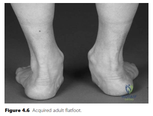
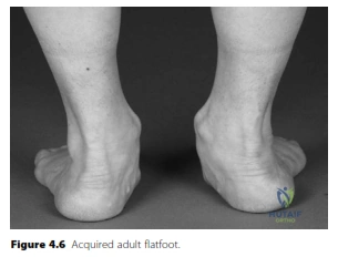
Candidate: The images demonstrate an acquired adult flatfoot deformity, clinically evident by the 'too many toes' sign and loss of the medial longitudinal arch. The most common underlying pathology is Posterior Tibial Tendon Dysfunction (PTTD). I classify this using the Johnson & Strom classification, later modified by Myerson:
- Stage I: Tenosynovitis, no deformity.
- Stage II: Flexible planovalgus deformity, correctable.
- Stage III: Fixed planovalgus deformity.
- Stage IV: Fixed deformity with valgus tilting of the talus in the ankle mortise (deltoid ligament failure).
Failing to mention the 'too many toes' sign or simply stating "flatfoot." Borderline candidates also often forget to explicitly distinguish between flexible (Stage II) and fixed (Stage III) deformity, which is the most critical clinical decision point for choosing between reconstruction and arthrodesis.
Start with a structured description (Heel valgus, forefoot abduction, medial arch loss). Immediately name the pathology (PTTD) and provide the classification system. Use the "Gold Standard" logic: The management is predicated on deformity flexibility and joint integrity. Explicitly mention that Stage IV adds talar tilt, which distinguishes it from Stage III.
You have a patient with a Stage II adult acquired flatfoot. You plan to perform a Flexor Digitorum Longus (FDL) transfer and a medializing calcaneal osteotomy. How do these procedures work, and what specific advice do you give the patient regarding the donor tendon harvest?
Candidate: The medializing calcaneal osteotomy shifts the calcaneal tuberosity medially. This shifts the Achilles insertion medially, transforming it from an everter to an inverter of the hindfoot, and improves the moment arm for the medial column. The FDL transfer acts as a tenodesis to replace the diseased Tibialis Posterior tendon. Regarding the harvest, I counsel the patient that they will maintain toe flexion because of the 'Knot of Henry'—the distal interconnections between the FDL and Flexor Hallucis Longus (FHL) tendons.
Failing to explain the biomechanics of the calcaneal shift. Simply saying "it fixes the heel" is insufficient. A common error is implying that FDL harvest results in loss of toe flexion; failing to mention the Knot of Henry demonstrates a lack of deep anatomical knowledge.
Explain the osteotomy's effect on the Achilles vector (medial shift = inversion) and its role in offloading the medial column. Mention passing the FDL graft from plantar to dorsal through the navicular. Clearly define the Knot of Henry as the fibrous connection allowing the FHL to compensate for FDL loss.
