Relieve Nerve Entrapment Pain with Arthroscopic Release
Key Takeaway
This article provides essential research regarding Relieve Nerve Entrapment Pain with Arthroscopic Release. Nerve entrapment arthroscopic refers to a minimally invasive surgical procedure to relieve pressure on a nerve, commonly the suprascapular nerve. This condition often results from constriction within the suprascapular or spinoglenoid notch, or from ganglion cyst compression. Arthroscopic techniques provide effective management for these constrictions, offering a direct solution when nonoperative treatments fail.
Relieve Nerve Entrapment Pain with Arthroscopic Release
Introduction and Epidemiology
Nerve entrapment syndromes around the shoulder girdle represent a challenging diagnostic and therapeutic dilemma for orthopedic surgeons. While numerous peripheral nerves course through this anatomically complex region, the suprascapular nerve (SSN) is the most frequently implicated in entrapment pathology, often presenting with insidious posterior shoulder pain, weakness, and muscular atrophy. These conditions can significantly impair shoulder function and quality of life, particularly in overhead athletes and individuals engaged in repetitive manual labor.
The incidence of suprascapular nerve entrapment is not precisely known but is increasingly recognized with advancements in diagnostic imaging and electrophysiological studies. It is commonly associated with specific anatomical constraints and biomechanical factors. Etiologies range from congenital variations in the suprascapular notch morphology, space-occupying lesions such as ganglion cysts, to dynamic compression from repetitive overhead activities, trauma, or chronic rotator cuff pathology. The evolution of arthroscopic techniques, pioneered by figures such as Thomas Samson and Laurent Lafosse, has revolutionized the surgical management of these entrapments, offering a minimally invasive approach to achieve effective decompression with reduced morbidity. This chapter provides a comprehensive review of suprascapular nerve entrapment, focusing on its surgical anatomy, pathogenesis, diagnosis, and the detailed arthroscopic surgical approach for its release.
Surgical Anatomy and Biomechanics
A thorough understanding of the suprascapular nerve's intricate course and its relationship to surrounding bony and ligamentous structures is paramount for effective diagnosis and safe surgical decompression. The suprascapular nerve receives its primary neural contributions from the C5 root, with additional, albeit minor, contributions from the C4 and C6 nerve roots. It originates from the upper trunk of the brachial plexus, traversing the supraclavicular fossa.
Suprascapular Notch and Supraglenoid Ligament Complex
The nerve then courses inferiorly and laterally, passing through the suprascapular notch. This notch, located on the superior border of the scapula at the base of the coracoid process, is a critical site of potential entrapment. The notch is typically bridged by the superior transverse scapular ligament (STSL), sometimes referred to as the suprascapular ligament, forming a fibrous tunnel through which the nerve passes. Variations in the morphology of this notch, such as a V-shaped configuration or a completely ossified foramen, are implicated in increasing the risk of compression. Just superior to the notch, the suprascapular artery typically courses, usually passing over the STSL, while the suprascapular nerve passes under it. However, anatomical variations exist, and the artery may also pass inferior to the ligament.
Upon exiting the suprascapular notch, beneath the STSL, the nerve typically divides into two major branches. One branch courses medially to innervate the supraspinatus muscle. The second, larger branch continues across the floor of the supraspinatus fossa of the scapula, traveling towards the junction of the scapular spine and the posterosuperior neck of the glenoid.
Spinoglenoid Notch and Inferior Transverse Scapular Ligament
This second branch then makes a short turn around the bone junction at the base of the scapular spine, specifically at the spinoglenoid notch. This region is another common site of entrapment, especially due to the presence of an inconsistently present, yet clinically significant, inferior transverse scapular ligament (ITSL), also known as the spinoglenoid ligament. This ligament can vary significantly in thickness and tension. After passing through or around the spinoglenoid notch, the nerve travels medially across the superior aspect of the infraspinatus fossa of the scapula, sending multiple branches into the infraspinatus muscle until terminating into its medial aspect.
``
The suprascapular nerve is purely motor to the supraspinatus and infraspinatus muscles, and provides sensory innervation to the posterior capsule of the shoulder, the acromioclavicular joint, and the subacromial bursa. Entrapment can thus manifest as motor weakness (abduction and external rotation) and sensory symptoms (posterior shoulder pain). The close proximity of the nerve to the glenohumeral joint also renders it susceptible to compression from intra-articular pathologies, such as large ganglion cysts originating from labral tears.
Pathogenesis of Entrapment
Nerve entrapment most commonly occurs at the suprascapular notch. Several factors contribute to this:
* Trauma: Direct trauma to the shoulder or indirect forces can lead to localized swelling, hematoma, or fibrosis, causing compression.
* Repetitive Overhead Use: Activities requiring hyper-retraction and protraction of the scapula, such as volleyball or tennis, can cause dynamic traction or compression of the nerve against the STSL, leading to microtrauma and subsequent fibrosis.
* Chronic Rotator Cuff Injuries: Massive or retracted rotator cuff tears, particularly involving the supraspinatus and infraspinatus, can lead to muscle atrophy and changes in scapular kinematics, potentially increasing tension on the nerve or altering the soft tissue environment around it. This is particularly relevant with ganglion cysts.
* Congenital Anomalies: As previously noted, a V-shaped or completely ossified suprascapular notch significantly predisposes individuals to entrapment.
Less common but equally important areas of entrapment include ganglion cyst compression, which can occur in the middle or posterior aspect of the supraspinatus fossa or at the spinoglenoid notch. These cysts are often associated with posterior labral tears, acting as a one-way valve to allow synovial fluid egress into the cyst. A thickened spinoglenoid ligament at the spinoglenoid notch can also independently cause entrapment, often exacerbated by arm movements that stretch the nerve.
Indications and Contraindications
The decision to proceed with arthroscopic suprascapular nerve release requires careful patient selection based on a comprehensive evaluation including clinical history, physical examination, imaging, and electrodiagnostic studies.
Indications for Operative Management
- Persistent Symptoms: Chronic, debilitating posterior shoulder pain, weakness, and/or atrophy of the supraspinatus and/or infraspinatus muscles, unresponsive to a minimum of 3-6 months of dedicated non-operative management.
- Electrodiagnostic Evidence: Confirmatory EMG/NCS findings demonstrating suprascapular nerve denervation or conduction block across the suprascapular or spinoglenoid notch.
- Imaging Findings: MRI demonstrating suprascapular or infraspinatus muscle atrophy, T2 signal changes consistent with denervation edema, or direct visualization of a compressive lesion such as a ganglion cyst or a thickened ligament. CT scan may further delineate bony anomalies of the suprascapular notch.
- Space-Occupying Lesions: Presence of a symptomatic ganglion cyst compressing the nerve, especially if it is large or expanding.
- Failed Non-Operative Management: Failure to improve with a structured rehabilitation program, activity modification, anti-inflammatory medications, and targeted injections.
Contraindications for Operative Management
- Active Infection: Any active local or systemic infection.
- Severe Medical Comorbidities: Uncontrolled medical conditions that significantly increase surgical risk.
- Unrealistic Patient Expectations: Patients with unrealistic expectations regarding pain relief or functional recovery.
- Lack of Clear Evidence of Entrapment: Absence of objective findings on EMG/NCS or imaging studies supporting suprascapular nerve entrapment.
- Acute Neuropathy: Some acute neuropathies may resolve spontaneously; observation may be warranted initially unless there is a clear, treatable compressive lesion.
- Diffuse Brachial Plexopathy: Symptoms attributable to a more proximal or widespread neurological disorder.
Operative vs. Non-Operative Indications
| Feature | Non-Operative Management | Operative Management |
|---|---|---|
| Duration of Symptoms | Acute onset, symptoms < 3 months | Chronic symptoms, > 3-6 months, or progressive neurological deficit |
| Severity of Symptoms | Mild to moderate pain, minimal weakness/atrophy | Severe pain, significant weakness, or progressive muscle atrophy |
| Imaging Findings | No definitive compressive lesion or small, asymptomatic cyst | Large ganglion cyst, clear ligamentous constriction, bony anomaly |
| Electrodiagnostics | Mild or absent denervation, normal conduction | Significant denervation, conduction block, or axon loss |
| Response to Treatment | Initial improvement with rest, PT, NSAIDs, or corticosteroid injection | Failed comprehensive non-operative protocol |
| Associated Pathology | Isolated nerve irritation, no major structural issues | Labral tear with cyst, massive rotator cuff tear, bony impingement |
Pre Operative Planning and Patient Positioning
Thorough preoperative planning is essential for optimizing surgical outcomes and minimizing complications. This includes a detailed review of all imaging, particularly MRI, to precisely localize the suspected site of compression (suprascapular notch, spinoglenoid notch, or ganglion cyst location). Electrodiagnostic studies confirm the diagnosis and provide baseline neurological status.
Anesthesia and Patient Positioning
The patient is typically placed under general anesthesia. Regional anesthesia, such as an interscalene block, can be a useful adjunct for postoperative pain control. Two primary positions are utilized for shoulder arthroscopy: the lateral decubitus position and the beach chair position. For suprascapular nerve decompression, both positions offer advantages.
-
Lateral Decubitus Position:
- The patient is positioned laterally, typically with the affected shoulder superior.
- The torso is secured to the operating table with a beanbag or suction mattress.
- The arm is suspended in approximately 45-70 degrees of abduction and 15 degrees of forward flexion, with 10-15 pounds of traction applied via a traction tower.
- This position allows excellent visualization of the posterior and superior aspects of the shoulder, making it favorable for posterior portals and access to the spinoglenoid notch. It also facilitates a gravitational effect on the brachial plexus, potentially reducing tension during certain maneuvers.
-
Beach Chair Position:
- The patient is positioned in a semi-recumbent, "beach chair" position, similar to a seated position, with the torso elevated 45-60 degrees.
- The head is secured to prevent excessive neck rotation or extension.
- The arm is free-draped, allowing for dynamic positioning throughout the case.
- This position allows for easy conversion to an open procedure if necessary and offers good ergonomic access for the surgeon. Some surgeons find access to the suprascapular notch easier in this position due to gravity's effect on soft tissues.
- Neurovascular monitoring, such as somatosensory evoked potentials (SSEPs), may be considered, especially in complex cases or when prolonged traction is anticipated, though its routine use for SSN release is not standard.
Regardless of positioning, careful padding of all pressure points is critical to prevent iatrogenic nerve compression.
Portal Placement
Standard posterior, anterosuperior, and lateral portals are generally used for diagnostic arthroscopy and initial management of intra-articular pathology. For suprascapular nerve decompression, specific accessory portals are often required to achieve optimal visualization and instrumentation:
- Standard Posterior Portal (Diagnostic): Approximately 2 cm medial and 1 cm inferior to the posterior corner of the acromion.
- Anterosuperior Portal: Established under direct visualization, anterior to the biceps tendon, approximately 1 cm medial to the lateral border of the acromion.
- Posterolateral Portal (Working): Often 2-3 cm lateral to the posterior portal, directly through the posterior deltoid. This portal is useful for instrumentation when working in the suprascapular notch area.
- Superior or Anterolateral Portal: May be used to facilitate specific angles for instrumentation.
- Neviaser Portal: This accessory portal, located slightly superior and medial to the posterior portal, can provide excellent access for viewing and instrumenting the suprascapular notch, especially when releasing the superior transverse scapular ligament. It typically passes just anterior to the scapular spine, offering a direct trajectory to the notch.
Detailed Surgical Approach and Technique
The arthroscopic release of the suprascapular nerve is a technically demanding procedure that requires meticulous attention to anatomical landmarks and neurovascular structures. The approach varies slightly depending on whether the entrapment is at the suprascapular notch or the spinoglenoid notch, or due to a ganglion cyst.
Diagnostic Arthroscopy
The procedure commences with a thorough diagnostic arthroscopy of the glenohumeral joint and subacromial space. This is critical to identify and address any coexisting intra-articular pathologies, such as labral tears, rotator cuff tears, or loose bodies, which may contribute to pain or cyst formation. If a ganglion cyst is present, its origin (e.g., posterior labral tear) should be identified and repaired.
Arthroscopic Suprascapular Notch Decompression
- Preparation of the Subacromial Space: Perform a complete bursectomy of the subacromial bursa to adequately visualize the superior aspect of the scapula and the underlying suprascapular notch.
 - Identification of Landmarks: Using a standard posterior portal for the arthroscope and an anterolateral or Neviaser portal for instrumentation, identify the suprascapular notch. Key landmarks include the base of the coracoid process anteriorly and the superior border of the scapula posteriorly. The coracoid process can be palpated extra-articularly and confirmed arthroscopically.
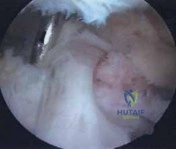
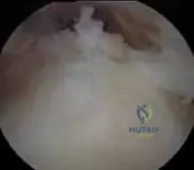
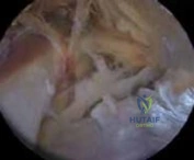 - Visualization of the Ligament: The superior transverse scapular ligament (STSL) typically appears as a dense, white fibrous band spanning the suprascapular notch. It can be challenging to visualize initially due to overlying soft tissues. Careful debridement with a shaver or radiofrequency wand helps expose it. The suprascapular nerve lies directly beneath this ligament. Great care must be taken to avoid damaging the suprascapular artery, which typically crosses superior to the ligament but can be variable.
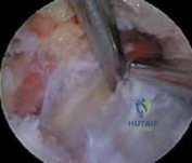
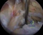
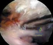
`` - Ligament Release: Using a blunt probe, palpate the ligament to confirm its tension and position. Then, with a hook knife or a specifically designed arthroscopic ligament release instrument, carefully divide the STSL. The release should begin on the anterior (coracoid) side of the ligament and proceed posteriorly. It is crucial to perform this under direct visualization, ensuring that the nerve is not inadvertently transected. Some surgeons prefer to use a nerve hook to gently elevate the nerve from the undersurface of the ligament prior to division. A blunt-tipped instrument or probe can be used to confirm complete release of the ligament by gently sweeping beneath the now-divided ligament, ensuring no residual tension on the nerve. The nerve should be seen pulsating freely once decompressed.
Arthroscopic Spinoglenoid Notch Decompression
- Access and Visualization: The spinoglenoid notch is located at the posterolateral aspect of the scapular spine, medial to the glenoid. This area is usually accessed with the arthroscope through a posterior or Neviaser portal, and instrumentation through a separate posterolateral portal. A thorough subacromial bursectomy provides the necessary working space.
- Identification of the Spinoglenoid Ligament: The inferior transverse scapular ligament (ITSL) or spinoglenoid ligament, when present, bridges the spinoglenoid notch. It lies deep to the infraspinatus muscle and can often be obscured by fatty tissue or inflammatory changes. Careful dissection and debridement with a shaver or radiofrequency wand are necessary.
- Ganglion Cyst Decompression: If a ganglion cyst is compressing the nerve at this location, it is typically decompressed first. This involves incising the cyst wall and aspirating its contents. The base of the cyst should then be debrided, and if a communication with a posterior labral tear is identified, the labral tear should be repaired to prevent recurrence of the cyst.
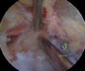
 - Ligament Release (if present): If a thickened spinoglenoid ligament is identified as a primary source of compression, it is carefully released using a hook knife. Similar to the STSL release, the nerve should be identified and protected throughout this maneuver. The suprascapular nerve often lies directly beneath this ligament as it enters the infraspinatus fossa.
``
Technical Pearls and Pitfalls
- Anatomical Variations: Always be prepared for anatomical variations in nerve course and ligamentous structures.
- Neurovascular Protection: The suprascapular artery and vein run in close proximity to the nerve at both notch locations. Meticulous technique, controlled shaver use, and careful electrosurgical application are crucial.
- Complete Release: Ensure complete release of the compressive structure. Incomplete release may lead to persistent symptoms.
- Cyst Management: For ganglion cysts, simply aspirating the cyst is often insufficient; identifying and addressing the communicating intra-articular pathology (e.g., labral tear) is paramount to prevent recurrence.
- Dynamic Assessment: After release, carefully probe the nerve to ensure it moves freely and is fully decompressed.
- Thermal Protection: When using radiofrequency devices, maintain irrigation flow and keep the tip away from the nerve to prevent thermal injury.
Complications and Management
While arthroscopic suprascapular nerve release is generally safe, potential complications can arise, necessitating vigilant surgical technique and thorough postoperative care.
Common Complications and Salvage Strategies
| Complication | Incidence (Approx.) | Salvage Strategies The arthroscopic release of the suprascapular nerve is a technically demanding procedure that requires meticulous attention to anatomical landmarks and neurovascular structures. The approach varies slightly depending on whether the entrapment is at the suprascapular notch or the spinoglenoid notch, or due to a ganglion cyst.
Diagnostic Arthroscopy
The procedure commences with a thorough diagnostic arthroscopy of the glenohumeral joint and subacromial space. This is critical to identify and address any coexisting intra-articular pathologies, such as labral tears, rotator cuff tears, or loose bodies, which may contribute to pain or cyst formation. If a ganglion cyst is present, its origin (e.g., posterior labral tear) should be identified and repaired.

Arthroscopic Suprascapular Notch Decompression
- Preparation of the Subacromial Space: Perform a complete bursectomy of the subacromial bursa to adequately visualize the superior aspect of the scapula and the underlying suprascapular notch. This step is crucial for clearing soft tissue that may obscure key anatomical landmarks and restrict instrument maneuverability.
 - Identification of Landmarks: Using a standard posterior portal for the arthroscope and an anterolateral or Neviaser portal for instrumentation, identify the suprascapular notch. Key landmarks include the base of the coracoid process anteriorly and the superior border of the scapula posteriorly. The coracoid process can be palpated extra-articularly and confirmed arthroscopically by tracing the anterior aspect of the scapular spine. The suprascapular notch lies at the medial aspect of the base of the coracoid.
 - Visualization of the Ligament: The superior transverse scapular ligament (STSL) typically appears as a dense, white fibrous band spanning the suprascapular notch. It can be challenging to visualize initially due to overlying soft tissues. Careful debridement with a shaver or radiofrequency wand helps expose it. The suprascapular nerve lies directly beneath this ligament. Great care must be taken to avoid damaging the suprascapular artery and vein, which typically cross superior to the ligament but can be variable. The identification of these vessels serves as an important guide to the location of the nerve.
`` - Ligament Release: Using a blunt probe, palpate the ligament to confirm its tension and position. Then, with a hook knife or a specifically designed arthroscopic ligament release instrument, carefully divide the STSL. The release should begin on the anterior (coracoid) side of the ligament and proceed posteriorly, ensuring that the entire width of the ligament is transected. It is crucial to perform this under direct visualization, ensuring that the nerve is not inadvertently transected. Some surgeons prefer to use a nerve hook to gently elevate the nerve from the undersurface of the ligament prior to division. A blunt-tipped instrument or probe can be used to confirm complete release of the ligament by gently sweeping beneath the now-divided ligament, ensuring no residual tension on the nerve. The nerve should be seen pulsating freely once decompressed, indicating successful release.

Arthroscopic Spinoglenoid Notch Decompression
- Access and Visualization: The spinoglenoid notch is located at the posterolateral aspect of the scapular spine, medial to the glenoid. This area is usually accessed with the arthroscope through a posterior or Neviaser portal, and instrumentation through a separate posterolateral portal. A thorough subacromial bursectomy provides the necessary working space. The infraspinatus muscle belly needs to be gently retracted or split to access the deep structures of the spinoglenoid notch.
- Identification of the Spinoglenoid Ligament and Cysts: The inferior transverse scapular ligament (ITSL) or spinoglenoid ligament, when present, bridges the spinoglenoid notch. It lies deep to the infraspinatus muscle and can often be obscured by fatty tissue or inflammatory changes. Careful dissection and debridement with a shaver or radiofrequency wand are necessary. Ganglion cysts are frequently found in this region, originating from posterior-superior labral tears.
- Ganglion Cyst Decompression: If a ganglion cyst is compressing the nerve at this location, it is typically decompressed first. This involves incising the cyst wall and aspirating its mucinous contents. The base of the cyst should then be debrided, and if a communication with a posterior labral tear is identified, the labral tear should be repaired using arthroscopic suture anchors to prevent recurrence of the cyst. Failure to address the intra-articular origin significantly increases recurrence rates.
 - Ligament Release (if present): If a thickened spinoglenoid ligament is identified as a primary source of compression, independent of or in conjunction with a cyst, it is carefully released using a hook knife. Similar to the STSL release, the suprascapular nerve should be identified and protected throughout this maneuver. The nerve often lies directly beneath this ligament as it enters the infraspinatus fossa. Confirmation of free nerve movement after release is essential.
Technical Pearls and Pitfalls
- Anatomical Variations: Always be prepared for anatomical variations in nerve course, ligamentous structures, and vascular anatomy. Preoperative MRI review is crucial for identifying these.
- Neurovascular Protection: The suprascapular artery and vein run in close proximity to the nerve at both notch locations. Meticulous technique, controlled shaver use with blunt tips, and careful electrosurgical application with low settings and constant irrigation are crucial to prevent thermal or mechanical injury. Identifying the artery and vein can often help locate the nerve.
- Complete Release: Ensure complete release of the compressive structure. Incomplete release, especially of the entire width of the STSL, may lead to persistent symptoms.
- Cyst Management: For ganglion cysts, simply aspirating the cyst is often insufficient; identifying and addressing the communicating intra-articular pathology (e.g., labral tear) is paramount to prevent recurrence. A thorough debridement of the cyst wall is also recommended.
- Dynamic Assessment: After release, carefully probe the nerve to ensure it moves freely within its newly decompressed canal and that there is no residual impingement with passive shoulder motion if possible.
- Thermal Protection: When using radiofrequency devices, maintain high irrigation flow, avoid direct contact with nerve tissue, and minimize power settings to prevent thermal injury.
- Portal Selection: Optimize portal placement to allow for direct visualization and instrument access without excessive angulation, which can hinder delicate maneuvers. The Neviaser portal can be particularly useful for direct visualization of the suprascapular notch.
Complications and Management
While arthroscopic suprascapular nerve release is generally considered a safe procedure, it is not without potential complications. Awareness of these risks and preparedness for their management are critical.
Common Complications and Salvage Strategies
| Complication | Incidence (Approx.) | Salvage Strategies ## Rotorestrictions
| Feature | Non-Operative Management | Operative Management |
| :-------------------------- | :-------------------------------------------------------------- | :----------------------------------------------------------- |
| Duration of Symptoms | Acute onset, symptoms < 3 months | Chronic symptoms, > 3-6 months, or progressive neurological deficit |
| Severity of Symptoms | Mild to moderate pain, minimal weakness/atrophy | Severe pain, significant weakness, or progressive muscle atrophy |
| Imaging Findings | No definitive compressive lesion or small, asymptomatic cyst | Large ganglion cyst, clear ligamentous constriction, bony anomaly |
| Electrodiagnostics | Mild or absent denervation, normal conduction | Significant denervation, conduction block, or axon loss |
| Response to Treatment | Initial improvement with rest, PT, NSAIDs, or corticosteroid injection | Failed comprehensive non-operative protocol |
| Associated Pathology | Isolated nerve irritation, no major structural issues | Labral tear with cyst, massive rotator cuff tear, bony impingement |
Pre Operative Planning and Patient Positioning
Thorough preoperative planning is essential for optimizing surgical outcomes and minimizing complications. This includes a detailed review of all imaging, particularly MRI, to precisely localize the suspected site of compression (suprascapular notch, spinoglenoid notch, or ganglion cyst location). Electrodiagnostic studies confirm the diagnosis and provide baseline neurological status, which is crucial for documenting improvement postoperatively.
Anesthesia and Patient Positioning
The patient is typically placed under general anesthesia. Regional anesthesia, such as an interscalene block, can be a useful adjunct for postoperative pain control, facilitating early rehabilitation and pain management. Two primary positions are utilized for shoulder arthroscopy: the lateral decubitus position and the beach chair position. For suprascapular nerve decompression, both positions offer distinct advantages.
-
Lateral Decubitus Position:
- The patient is positioned laterally, typically with the affected shoulder superior.
- The torso is secured to the operating table with a beanbag or suction mattress, ensuring stability.
- The arm is suspended in approximately 45-70 degrees of abduction and 15 degrees of forward flexion, with 10-15 pounds of traction applied via a traction tower. This provides distraction of the glenohumeral joint and opens the subacromial space.
- This position allows excellent visualization of the posterior and superior aspects of the shoulder, making it favorable for posterior portals and access to the spinoglenoid notch. It also facilitates a gravitational effect on the brachial plexus, potentially reducing tension during certain maneuvers and allowing the arm to be manipulated by an assistant to tension or relax surrounding structures.
-
Beach Chair Position:
- The patient is positioned in a semi-recumbent, "beach chair" position, similar to a seated position, with the torso elevated 45-60 degrees.
- The head is secured in a neutral position to prevent excessive neck rotation or extension, which could stretch the brachial plexus.
- The arm is free-draped, allowing for dynamic positioning throughout the case without the need for a traction tower.
- This position allows for easy conversion to an open procedure if necessary and offers good ergonomic access for the surgeon, especially for anterior and superior work. Some surgeons find access to the suprascapular notch easier in this position due to gravity's effect on soft tissues, which can retract them inferiorly.
- Neurovascular monitoring, such as somatosensory evoked potentials (SSEPs), may be considered, especially in complex cases or when prolonged traction is anticipated, though its routine use for SSN release is not universally standard.
Regardless of positioning, careful padding of all pressure points, including the axilla, elbow, and peroneal nerve at the fibular head, is critical to prevent iatrogenic nerve compression or injury.
Portal Placement
Standard posterior, anterosuperior, and lateral portals are generally used for diagnostic arthroscopy and initial management of intra-articular pathology. For suprascapular nerve decompression, specific accessory portals are often required to achieve optimal visualization and instrumentation:
- Standard Posterior Portal (Diagnostic): Approximately 2 cm medial and 1 cm inferior to the posterior corner of the acromion. This provides the initial viewing portal for glenohumeral and subacromial diagnostics.
- Anterosuperior Portal: Established under direct visualization from the posterior portal, anterior to the biceps tendon, approximately 1 cm medial to the lateral border of the acromion. Useful for instrumentation within the joint.
- Posterolateral Portal (Working): Often 2-3 cm lateral to the posterior portal, directly through the posterior deltoid. This portal is particularly useful for instrumentation when working in the suprascapular notch area and for managing spinoglenoid cysts.
- Superior or Anterolateral Portal: May be used to facilitate specific angles for instrumentation, particularly when working in the subacromial space towards the suprascapular notch.
- Neviaser Portal: This accessory portal, located slightly superior and medial to the standard posterior portal, often just anterior to the scapular spine, can provide excellent access for viewing and instrumenting the suprascapular notch directly. It offers a more direct trajectory to release the superior transverse scapular ligament. It is typically established by bringing a spinal needle from outside-in to confirm optimal placement under arthroscopic guidance from the posterior portal.
Detailed Surgical Approach and Technique
The arthroscopic release of the suprascapular nerve is a technically demanding procedure that requires meticulous attention to anatomical landmarks and neurovascular structures. The approach varies slightly depending on whether the entrapment is at the suprascapular notch or the spinoglenoid notch, or due to a ganglion cyst.
Diagnostic Arthroscopy
The procedure commences with a thorough diagnostic arthroscopy of the glenohumeral joint and subacromial space. This is critical to identify and address any coexisting intra-articular pathologies, such as labral tears, rotator cuff tears, or loose bodies, which may contribute to pain or cyst formation. If a ganglion cyst is present, its origin (e.g., posterior labral tear) should be identified and repaired.

Arthroscopic Suprascapular Notch Decompression
- Preparation of the Subacromial Space: Perform a complete diagnostic subacromial bursectomy to adequately visualize the superior aspect of the scapula and the underlying suprascapular notch. This initial debridement is crucial for clearing obscuring soft tissue and creating a sufficient working space for instrument manipulation.
 - Identification of Landmarks: Using a standard posterior portal for the arthroscope and an anterolateral or Neviaser portal for instrumentation, carefully identify the suprascapular notch. Key anatomical landmarks include the base of the coracoid process anteriorly and the superior border of the scapula posteriorly. The coracoid process can be palpated extra-articularly and then confirmed arthroscopically by tracing the anterior aspect of the scapular spine to its base. The suprascapular notch lies just medial to the base of the coracoid process, along the superior border of the scapula.
 - Visualization of the Ligament: The superior transverse scapular ligament (STSL) typically presents as a dense, white fibrous band spanning the suprascapular notch. Initial visualization can be challenging due to overlying fat, fibrous tissue, or the suprascapular artery and vein. Careful debridement of these superficial soft tissues using a shaver or a radiofrequency wand (with extreme caution to avoid thermal injury to vascular or neural structures) helps to expose the STSL. It is imperative to identify the suprascapular artery and vein, which usually cross superior to the STSL, serving as critical guides to the underlying nerve. However, anatomical variations can occur where these vessels pass with the nerve. The suprascapular nerve lies directly beneath this ligament.
`` - Ligament Release: Using a blunt probe or a nerve hook, gently palpate the ligament to confirm its tension and position relative to the underlying nerve. A nerve hook can be introduced through a separate portal to gently elevate the nerve from the undersurface of the ligament, providing an added layer of protection. Then, with a hook knife or a specifically designed arthroscopic ligament release instrument, carefully divide the STSL. The release should begin on the anterior (coracoid) side of the ligament and proceed posteriorly, ensuring that the entire width of the ligament is transected to achieve complete decompression. It is crucial to perform this under direct visualization, maintaining absolute certainty of the instrument's position relative to the nerve. After transection, a blunt-tipped instrument can be swept beneath the now-divided ligament to ensure no residual tension or fibrous bands remain. The suprascapular nerve should be observed to pulsate freely once successfully decompressed, indicating a successful release.

Arthroscopic Spinoglenoid Notch Decompression
- Access and Visualization: The spinoglenoid notch is located at the posterolateral aspect of the scapular spine, medial to the posterior glenoid rim. This area is typically accessed with the arthroscope through a posterior or Neviaser portal, and instrumentation via a separate posterolateral portal. A thorough subacromial bursectomy is performed to provide adequate working space. The infraspinatus muscle belly needs to be gently retracted or, if necessary, carefully split along its fibers to visualize the deep structures of the spinoglenoid notch.
- Identification of the Spinoglenoid Ligament and Cysts: The inferior transverse scapular ligament (ITSL), also known as the spinoglenoid ligament, when present, bridges the spinoglenoid notch. It lies deep to the infraspinatus muscle and can often be obscured by fatty tissue, inflammatory changes, or the presence of a ganglion cyst. Careful and precise dissection and debridement with a blunt shaver or radiofrequency wand are necessary to expose the nerve and any compressive structures. Ganglion cysts are frequently encountered in this region, typically originating from posterior-superior labral tears.
- Ganglion Cyst Decompression: If a ganglion cyst is compressing the nerve at this location, it is the primary target for decompression. This involves carefully incising the cyst wall and aspirating its mucinous contents. The base of the cyst should then be thoroughly debrided, and if a communicating posterior labral tear is identified, it must be repaired using arthroscopic suture anchors. Addressing the intra-articular origin is paramount to preventing recurrence of the cyst and subsequent nerve compression.
 - Ligament Release (if present): If a thickened spinoglenoid ligament is identified as a primary source of compression, either independently or in conjunction with a cyst, it is carefully released. A hook knife is typically used, with strict attention to protecting the underlying suprascapular nerve. The nerve often lies directly beneath this ligament as it enters the infraspinatus fossa to innervate the infraspinatus muscle. Confirmation of free nerve movement after the release is essential to ensure complete decompression.

Technical Pearls and Pitfalls
- Anatomical Variations: Surgeons must be prepared for the wide range of anatomical variations in nerve course, ligamentous structures, and vascular anatomy. Comprehensive preoperative MRI review is crucial for identifying these individual patient specificities.
- Neurovascular Protection: The suprascapular artery and vein run in close proximity to the nerve at both notch locations. Meticulous technique, controlled shaver use with blunt tips, and judicious radiofrequency application with low settings and constant irrigation are paramount to prevent thermal or mechanical injury. Identifying the artery and vein often serves as a critical guide to the underlying nerve.
- Complete Release: Ensure complete release of the compressive structure. Incomplete release, particularly of the entire width of the STSL, may lead to persistent or recurrent symptoms.
- Cyst Management: For ganglion cysts, simple aspiration of the cyst is often insufficient. Identifying and addressing the communicating intra-articular pathology (e.g., labral tear) is paramount to prevent recurrence. Thorough debridement of the cyst wall is also recommended.
- Dynamic Assessment: After release, carefully probe the nerve to ensure it moves freely within its newly decompressed canal and that there is no residual impingement with passive shoulder motion, if feasible.
- Thermal Protection: When using radiofrequency devices, maintain high irrigation flow, avoid direct contact with nerve tissue, and minimize power settings to prevent thermal injury.
- Portal Selection: Optimize portal placement to allow for direct visualization and instrument access without excessive angulation. The Neviaser portal can be particularly useful for direct visualization of the suprascapular notch due to its favorable trajectory.
Complications and Management
While arthroscopic suprascapular nerve release is generally considered a safe procedure with a high success rate, it is not without potential complications. Awareness of these risks and preparedness for their management are critical for orthopedic surgeons.
Common Complications and Salvage Strategies
| Complication | Incidence (Approx.) | Salvage Strategies | Nerve Injury (Suprascapular nerve) | Rare (direct trauma during release) | Acute: Re-exploration, nerve repair/grafting if transected. If only neuropraxia, watchful waiting, electrodiagnostic follow-up.
Chronic: Nerve grafting or palliative tendon transfer if no recovery after sufficient time. |
| Vascular Injury | Rare (suprascapular artery/vein) | Direct pressure with instruments. If bleeding persists, consider electrocautery or packing. Open exploration for larger hematomas or uncontrolled bleeding. |
| Inadequate Decompression | 5-10% (persistent symptoms) | Careful re-evaluation with imaging and electrodiagnostics. Revision arthroscopy to ensure complete ligamentous release, address missed cyst, or new area of compression. Consider open conversion if arthroscopic limitations. |
| Recurrence of Ganglion Cyst | 10-20% (if primary cause, e.g., labral tear, not addressed) | Re-evaluation for underlying labral pathology. Revision arthroscopy for cyst decompression and definitive labral repair. |
| Infection (Superficial/Deep) | <1% | Superficial: Oral antibiotics, local wound care.
Deep: Arthroscopic irrigation and debridement, targeted intravenous antibiotics based on culture, hardware removal if present and infected. |
| Shoulder Stiffness | 5-15% | Aggressive physical therapy, ROM exercises. Consideration of manipulation under anesthesia or arthroscopic capsular release if conservative measures fail. |
| Persistent Pain | Variable (multifactorial) | Thorough diagnostic workup for other pain generators (e.g., missed pathology, nerve irritation from scar tissue, psychological factors). Pain management referral. Revision surgery if clear treatable cause. |
| Axillary Nerve Injury | Rare (from portal placement) | If identified acutely, nerve repair/grafting. If delayed, neurolysis, watchful waiting, or palliative tendon transfer (e.g., latissimus dorsi transfer) for chronic deficits. |
| Brachial Plexus Injury | Rare (from traction or positioning) | Remove traction immediately, reposition patient. Postoperative neurological evaluation, electrodiagnostics, neurological consultation. Supportive care. |
| CRPS (Complex Regional Pain Syndrome) | Rare (<1%) | Multidisciplinary approach: early recognition, pain management specialist referral, physical therapy, pharmacological agents (gabapentin, tricyclics), stellate ganglion blocks. |
General Management Principles for Complications
- Prevention: Meticulous surgical technique, precise portal placement, careful anatomical identification, and appropriate instrumentation are the cornerstones of complication prevention.
- Early Recognition: Prompt identification of complications is critical. Surgeons and surgical teams should be vigilant for signs such as unexpected neurological deficits, excessive bleeding, or signs of infection.
- Thorough Workup: When a complication is suspected, a thorough diagnostic workup is necessary. This may involve repeat imaging (MRI, CT, ultrasound), electrodiagnostic studies (EMG/NCS), laboratory tests (CBC, CRP, ESR), or neurological consultation.
- Patient Communication: Open and honest communication with the patient regarding any complications, their implications, and the management plan is paramount for maintaining trust and facilitating recovery.
- Multidisciplinary Approach: Management of complex complications, particularly nerve injuries or chronic pain, often benefits from a multidisciplinary approach involving neurologists, pain management specialists, and rehabilitation therapists.
Post Operative Rehabilitation Protocols
Postoperative rehabilitation is an integral component of successful outcomes following arthroscopic suprascapular nerve release. The protocol aims to minimize pain, protect the decompressed nerve, restore range of motion, and gradually strengthen the shoulder musculature, ultimately facilitating a return to function and activity. The protocol must be individualized based on the patient's preoperative deficits, concomitant procedures (e.g., labral repair, rotator cuff repair), and the surgeon's preference.
Phase I: Protection and Early Motion (Weeks 0-4)
- Immobilization: The arm is typically placed in a sling for comfort and protection for 1-2 weeks, particularly if concomitant procedures such as labral repair were performed. For isolated nerve release, immobilization may be shorter or omitted, with emphasis on pain control.
- Pain and Swelling Management: Cryotherapy, NSAIDs (if not contraindicated), and appropriate analgesics.
- Passive Range of Motion (PROM): Gentle passive range of motion exercises for the shoulder within pain-free limits. Emphasis on forward flexion and external rotation. Avoidance of positions that may place undue tension on the suprascapular nerve, such as excessive horizontal adduction combined with internal rotation (though this is less common for SSN).
- Pendulum Exercises: Gentle pendulum exercises to encourage early motion and reduce stiffness.
- Elbow, Wrist, Hand ROM: Active range of motion for the elbow, wrist, and hand to prevent stiffness and maintain circulation.
- Scapular Stabilization: Initiate gentle isometric scapular setting exercises, focusing on periscapular muscle activation without significant shoulder motion.
Phase II: Gradual Strengthening and Active Range of Motion (Weeks 4-12)
- Discontinue Sling: As comfort allows and with satisfactory pain control.
- Active Range of Motion (AROM): Progress from PROM to AROM for the shoulder, gradually increasing the arc of motion.
- Isometrics: Initiate pain-free isometric exercises for the rotator cuff (supraspinatus, infraspinatus) and deltoid.
- Light Resistance Exercises: Begin light resistance exercises with elastic bands or light weights, targeting the supraspinatus (empty can/full can), infraspinatus (external rotation), and scapular stabilizers (rows, prone extension).
- Neuromuscular Control: Focus on exercises that improve proprioception and dynamic stability of the shoulder girdle.
- Cardiovascular Conditioning: Maintain general fitness with activities that do not involve the affected shoulder.
Phase III: Advanced Strengthening and Return to Activity (Weeks 12-24)
- Progressive Resistance: Gradually increase the intensity and resistance of strengthening exercises for the rotator cuff, deltoid, and periscapular musculature.
- Functional Exercises: Incorporate functional, sport-specific, or work-specific exercises that mimic required movements.
- Plyometrics (if applicable): For athletes, begin progressive plyometric drills and overhead throwing mechanics (if appropriate for their sport).
- Return to Activity: Gradual return to light activities and work, progressing to full activities and sports as strength, motion, and confidence improve. This phase is highly individualized and requires close collaboration between the surgeon, physical therapist, and patient. Electrodiagnostic studies may be repeated at 6-12 months post-op to document nerve recovery.
Key Considerations
- Concomitant Procedures: If a rotator cuff repair or labral repair was performed, the rehabilitation protocol will be modified to protect the repaired tissues, often requiring a slower progression of motion and strengthening.
- Nerve Recovery: Nerve recovery is typically slow and can take several months to a year, or even longer, depending on the severity of the preoperative compression and the extent of nerve damage. Patients should be counseled on realistic expectations regarding the timeline for symptom improvement and functional recovery. Muscle atrophy may take substantial time to resolve, and in some cases, may not fully recover.
- Pain Monitoring: Rehabilitation should always be guided by the patient's pain levels. Any exacerbation of pain, particularly nerve-related symptoms, should prompt a reassessment and potential modification of the protocol.
Summary of Key Literature and Guidelines
The understanding and management of suprascapular nerve entrapment have evolved significantly with advancements in diagnostic capabilities and arthroscopic techniques. Current literature supports arthroscopic release as a safe and effective treatment option for symptomatic cases refractory to conservative management.
Early studies primarily focused on open decompression, demonstrating good results but with higher morbidity. The transition to arthroscopic techniques, particularly popularized by authors like Lafosse and Samson, has shown comparable efficacy with the added benefits of minimally invasive surgery, including reduced postoperative pain, shorter hospital stays, and quicker recovery.
Key findings from the literature include:
- Efficacy: Multiple case series and retrospective reviews consistently report good to excellent outcomes with arthroscopic SSN decompression, with significant improvement in pain, function, and reduction in muscle atrophy in a majority of patients. Clinical improvement in pain is often seen earlier than objective neurological recovery.
- Concomitant Pathology: The arthroscopic approach allows for simultaneous treatment of associated intra-articular pathologies, such as labral tears (which can lead to ganglion cysts) and rotator cuff pathology, which is a significant advantage. Addressing the underlying cause of a ganglion cyst (e.g., repairing a posterior labral tear) is crucial for preventing recurrence.
- Predictors of Outcome: Factors associated with better outcomes generally include shorter duration of symptoms, younger patient age, absence of severe muscle atrophy, and less severe nerve damage on electrodiagnostic studies. Conversely, long-standing symptoms with severe, irreversible muscle atrophy may lead to less complete functional recovery, although pain relief is often still achievable.
- Comparison to Open Surgery: While direct randomized controlled trials comparing arthroscopic and open techniques are limited due to the rarity of the condition, arthroscopic techniques are generally favored due to their less invasive nature, excellent visualization, and ability to address associated intra-articular pathology. Outcomes appear comparable or superior due to reduced surgical trauma.
- Diagnostic Modalities: MRI is highly valuable for identifying compressive lesions (cysts, thickened ligaments) and assessing muscle edema/atrophy. Electrodiagnostic studies (EMG/NCS) are considered the gold standard for confirming nerve involvement and localization of the lesion, providing objective evidence of denervation or conduction block.
- Long-Term Results: Long-term follow-up studies typically show sustained improvements, though some patients may experience residual weakness or mild pain, especially in cases of severe preoperative atrophy. The potential for nerve regeneration, even in chronic cases, underscores the importance of decompression.
Current guidelines generally advocate for a trial of conservative management for at least 3-6 months. Surgical intervention is reserved for patients with persistent, debilitating symptoms, progressive neurological deficits, or clear evidence of a compressive lesion (e.g., a large ganglion cyst or bony impingement) confirmed by imaging and electrophysiological studies. Arthroscopic techniques are now the preferred method for surgical decompression, offering a comprehensive solution for both intra-articular and extra-articular sources of suprascapular nerve entrapment. Future research will likely continue to refine surgical techniques, improve patient selection criteria, and further delineate long-term neurological recovery patterns.
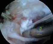
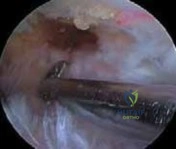
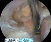
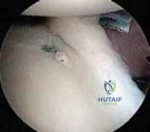
Clinical & Radiographic Imaging