Superficial Radial Nerve Injury: A Clinical Case Study on Diagnosis in the Anatomical Snuffbox
Key Takeaway
Superficial radial nerve (SRN) injury is diagnosed via meticulous sensory assessment in its distribution, especially the dorsal thumb web space. Intact radial nerve motor function rules out deeper involvement. Key diagnostic tests include two-point discrimination, light touch, and pinprick sensation, comparing with the contralateral hand to confirm isolated sensory loss after wrist laceration.
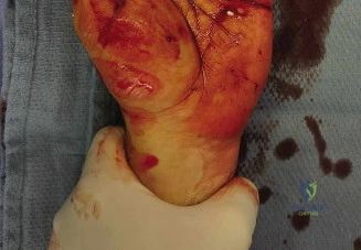
A 34-year-old male presents with a laceration to the dorsoradial aspect of the wrist following a glass injury. You note complete anesthesia over the dorsal thumb and first web space. Given this clinical picture, what is your immediate diagnostic priority, and how would you document the neurological deficit?
Candidate: I would immediately perform a detailed neurological exam before any local anesthesia is administered. I would test for Tinel’s sign at the site and map the sensory loss. I would document this as a Sunderland Grade V (neurotmesis) of the superficial radial nerve based on the loss of sensation in its autonomous distribution.
Candidates often jump straight to "I would order an MRI to confirm the nerve injury" or perform a rapid assessment after giving local lidocaine, which renders the sensory exam invalid. They also often fail to mention checking for concomitant tendon or arterial injuries.
The candidate must emphasize: 1) Clinical assessment PRIOR to any regional/local block. 2) Systematic exclusion of motor deficits (PIN injury) and vascular status (Allen's test). 3) Precise sensory mapping (monofilament/2-point discrimination). 4) Classification of the nerve injury (Sunderland V) and a high index of suspicion for associated structure damage (cephalic vein, radial artery, extensor tendons).
You have decided to proceed to the operating theatre for exploration and repair. Describe your technical approach to the nerve, focusing on stump preparation and coaptation strategy.
Candidate: I would use loupe magnification and then the operating microscope. I must resect the nerve ends back to healthy fascicles until I see "pouting." I will perform an epineurial repair using 9-0 nylon, ensuring the repair is tension-free, and confirm topographical alignment using vasa nervorum as landmarks.
Failing to emphasize the importance of identifying healthy, bleeding (pouting) fascicles. Many candidates also forget to mention the critical importance of keeping the repair tension-free, which often leads to using interpositional grafting if primary closure would be under tension.
A structured answer: 1) Exposure via "lazy-S" incision to avoid scar contracture. 2) Identification of healthy proximal/distal stumps. 3) Serial trimming to healthy fascicular architecture. 4) Microsurgical epineurial neurorrhaphy (9-0 monofilament) with 180-degree stay sutures. 5) Mention of tension-free coaptation and fibrin glue adjuncts.
