Mastering Peripheral Nerve Repair: Primary, Delayed Primary, and Secondary Strategies
Key Takeaway
The timing of peripheral nerve repair is a critical determinant of functional recovery. Primary repair is indicated for clean, sharp transections, while secondary repair is reserved for crush, traction, or highly contaminated injuries requiring scar demarcation. This guide details the evidence-based indications, biomechanical principles of tension-free coaptation, step-by-step surgical techniques, and postoperative rehabilitation protocols essential for optimizing motor and sensory outcomes in peripheral nerve surgery.
Comprehensive Introduction and Patho-Epidemiology
The management of peripheral nerve injuries remains one of the most technically demanding, biologically complex, and prognostically unpredictable challenges in operative orthopaedics and reconstructive microsurgery. The ultimate goal of peripheral nerve repair is to restore functional motor execution and protective sensory feedback by facilitating the precise, topographically accurate regeneration of axons across the zone of injury into the distal endoneurial tubes. Achieving this requires not only meticulous microsurgical technique but also a profound understanding of the cellular and molecular events that govern neurodegeneration and subsequent regeneration. The surgeon is constantly racing against a biological clock, balancing the optimal local wound environment against the progressive, irreversible degradation of distal target organs.
The pathophysiology of peripheral nerve transection initiates a highly orchestrated cascade of cellular events both proximal and distal to the zone of injury. Distally, the severed axon undergoes Wallerian degeneration, a process characterized by the granular disintegration of the axonal cytoskeleton and myelin sheath within 48 to 96 hours post-injury. Concurrently, resident Schwann cells dedifferentiate, proliferate, and phagocytose the myelin debris in conjunction with recruited macrophages. These activated Schwann cells then align longitudinally to form the Bands of Büngner, which serve as the critical biological conduits that guide regenerating proximal axons toward their designated end-organs. Proximally, the neuronal cell body undergoes chromatolysis, an anabolic shift characterized by the dispersion of Nissl substance and upregulation of structural proteins (e.g., tubulin, actin) and neurotrophic factors required to drive axonal sprouting.
Epidemiologically, peripheral nerve injuries are a significant source of long-term morbidity, affecting approximately 2% to 5% of all patients presenting with extremity trauma. The upper extremity is disproportionately affected, accounting for over 70% of all peripheral nerve lesions, with the radial, ulnar, and median nerves most frequently implicated. Mechanisms of injury range from low-energy sharp lacerations (e.g., glass, scalpel) to high-energy ballistic trauma, severe crush injuries, and traction-avulsion mechanisms typically seen in high-speed motor vehicle collisions or industrial accidents. The socioeconomic burden of these injuries is staggering, as they predominantly afflict young, working-age individuals, often resulting in prolonged disability, loss of manual dexterity, and chronic neuropathic pain syndromes.
The controversy regarding the optimal timing of nerve repair has historically divided surgical opinion, but modern consensus has synthesized these divergent views into a highly stratified approach. The nomenclature applied to the timing of nerve repair is strictly defined by the interval between the injury and the surgical intervention. Primary repair is performed immediately after the injury, typically within the first 6 to 12 hours. Delayed primary repair is executed within the first 2 to 2.5 weeks post-injury, allowing for soft-tissue stabilization while avoiding significant biological degradation. Secondary repair is performed after 3 weeks, often extending up to 3 to 6 months post-injury, and is specifically reserved for high-energy injuries where the true longitudinal extent of intraneural damage (the zone of injury) cannot be accurately delineated in the acute setting.
Detailed Surgical Anatomy and Biomechanics
A masterful peripheral nerve repair demands an intimate, three-dimensional understanding of intraneural microanatomy. The peripheral nerve is a highly organized, hierarchical structure designed to protect delicate axonal cylinders from mechanical deformation while facilitating rapid action potential propagation. The individual axon, surrounded by its Schwann cell investment, is encased within the endoneurium, a loose connective tissue matrix rich in collagen and glycosaminoglycans. Bundles of endoneurial tubes are grouped into fascicles, which are tightly bound by the perineurium. The perineurium is a lamellated, metabolically active structure composed of specialized perineurial cells and tight junctions that form the critical blood-nerve barrier, maintaining the specialized endoneurial fluid microenvironment essential for axonal conduction.
Fascicles are further bundled together and protected by the epineurium, which is divided into two distinct anatomical zones. The interfascicular epineurium fills the spaces between the fascicles, providing a gliding layer that dissipates compressive and tensile forces. The epifascicular (or external) epineurium forms the robust, outermost sheath of the nerve trunk. Surrounding the entire nerve is the mesoneurium (or paraneurium), a specialized layer of adventitial tissue that loosely tethers the nerve to the surrounding fascial bed, allowing for the longitudinal excursion required during joint movement. The vascular supply to the nerve, the vasa nervorum, consists of a highly redundant, anastomotic network of extrinsic segmental vessels that penetrate the epineurium to form an intrinsic longitudinal plexus within the perineurial and endoneurial spaces.
The intraneural topography, or the spatial arrangement of fascicles within the nerve trunk, is a critical concept that dictates the surgical approach to coaptation. As famously described by Sunderland, the fascicular pattern is not static; rather, fascicles repeatedly divide, anastomose, and interweave as they travel distally down the extremity. In proximal nerve segments (e.g., the brachial plexus or proximal sciatic nerve), the fascicular arrangement is highly complex and polyfascicular, making precise motor-to-motor and sensory-to-sensory alignment exceedingly difficult. Conversely, in distal nerve segments (e.g., the median nerve at the wrist), the fascicles segregate into distinct, functionally specific groups (oligofascicular or grouped fascicular patterns), allowing the microsurgeon to perform targeted, anatomically precise grouped fascicular repairs.
Biomechanically, the peripheral nerve exhibits viscoelastic properties, allowing it to withstand a certain degree of physiological stretch without sustaining structural or ischemic damage. However, the threshold for iatrogenic injury during surgical repair is remarkably low. Foundational studies by Lundborg and Rydevik demonstrated that an elongation of merely 8% of the nerve's resting length results in significant venular occlusion and intraneural venous congestion. When elongation reaches 15%, the intrinsic microcirculation is completely obliterated, leading to profound intraneural ischemia. This ischemia triggers a cascade of fibroblast proliferation, collagen deposition, and ultimately, dense intraneural scarring that acts as an impenetrable physical barrier to regenerating axons. Therefore, the absolute avoidance of tension at the coaptation site is the most critical biomechanical principle in peripheral nerve surgery.
Exhaustive Indications and Contraindications
The decision-making algorithm for the timing and technique of peripheral nerve repair is governed by the mechanism of injury, the condition of the soft-tissue envelope, the presence of concomitant skeletal or vascular trauma, and the physiological status of the patient. Primary or delayed primary repair is the unequivocal gold standard for clean, sharp nerve transections. In these specific scenarios, the zone of trauma is highly localized to the immediate cut ends of the nerve, and the extent of intraneural damage is minimal and macroscopically apparent. Immediate, tension-free epineurial coaptation in these cases capitalizes on a pristine local wound environment, minimizes the retraction of the nerve stumps, and prevents the progressive anatomical mismatch caused by distal endoneurial shrinkage.
Conversely, primary repair is strictly contraindicated in the setting of crush, traction, avulsion, or high-energy blast injuries. These mechanisms impart severe, longitudinal viscoelastic trauma to the nerve trunk, causing intraneural hemorrhage, fascicular disruption, and microvascular thrombosis that extends centimeters proximal and distal to the macroscopic site of rupture. At the time of acute injury, it is visually and microscopically impossible to differentiate between healthy, viable fascicles and those that have suffered irreversible ultrastructural damage. Attempting primary repair on a stretched or crushed nerve inevitably results in suturing necrotic, ischemic tissue to necrotic, ischemic tissue, absolutely guaranteeing the formation of a dense neuroma-in-continuity and the complete failure of axonal regeneration.
In high-energy scenarios, the surgeon must actively choose to delay the repair, employing the principle of "scar demarcation." By waiting 3 to 6 weeks, the biological processes of Wallerian degeneration and intraneural fibrosis run their course, allowing the zone of proximal and distal trauma to declare itself as a palpable, fibrotic neuroma. During a secondary repair, the surgeon can serially section (bread-loaf) the nerve stumps until healthy, well-vascularized, pouting fascicles are visualized, ensuring that the coaptation is performed in healthy, viable tissue. Furthermore, severe wound contamination, active infection, or the lack of a well-vascularized soft-tissue bed (requiring local or free flap coverage) are absolute contraindications to primary nerve repair, as the resulting inflammatory milieu will inevitably destroy the delicate micro-coaptation.
| Clinical Scenario / Factor | Primary Repair (<12 hours) | Delayed Primary (2-3 weeks) | Secondary Repair (>3 weeks) |
|---|---|---|---|
| Mechanism of Injury | Clean, sharp laceration (glass, knife) | Sharp laceration with delayed presentation | Crush, traction, avulsion, blast, gunshot |
| Zone of Injury | Highly localized, minimal intraneural trauma | Localized, minimal intraneural trauma | Extensive, longitudinal, undefined acutely |
| Soft Tissue Envelope | Clean, viable, well-vascularized | Viable, recovering from mild trauma | Severe damage requiring flap coverage/grafting |
| Wound Contamination | Clean, uncontaminated | Mild contamination, cleared after debridement | Gross contamination, active infection |
| Polytrauma Status | Hemodynamically stable, isolated injury | Stabilized after initial damage control | Unstable acutely, requiring delayed intervention |
| Surgical Advantage | Prevents retraction, optimal endoneurial match | Allows logistical planning, team availability | Allows scar demarcation, ensures healthy margins |
A critical biological parameter that strictly limits the indications for delayed or secondary repair is the viability of the distal end-organs. While sensory receptors (e.g., Meissner's corpuscles, Pacinian corpuscles) can survive and successfully accept reinnervation for up to 2 to 3 years post-injury, the motor endplates undergo progressive, irreversible apoptosis and fibrosis. According to Sunderland's seminal observations, denervated muscle fibers undergo irreversible fibrotic replacement after 12 to 18 months, rendering functional motor recovery impossible even if axonal reinnervation eventually occurs. Therefore, while secondary repairs are necessary for high-energy injuries, the surgeon must prioritize and expedite the reconstruction of critical motor nerves to ensure regenerating axons reach the motor endplates before the onset of irreversible muscle fibrosis.
Pre-Operative Planning, Templating, and Patient Positioning
Thorough preoperative planning is the cornerstone of successful peripheral nerve reconstruction, particularly in the setting of delayed or secondary repairs. The clinical examination remains the most critical diagnostic tool. The surgeon must meticulously document the exact anatomical distribution of sensory deficits using static and moving two-point discrimination, Semmes-Weinstein monofilament testing, and mapping of the advancing Tinel's sign. Motor function must be rigorously graded using the Medical Research Council (MRC) scale (M0 to M5) for every individual muscle innervated by the injured nerve. This baseline clinical map not only confirms the anatomical level of the lesion but also serves as the absolute reference point for monitoring postoperative recovery.
Electrodiagnostic studies, comprising Electromyography (EMG) and Nerve Conduction Studies (NCS), are indispensable adjuncts, but their timing is critical. In the acute setting (within the first 7 to 10 days), EMG/NCS is of limited utility for localizing the lesion because the distal axonal segments have not yet undergone complete Wallerian degeneration and may still conduct action potentials. However, at 3 to 4 weeks post-injury, EMG will reliably demonstrate fibrillation potentials and positive sharp waves in the denervated musculature, confirming complete axonal disruption. High-resolution ultrasound (HRUS) and Magnetic Resonance Neurography (MRN) have emerged as revolutionary imaging modalities in modern nerve surgery. MRN, utilizing heavily T2-weighted, fat-suppressed sequences (e.g., STIR, SPAIR), can exquisitely visualize intraneural edema, neuroma formation, and the exact longitudinal extent of the zone of injury, allowing the surgeon to accurately template the required length of nerve grafts preoperatively.
Patient positioning and anesthetic management must be meticulously coordinated with the anesthesia team. General anesthesia is strongly preferred to ensure absolute patient immobility during the delicate, high-magnification microsurgical coaptation. However, a critical anesthetic caveat is the strict avoidance of long-acting non-depolarizing neuromuscular blocking agents (NMBAs). If intraoperative nerve stimulation is planned to identify motor fascicles or assess the continuity of a neuroma-in-continuity, the patient must not be paralyzed. Short-acting paralytics may be used for intubation, but the train-of-four must be fully recovered prior to the commencement of surgical exploration. The patient is positioned to allow extensile exposure of the injured extremity, and potential autologous nerve graft donor sites (most commonly the bilateral lower extremities for sural nerve harvest) must be prepped and draped into the sterile field.
The use of a pneumatic tourniquet is highly recommended during the initial surgical exposure and neurolysis to maintain a bloodless field and prevent iatrogenic injury to the distorted neurovascular structures. However, the tourniquet must be deflated prior to the final preparation of the nerve stumps and the microsurgical coaptation. Deflation serves two critical purposes: first, it allows the surgeon to achieve absolute, meticulous hemostasis of the surrounding soft tissue bed, preventing postoperative hematoma formation which can compress the repair. Second, and most importantly, it allows the surgeon to visualize the brisk, pulsatile bleeding from the epineurial and interfascicular vessels (vasa nervorum) at the cut ends of the nerve, which is the ultimate clinical indicator of tissue viability and the prerequisite for a successful repair.
Step-by-Step Surgical Approach and Fixation Technique
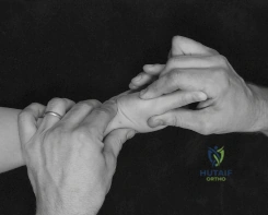
The surgical approach to peripheral nerve repair demands an uncompromising adherence to microsurgical principles, utilizing extensile exposures, specialized instrumentation, and high-quality optical magnification. Operating loupes providing 3.5x to 4.5x magnification are sufficient for the initial macroscopic exposure and neurolysis, but an operating microscope is absolutely mandatory for the meticulous preparation of the nerve stumps, fascicular alignment, and the placement of micro-sutures. The incision must be extensile and should never be placed directly over the anticipated zone of injury to avoid placing the surgical scar directly over the nerve repair. The fundamental principle of exposure is the "outside-in" approach: the surgeon must first identify the injured nerve in pristine, unscarred anatomical planes both proximally and distally, and then carefully trace the nerve toward the central zone of trauma.
Once the injured nerve ends are identified and mobilized, the critical step of stump preparation begins. The nerve must be handled exclusively by the external epineurium using fine, non-toothed micro-forceps; the delicate internal fascicles must never be grasped or crushed. In secondary repairs, the proximal neuroma and the distal glioma must be radically resected. Utilizing a fresh, specialized microsurgical nerve blade or sharp nerve scissors, the surgeon performs serial 1-millimeter step-cuts (bread-loafing) through the fibrotic nerve ends. This serial sectioning continues proximally and distally until healthy tissue is encountered. The visual cues of viability are unmistakable under the microscope: healthy fascicles will "pout" or "mushroom" out of the investing epineurial sheath, the intraneural architecture will appear distinct and glistening, and there will be brisk, punctate bleeding from the intrinsic vasa nervorum.
Achieving accurate topographical alignment is the most technically demanding and biologically critical step of the coaptation. If motor axons regenerate into sensory endoneurial tubes, or vice versa, the functional outcome will be zero, regardless of the quality of the micro-suturing. The surgeon must utilize multiple anatomical landmarks to ensure correct rotational alignment. Surface landmarks, such as the longitudinal epineurial blood vessels, provide the initial orientation. Cross-sectional fascicular mapping, matching the size, geometry, and spatial arrangement of the fascicular bundles between the proximal and distal stumps, refines the alignment. In complex mixed nerves (e.g., the ulnar or median nerve), intraoperative nerve stimulation of the distal stump (if performed within 72 hours of injury before complete Wallerian degeneration) or awake patient stimulation can definitively segregate motor from sensory fascicles.
The coaptation itself is typically performed using an epineurial repair technique, which is the standard for most peripheral nerves. Using 8-0 or 9-0 non-absorbable monofilament sutures (e.g., Nylon) on a spatulated micro-needle, the first two sutures are placed exactly 180 degrees apart in the external epineurium to establish rotational alignment and act as gentle traction sutures. Intermediate sutures are then placed to accurately approximate the epineurial edges, ensuring that the fascicles are gently contained without being buckled or strangulated. The surgeon must place the minimum number of sutures required to close the gap; excessive suturing incites a robust foreign body giant cell reaction and promotes detrimental intraneural scarring. Fibrin glue may be used as an adjunct to augment the repair and reduce the required suture burden.
If, after adequate mobilization, the nerve ends cannot be approximated without tension—defined clinically as the ability to hold the nerve ends together with a single 8-0 suture—a nerve graft is absolutely and unequivocally indicated. The reversed sural nerve autograft remains the workhorse donor due to its length, favorable fascicular architecture, and minimal donor site morbidity. Because the donor nerve is typically smaller in diameter than the recipient nerve, multiple strands are cut and sutured together to create a "cable graft" that matches the cross-sectional area of the injured nerve. Crucially, the autograft segments must be anatomically reversed; this prevents regenerating proximal axons from escaping through the transected lateral branches of the donor nerve, ensuring they are directed linearly toward the distal stump. For non-critical sensory nerve gaps less than 3 centimeters, processed nerve allografts or synthetic bioabsorbable conduits may serve as acceptable alternatives, sparing the patient donor site morbidity.
Complications, Incidence Rates, and Salvage Management
Despite flawless microsurgical execution, peripheral nerve repair is fraught with potential complications, primarily due to the inherent biological limitations of axonal regeneration and the volatile nature of the wound healing cascade. The most devastating and functionally limiting complication is the development of a neuroma-in-continuity or a painful terminal neuroma. This occurs when regenerating axons escape the coaptation site due to excessive tension, malalignment, or a hostile, scarred soft-tissue bed, resulting in a disorganized, highly sensitized mass of axons and fibrous tissue. Patients present with excruciating neuropathic pain, severe hyperalgesia, and a violently positive Tinel's sign localized to the repair site, often completely precluding the use of the affected extremity. Prevention is entirely dependent on achieving a tension-free repair and ensuring the coaptation is wrapped in a healthy, well-vascularized soft-tissue envelope.
Failure of axonal regeneration and the subsequent absence of motor or sensory recovery is a profound complication that demands a rigorous diagnostic workup. The etiology is often multifactorial, including unrecognized tension at the repair site, subclinical infection, severe fascicular malalignment, or an excessive delay in surgical intervention resulting in irreversible end-organ apoptosis. Clinically, this manifests as a static Tinel's sign that fails to advance distally over serial monthly examinations, coupled with profound, progressive muscle atrophy and the absence of reinnervation potentials on serial EMG studies. When primary or secondary nerve repair fails, or when a patient presents with an injury that has already exceeded the 12-to-18-month biological window for motor endplate viability, traditional nerve repair is futile, and the surgeon must pivot to salvage reconstructive strategies.
Joint contractures and fixed skeletal deformities are insidious complications resulting from prolonged muscular imbalance during the extended denervation period. As the denervated agonist muscles atrophy, the unopposed pull of the intact antagonist muscles leads to rapid tightening of the joint capsule and collateral ligaments. If a joint becomes rigidly contracted, even a perfectly successful nerve regeneration will yield a functionally useless limb. Therefore, the prevention of contractures through aggressive, daily passive range-of-motion therapy is as critical to the final outcome as the microsurgery itself.
| Complication | Estimated Incidence | Pathophysiology & Risk Factors | Salvage & Management Strategies |
|---|---|---|---|
| Neuroma-in-Continuity / Painful Neuroma | 5% - 15% | Axonal escape due to tension, poor soft tissue bed, or fascicular mismatch. | Excision and interposition nerve grafting; targeted muscle reinnervation (TMR); burying stump in muscle/bone. |
| Failure of Motor Recovery | 15% - 30% (varies by nerve/level) | Delayed timing (>18 months), severe crush mechanism, long regeneration distance. | Tendon transfers (e.g., radial nerve palsy transfers); Free functioning muscle transfer (FFMT); Regional nerve transfers. |
| Joint Contractures | 20% - 40% | Prolonged muscular imbalance, lack of passive ROM therapy during denervation. | Aggressive physical therapy, dynamic splinting, surgical capsulotomy/tenolysis prior to tendon transfers. |
| Complex Regional Pain Syndrome (CRPS) | 2% - 10% | Abnormal sympathetic nervous system response to nerve trauma and surgery. | Multidisciplinary pain management, sympathetic nerve blocks, gabapentinoids, aggressive desensitization therapy. |
| Donor Site Morbidity (Sural Nerve) | 10% - 20% | Numbness over lateral foot, painful neuroma at the proximal transection site. | Preoperative patient counseling; burying the proximal transected end of the sural nerve deep into the calf musculature. |
In the modern era of peripheral nerve surgery, the paradigm for managing delayed presentations and failed proximal repairs has shifted dramatically toward regional nerve transfers (neurotization). Nerve transfers involve taking an expendable, healthy donor motor fascicle in close anatomical proximity to the paralyzed target muscle and coapting it directly to the distal motor nerve of that muscle. By bypassing the proximal zone of injury and the long anatomical distance required for regeneration, nerve transfers drastically reduce the regeneration time, allowing axons to reach the motor endplates before irreversible fibrosis occurs. Classic examples include the Oberlin transfer (ulnar nerve fascicles to the musculocutaneous nerve for biceps restoration) and spinal accessory to suprascapular nerve transfers for brachial plexus avulsions. When local nerve transfers are not feasible, traditional tendon transfers or free functioning muscle transfers (e.g., gracilis transfer) remain the definitive salvage procedures to restore basic mechanical function to the extremity.
Phased Post-Operative Rehabilitation Protocols
The ultimate functional success of a meticulously performed peripheral nerve repair relies just as heavily on the execution of a rigorous, phased postoperative rehabilitation protocol as it does on the surgical technique. The rehabilitation program is designed to protect the fragile micro-coaptation during the initial inflammatory and proliferative phases of wound healing, prevent the formation of restrictive soft-tissue adhesions, and ultimately retrain the central nervous system to interpret the altered sensory input and recruit newly reinnervated motor units. This requires a highly synchronized, multidisciplinary approach involving the orthopedic microsurgeon, specialized certified hand therapists (CHTs), and the patient.
Phase 1 encompasses the initial Immobilization period, extending from postoperative day zero to week three. The primary objective during this phase is the absolute protection of the repair site from tensile forces. The injured extremity is immediately immobilized in a custom-fabricated, well-padded orthosis. The specific position of immobilization is dictated by the anatomical location of the repair; for example, a median or ulnar nerve repair at the volar wrist requires the wrist to be immobilized in 20 to 30 degrees of flexion to remove all tension from the coaptation. Strict elevation of the extremity is mandated to minimize postoperative edema, which can cause detrimental compression of the delicate intraneural microcirculation. During this phase, active and passive range of motion is strictly limited to the joints completely uninvolved and distal/proximal to the immobilized segment to prevent secondary stiffness.
Phase 2, the Early Mobilization phase, typically spans from week three to week six postoperatively. At this juncture, the epineurial repair has gained sufficient tensile strength to withstand gentle, controlled mechanical stress. The orthosis is gradually and serially adjusted to bring the immobilized joint back to a neutral anatomical position, typically at a rate of 10 degrees per week. Concurrently, highly controlled, protected nerve gliding exercises are introduced. These specific, synergistic joint movements are designed to induce longitudinal excursion of the nerve trunk within its fascial bed, preventing the repair site from adhering to the surrounding soft tissues (extraneural scarring) while strictly avoiding any tension across the coaptation itself.
Phase 3, the Re-education and Strengthening phase, begins after week six and continues indefinitely as axonal regeneration progresses. The surgeon and therapist must closely monitor the advancing front of the Tinel's sign, which serves as the clinical biomarker for regenerating axons, typically advancing at a physiological rate of 1 millimeter per day (or approximately 1 inch per month). As these regenerating sensory axons reach the distal cutaneous targets, patients frequently experience severe hyperesthesia or altered, dysesthetic sensation due to the immaturity of the reinnervation and altered cortical mapping. Aggressive sensory re-education and desensitization protocols—utilizing a progression of various textures, fluidotherapy, and vibration—are critical to help the somatosensory cortex recalibrate and interpret these new signals as normal touch rather than pain. Simultaneously, as motor reinnervation begins, electromyographic (EMG) biofeedback and gravity-eliminated exercises are employed to assist the patient in consciously recruiting and strengthening the newly reinnervated, highly fatigable motor units.
Summary of Landmark Literature and Clinical Guidelines
The contemporary principles of peripheral nerve surgery are built upon a foundation of landmark anatomical studies, biomechanical research, and wartime clinical observations. The foundational classification systems proposed by Seddon (1943) and Sunderland (1951) remain the absolute bedrock of peripheral nerve pathology. Seddon's classification of neuropraxia, axonotmesis, and neurotmesis provided the first clinical framework for predicting spontaneous recovery versus the need for surgical intervention. Sunderland expanded upon this by detailing the specific microanatomical structures (myelin, axon, endoneurium, perineurium, epineurium) disrupted in varying degrees of traction and crush injuries, establishing the biological rationale for why high-energy injuries fail to regenerate spontaneously and require delayed secondary resection and grafting.
The surgical technique of nerve repair underwent a paradigm shift in the 1970s due to the seminal work of Hanno Millesi. Prior to Millesi, surgeons frequently utilized extreme joint positioning and extensive nerve mobilization to achieve primary end-to-end repairs, often under significant tension. Millesi's groundbreaking experimental and clinical studies definitively proved that a tension-free interfascicular nerve graft yields vastly superior functional outcomes compared to a primary repair performed under tension. He demonstrated that the biological barrier of tension-induced intraneural ischemia and scarring is far more detrimental to axonal regeneration than the biological disadvantage of requiring regenerating axons to
