
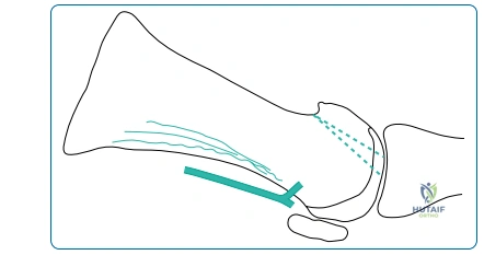
A 55-year-old active school teacher presents with a 2-year history of worsening dorsal foot pain. She describes "stiffness" in her big toe, worse with walking in flat shoes, and notes a visible prominence on the dorsum of the MTPJ. She has failed conservative management including shoe modifications, rocker-bottom insoles, and NSAIDs. Examination reveals limited dorsiflexion of the first MTPJ (20 degrees) with terminal pain. What is your initial assessment and how would you classify this?
Candidate: This patient has hallux rigidus. I would use the Coughlin and Shurnas classification to grade it based on clinical motion and radiographic joint space narrowing. She likely has a Grade 2 lesion given the osteophytes and restricted motion. I would discuss potential surgical options starting with a cheilectomy.
Failure to mention the "Windlass mechanism" or the biomechanical consequences of the pathology. Candidates often jump straight to "cheilectomy" without mentioning the differential diagnosis (e.g., sesamoiditis or inflammatory arthropathy) or the importance of assessing the IP joint and plantar articular cartilage.
Define this as Hallux Rigidus. State clearly that the Coughlin and Shurnas classification is the standard. Acknowledge that the goal is to assess if the joint is "salvageable" (Grade 1-2) or "end-stage" (Grade 3-4). Mention that the clinical examination must exclude global joint pain (which would contraindicate cheilectomy). Finally, link the biomechanical deficit—loss of the Windlass mechanism—to her compensatory gait and potential transfer metatarsalgia.
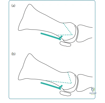
You decide to proceed with a first MTPJ arthrodesis for a patient with end-stage hallux rigidus. What are the critical technical objectives for this procedure, and what is your preferred position for the fusion?
Candidate: The goal is a solid, pain-free fusion. I aim for 10-15 degrees of valgus and 10-15 degrees of dorsiflexion relative to the floor. I prefer a compression screw supplemented with a dorsal plate for stability.
Confusing the degrees of dorsiflexion relative to the metatarsal vs. the floor. Forgetting to mention the importance of rotational alignment (toenail should face directly upwards). Neglecting to discuss the use of cup-and-cone reamers which preserve bone stock.
Start with the biomechanical goal: load-bearing without impingement. 1. Position: 10-15° valgus, 15-20° dorsiflexion relative to the first metatarsal axis. 2. Rotation: Neutral. 3. Fixation: Lag screw for interfragmentary compression, dorsal neutralization plate for bending/torsional rigidity. 4. Key Technicality: Use of cup-and-cone reamers to maximize surface area and allow subtle adjustments in position before final fixation.
A patient returns 6 months post-arthrodesis complaining of pain under the second metatarsal head. What is the most likely cause, and how do you evaluate it?
Candidate: This sounds like transfer metatarsalgia. It happens if the fusion is in too much dorsiflexion or if the first ray has been significantly shortened. I would examine their gait and check the radiographs for the position of the fusion.
Ignoring the role of excessive shortening during joint preparation. Failing to offer a management strategy—it's not always surgical; mention orthotics and metatarsal pads before jumping to revision surgery.
Identify this as Transfer Metatarsalgia secondary to a shortened or dorsiflexed first ray. Evaluation: Clinical gait analysis, weight-bearing radiographs to assess fusion angle, and physical exam to assess lesser MTPJ synovitis. Management: 1st line is non-operative: custom orthotics with a metatarsal pad to redistribute weight. 2nd line: if conservative measures fail, revision osteotomy to correct the position or (rarely) distal metatarsal osteotomies (Weil) on the lesser rays.
Detailed Chapters & Topics
Dive deeper into specialized chapters regarding surgery-of-the-foot