First Metatarsal Surgery: Expert Solutions for Foot & Ankle Pain

Key Takeaway
Looking for accurate information on First Metatarsal Surgery: Expert Solutions for Foot & Ankle Pain? The midfoot bridges the hindfoot and forefoot, facilitating adduction and abduction. It includes intercuneiform, naviculocuneiform, and tarsalmetatarsal (TMT) joints. These joints are vital for foot biomechanics, with the fi rst metatarsal playing a significant role in forefoot stability and weight-bearing during the gait cycle, particularly at the first TMT joint.
A 55-year-old female presents with progressive forefoot pain and deformity. Clinical examination reveals a prominent medial eminence and a laterally deviated hallux. You suspect hallux valgus. What are the key radiographic parameters you must evaluate to determine the appropriate surgical plan?
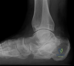
Candidate: I would measure the Hallux Valgus Angle (HVA), the Intermetatarsal Angle (IMA), and the Distal Metatarsal Articular Angle (DMAA). I would also look at the lateral view for Meary's angle to check for midfoot collapse and a sesamoid axial view to assess sesamoid position and crista erosion.
Listing angles without defining normal ranges or explaining their clinical significance. Failing to mention the sesamoid axial view is a major oversight, as sesamoid subluxation is a key driver of recurrence and indicates the severity of the frontal plane deformity.
Start with the HVA (<15°), IMA (<9°), and DMAA to define the magnitude of the angular deformity. Critically, emphasize the importance of the sesamoid axial view to assess the sesamoid-metatarsal relationship, which dictates whether a soft-tissue release alone is sufficient or if a bony realignment (osteotomy/fusion) is required. Finally, mention assessing joint congruency and the sagittal alignment (Meary's) to rule out midfoot instability which might necessitate a Lapidus.
You have decided to proceed with a first TMT fusion (Lapidus procedure). A primary concern is the prevention of complications, specifically non-union and hardware prominence. What is the modern biomechanical strategy to optimize the success of this procedure?
Candidate: The focus should be on achieving multiplanar correction—specifically addressing pronation, plantarflexion, and the IMA. Biomechanically, the use of a plantar-lateral locking plate is superior to traditional screw-only fixation as it provides a tension-side construct, allowing for earlier weight-bearing and reducing non-union rates.
Focusing only on "fusing the joint" without mentioning the restoration of the windlass mechanism. A common failing is neglecting to mention the importance of addressing frontal plane rotation (supination of the first metatarsal) during the correction.
Structure the answer around the three pillars of a successful Lapidus: 1) Multiplanar Correction: Aggressive derotation and plantarflexion of the first metatarsal to restore the longitudinal arch. 2) Bone Preparation: Meticulous cartilage removal and subchondral bone preparation to create an optimal biological environment. 3) Mechanical Construct: The move toward plantar/medial locking plates which function as internal fixation, providing superior stability in the tension band zone compared to cross-screws alone, thereby minimizing the risk of non-union and hardware irritation.
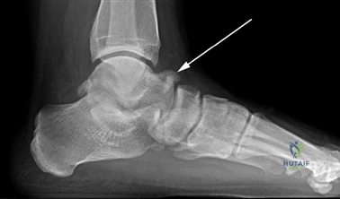
A patient returns 6 months post-distal chevron osteotomy complaining of persistent lateral forefoot pain and difficulty with push-off. Radiographs show a healed osteotomy, but the first metatarsal appears shorter than the second. What is your diagnosis and management plan?

Candidate: The patient has transfer metatarsalgia caused by excessive shortening or elevation of the first ray. I would initially manage this conservatively with orthotics, such as a metatarsal pad, to redistribute pressure. If this fails, I might consider a Weil osteotomy of the lesser metatarsals or a revision of the first ray to restore its load-bearing capacity.
Immediately suggesting surgical revision without emphasizing a trial of conservative therapy. In the FRCS exam, you must show you are a "conservative" surgeon who exhausts non-operative options for secondary complications before rushing back to the theatre.
Identify the pathophysiology: The first metatarsal is no longer functioning as the primary weight-bearing axis due to the "shortening/elevation effect." Emphasize a hierarchical management approach: 1) Clinical/Radiographic confirmation of the "short first ray." 2) Conservative (Primary): Offloading with custom orthotics/metatarsal pads. 3) Surgical (Salvage): If refractory, define the goal—either shortening the lesser rays (Weil osteotomy) to balance the parabola, or, in severe cases, correcting the first ray elevation through a corrective revision osteotomy or fusion to restore the windlass effect.
