
Pediatric Orthopaedics: Comprehensive MCQ Question Bank & Exam Prep
Comprehensive 100-Question Exam
00:00
Start Quiz
Question 1
A 13-year-old obese male presents with left groin and knee pain that has been worsening over 6 months. Radiographs reveal a severe chronic Slipped Capital Femoral Epiphysis (SCFE). He undergoes in situ percutaneous pinning. What is the most significant recognized mechanical risk factor for developing chondrolysis in this patient?
Explanation
Chondrolysis is a devastating complication of SCFE characterized by rapid destruction of the articular cartilage. Unrecognized penetration of the pin/screw into the hip joint space is the most significant mechanical risk factor for chondrolysis. To prevent this, careful fluoroscopic evaluation (e.g., the 'approach-withdraw' technique) is critical.

Question 2
A 4-week-old infant with idiopathic clubfoot is being treated with the Ponseti method. After 5 weeks of serial casting, the cavus, adductus, and varus deformities have completely resolved. However, passive ankle dorsiflexion is only 5 degrees. What is the next most appropriate step in management?
Explanation
In the Ponseti method, after the cavus, adductus, and varus deformities are corrected (which relies on abduction around the talar head), the equinus is addressed. If ankle dorsiflexion is less than 10 to 15 degrees, a percutaneous Achilles tenotomy is indicated. This is required in 80% to 90% of patients with idiopathic clubfoot.
Question 3
A 7-year-old child with spastic quadriplegic cerebral palsy (GMFCS Level V) is evaluated for hip pain and difficulty with perineal hygiene. An AP pelvis radiograph demonstrates a Reimers' migration index of 65% bilaterally. What is the most appropriate surgical intervention to stabilize the hips?
Explanation
In a child with cerebral palsy, a Reimers' migration percentage > 40-50% indicates significant hip subluxation with secondary acetabular dysplasia. Soft-tissue releases alone are inadequate. A reconstructive procedure combining a proximal femoral varus derotational osteotomy (VDRO) and a pelvic volume-reducing osteotomy (e.g., Dega, San Diego, or Pemberton) is the gold standard for reconstruction.
Question 4
A 6-week-old female infant, born in a breech presentation, is evaluated for Developmental Dysplasia of the Hip (DDH). A coronal ultrasound of the hip is performed. The alpha angle is measured at 40 degrees and the beta angle at 80 degrees. According to the Graf classification, what is the diagnosis and the most appropriate management?
Explanation
According to Graf's ultrasound classification, a Type III hip has an alpha angle < 43 degrees and a beta angle > 77 degrees, indicating an eccentrically located (subluxated) femoral head with no structural alteration of the acetabular cartilage rim. The standard initial non-operative treatment for a Graf III hip in an infant < 6 months is a Pavlik harness.
Question 5
A 13-year-old male presents with rigid, painful flat feet and recurrent ankle sprains. On examination, he demonstrates restricted subtalar motion and peroneal spasticity. Radiographs demonstrate an 'anteater nose' sign. Which of the following is the most likely diagnosis?
Explanation
The 'anteater nose' sign is seen on the 45-degree internal oblique radiograph of the foot and is pathognomonic for a calcaneonavicular coalition. It represents an elongation of the anterior process of the calcaneus attempting to bridge to the navicular. In contrast, the 'C-sign' on a lateral radiograph is associated with a talocalcaneal coalition.
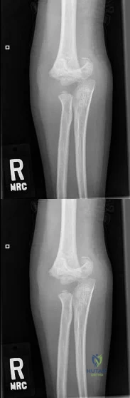
Question 6
A 6-year-old child sustains a widely displaced, extension-type supracondylar humerus fracture. On presentation to the emergency department, the hand is pink, warm, and has a brisk capillary refill, but the radial pulse is entirely non-palpable. What is the most appropriate initial step in the management of this patient?
Explanation
This is a classic 'pulseless, pink hand' scenario in a pediatric supracondylar humerus fracture. The hand remains perfused via collateral circulation despite brachial artery entrapment or kinking at the fracture site. The recommended initial management is urgent closed reduction and percutaneous pinning (CRPP). Pulse typically returns after realignment; if the hand becomes cold/white post-reduction, vascular exploration is indicated.
Question 7
An 8-year-old boy is diagnosed with Legg-Calvé-Perthes disease. During the fragmentation stage, the treating orthopaedist assesses the radiographs for 'head-at-risk' signs described by Catterall. Which of the following is considered a Catterall 'head-at-risk' sign?
Explanation
Catterall described five 'head-at-risk' signs indicating a poorer prognosis in Perthes disease: 1) Gage's sign (a V-shaped radiolucency in the lateral portion of the epiphysis/metaphysis), 2) calcification lateral to the epiphysis, 3) lateral subluxation of the femoral head, 4) a horizontal growth plate, and 5) metaphyseal cysts.
Question 8
A 4-year-old child with achondroplasia presents with progressive weakness in all four extremities, new-onset hyperreflexia, and frequent episodes of central sleep apnea. What is the most appropriate imaging study to evaluate the underlying cause of these symptoms?
Explanation
The symptoms described point to high cervical myelopathy and lower brainstem compression, which in young children with achondroplasia is typically caused by foramen magnum stenosis. MRI of the craniovertebral junction is the imaging modality of choice to evaluate the extent of neural compression.
Question 9
A 3-year-old obese female presents with bilateral bowing of the legs. Standing AP radiographs show a sharp varus angulation at the proximal tibial metaphyses. The metaphyseal-diaphyseal angle (Drennan's angle) is measured at 18 degrees bilaterally. What is the most appropriate management?
Explanation
A metaphyseal-diaphyseal angle (Drennan's angle) > 16 degrees strongly suggests infantile Blount disease rather than physiologic bowing. In children under 3 to 4 years of age with early-stage infantile Blount disease (Langenskiöld stage I or II), bracing with knee-ankle-foot orthoses (KAFOs) is the initial non-operative treatment of choice.
Question 10
A 5-year-old child is evaluated for a painless waddling gait. AP pelvis radiographs reveal a decreased neck-shaft angle and a Hilgenreiner Epiphyseal Angle (HEA) of 65 degrees. A triangular fragment of bone is visible in the inferior aspect of the femoral neck. What is the recommended treatment for this condition?
Explanation
This patient has developmental coxa vara, characterized by a 'fairbank' triangle and a high Hilgenreiner Epiphyseal Angle (HEA). An HEA > 60 degrees has a high rate of progressive deformity and is an absolute indication for surgery. The treatment of choice is a valgus-producing proximal femoral osteotomy to correct the HEA to < 35-40 degrees, thereby converting shear forces into compressive forces.
Question 11
A 6-year-old girl complains of a painful, audible 'popping' sensation in her lateral knee when walking. MRI confirms the presence of a discoid lateral meniscus. Which specific variant of a discoid meniscus lacks normal posterior coronary ligament attachments, directly causing this 'snapping knee' syndrome?
Explanation
The Wrisberg variant of a discoid lateral meniscus lacks normal posterior capsular attachments (the coronary ligaments) and is anchored solely by the meniscofemoral ligament of Wrisberg. This hypermobility allows the meniscus to subluxate into the intercondylar notch during extension, causing a loud and painful 'snap'.
Question 12
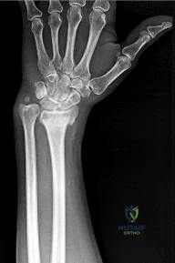
A newborn infant presents with severe radial deviation of both wrists. Radiographs confirm bilateral absent radii. Physical examination reveals that the thumbs are present bilaterally. Laboratory evaluation shows a profoundly low platelet count. Which of the following is the most likely diagnosis?
Explanation
TAR (Thrombocytopenia-Absent Radius) syndrome is classically characterized by the absence of the radius with the preservation of the thumb. This critical clinical feature distinguishes TAR syndrome from Fanconi anemia and Holt-Oram syndrome, where an absent radius is almost universally accompanied by an absent or severely hypoplastic thumb.
Question 13
A 14-year-old boy sustains an ankle injury. Radiographs show a Salter-Harris III fracture of the anterolateral aspect of the distal tibial epiphysis. This fracture pattern (juvenile Tillaux fracture) is caused by an avulsion force from which of the following ligaments?
Explanation
A juvenile Tillaux fracture occurs because the distal tibial physis closes in a predictable pattern: centrally, then anteromedially, then posteromedially, and finally anterolaterally. External rotation of the foot causes the anterior inferior tibiofibular ligament (AITFL) to avulse the still-open anterolateral portion of the epiphysis.
Question 14
A 10-year-old boy falls off his bicycle and presents with knee hemarthrosis. Radiographs reveal a Meyers and McKeever Type II fracture of the tibial eminence. Which of the following best describes the anatomical displacement in this fracture pattern?
Explanation
The Meyers and McKeever classification for tibial eminence fractures is: Type I (non-displaced), Type II (anterior elevation with an intact posterior bony hinge, resembling a 'bird's beak'), Type III (completely displaced; IIIa without rotation, IIIb with rotation), and Type IV (comminuted, added later by Zaricznyj).
Question 15
A 12-year-old competitive gymnast presents with severe low back pain and radicular pain radiating down her posterior left leg. Lateral radiographs demonstrate a Meyerding Grade IV isthmic spondylolisthesis at L5-S1 with a slip angle of 55 degrees. What is the most appropriate surgical treatment?
Explanation
High-grade spondylolisthesis (Meyerding Grade III-V) with radicular symptoms and a high slip angle (>40-50 degrees) carries a high risk of progression and pseudarthrosis if treated with in situ fusion. The standard of care is decompression of the compressed nerve roots, partial reduction of the slip angle, and posterior instrumented fusion, typically extending from L4 to S1.
Question 16
A 10-year-old boy with known mutations in the EXT1 gene is followed for multiple bone lesions. He presents with a rapidly enlarging, painful mass on his proximal tibia. He is at highest risk for developing which of the following malignancies?
Explanation
Mutations in the EXT1 and EXT2 genes cause Multiple Hereditary Exostoses (osteochondromatosis). The most significant complication is the malignant transformation of an osteochondroma into a secondary peripheral chondrosarcoma, which occurs in 1% to 5% of patients, typically marked by rapid growth or a new onset of pain.
Question 17
A 4-year-old boy presents with a 2-day history of right hip pain and a limp, now refusing to bear weight completely. His temperature is 38.6°C (101.5°F), WBC count is 14,500/mm³, ESR is 45 mm/hr, and CRP is elevated. According to the Kocher criteria, what is the approximate predictive probability of septic arthritis in this patient?
Explanation
The Kocher criteria to differentiate septic arthritis from transient synovitis in children are: 1) Non-weight-bearing, 2) Temperature > 38.5°C, 3) ESR > 40 mm/hr, and 4) WBC > 12,000/mm³. The probability of septic arthritis is approximately 3% for 1 predictor, 40% for 2 predictors, 93% for 3 predictors, and 99% when all 4 predictors are present.
Question 18
A 6-month-old infant presents with multiple fractures of varying ages, blue sclerae, and generalized osteopenia. Genetic testing reveals a mutation in the COL1A1 gene. Which of the following is the primary mechanism of action of the most commonly prescribed class of medications (bisphosphonates) used to treat this condition?
Explanation
Osteogenesis Imperfecta (OI) is treated medically with bisphosphonates (e.g., pamidronate or zoledronic acid). Bisphosphonates are analogues of inorganic pyrophosphate that bind to hydroxyapatite and function primarily by inhibiting osteoclast-mediated bone resorption, thereby increasing bone mineral density and decreasing the fracture rate.
Question 19
A 2-year-old child presents with an anterolateral bowing deformity of the left tibia. Radiographs demonstrate a diaphyseal fracture with sclerotic, tapered ends that has failed to heal. This condition, congenital pseudarthrosis of the tibia (CPT), is most highly associated with which of the following systemic disorders?
Explanation
Anterolateral bowing of the tibia is the hallmark precursor to Congenital Pseudarthrosis of the Tibia (CPT). CPT is highly associated with Neurofibromatosis type 1 (NF1); approximately 50% of patients diagnosed with CPT will have or develop clinical signs of NF1.
Question 20
A 12-year-old male presents with acute severe groin pain after jumping off a swing. He is completely unable to bear weight, even with crutch assistance. Radiographs demonstrate a Slipped Capital Femoral Epiphysis (SCFE). What is the approximate risk of avascular necrosis (AVN) in this type of SCFE compared to a 'stable' slip?
Explanation
Loder classified SCFE into 'stable' (able to bear weight with or without crutches) and 'unstable' (unable to bear weight). Unstable SCFE is an acute emergency that carries a high risk of avascular necrosis (AVN), historically ranging from 25% to 50% (often cited around 47%), whereas stable SCFE has an AVN risk of nearly 0%.
Question 21
What radiographic parameter is considered the most reliable and predictive marker for the risk of hip dislocation in a non-ambulatory child with Cerebral Palsy (GMFCS Level IV or V)?
Explanation
Reimers' migration percentage is the primary tool used in hip surveillance programs for children with cerebral palsy. It measures the percentage of the ossified femoral head outside the lateral margin of the acetabulum (Perkin's line). A migration percentage greater than 30% indicates subluxation and an increased risk for progression to dislocation, particularly in non-ambulatory children (GMFCS IV and V).
Question 22
An 18-month-old female presents with an untreated, late-diagnosed Developmental Dysplasia of the Hip (DDH). Open reduction and a pelvic osteotomy are planned to improve anterolateral acetabular coverage. Which of the following pelvic osteotomies utilizes the pubic symphysis as its primary hinge?
Explanation
The Salter innominate osteotomy is a complete cut through the ilium extending to the greater sciatic notch. It redirects the entire acetabulum using the pubic symphysis as its hinge. The Pemberton osteotomy hinges on the triradiate cartilage, and the Dega osteotomy hinges on the intact posterior cortex of the ilium.
Question 23
A 7-year-old boy is diagnosed with Legg-Calvé-Perthes disease. According to the Herring Lateral Pillar Classification, which of the following findings places him in Group C and predicts the poorest clinical outcome?
Explanation
The Herring Lateral Pillar Classification is highly prognostic in Legg-Calvé-Perthes disease. Group A involves no lateral pillar involvement. Group B involves >50% maintenance of lateral pillar height. Group C involves <50% maintenance of lateral pillar height, which portends a poor outcome with a high risk of developing an aspherical femoral head and early osteoarthritis.
Question 24
Which of the following patients presenting with a unilateral Slipped Capital Femoral Epiphysis (SCFE) has the strongest absolute indication for prophylactic in situ pinning of the contralateral hip?
Explanation
Endocrine disorders (such as hypothyroidism) and metabolic bone diseases (like renal osteodystrophy) or a history of radiation therapy carry an exceptionally high risk (up to 50-100%) for the development of a bilateral SCFE. While obesity and delayed skeletal age are risk factors, a diagnosed underlying systemic endocrinopathy or metabolic disorder is considered a strong and almost absolute indication for prophylactic contralateral pinning.
Question 25
A 2-year-old girl presents with progressive bowing of her left leg. Standing full-length radiographs demonstrate a marked varus deformity isolated to the proximal tibia. Which of the following radiographic parameters is most strongly predictive of progression to true infantile Blount's disease rather than physiological bowing?
Explanation
The metaphyseal-diaphyseal angle (Drennan's angle) is the most reliable early radiographic predictor for infantile Blount's disease. An angle > 16 degrees indicates a high likelihood (up to 95%) of progression to Blount's disease, whereas an angle < 10 degrees strongly suggests physiological bowing. Angles between 10 and 16 degrees require close observation.
Question 26
A 4-month-old infant with molecularly confirmed achondroplasia is being evaluated. What is the most common cause of sudden, unexpected infant death in patients with this skeletal dysplasia?
Explanation
Infants with achondroplasia have a narrow skull base, which can lead to severe foramen magnum stenosis. This stenosis can compress the cervicomedullary junction, resulting in central apnea and sudden infant death syndrome (SIDS). Evaluation typically involves MRI and polysomnography. Suboccipital decompression is indicated if severe compression or apnea is identified.
Question 27
In the evaluation of pediatric developmental (isthmic) spondylolisthesis at L5-S1, which of the following spinopelvic parameters is most strongly associated with an increased risk of severe slip progression?
Explanation
Pelvic Incidence (PI) is an anatomical spinopelvic parameter that is fixed after skeletal maturity. A high PI results in a high shear stress at the lumbosacral junction. Patients with developmental, high-grade spondylolisthesis almost universally have a significantly higher pelvic incidence compared to the general population, which drives slip progression.
Question 28
A 2.5-year-old boy treated successfully in infancy for a right idiopathic clubfoot using the Ponseti method returns to the clinic. His parents report worsening of his foot shape over the last 3 months, and admit to discontinuing the foot abduction orthosis. Examination reveals dynamic supination and recurrent equinus. What is the most appropriate initial management?
Explanation
Relapse of clubfoot deformities after Ponseti treatment is most commonly due to poor compliance with bracing. Regardless of the child's age or the presence of a dynamic supination component, the first-line treatment for a relapse is always repeat manipulation and serial long-leg casting to correct the deformities. Once the foot is supple and plantigrade, a surgical procedure such as an Anterior Tibial Tendon Transfer (ATTT) may be indicated to prevent further relapse, but it should not be performed before recasting.
Question 29
A 12-year-old boy complains of recurrent left lateral ankle sprains and a painful, rigid flatfoot. Physical examination reveals spasm of the peroneal tendons. Which radiographic view is best to properly identify a calcaneonavicular coalition (the 'anteater nose' sign)?
Explanation
A calcaneonavicular coalition is best visualized on a 45-degree internal oblique radiograph of the foot, which throws the anterior process of the calcaneus into profile, demonstrating the 'anteater nose' sign. Talocalcaneal coalitions are best seen on a Harris axial heel view or a CT scan (and suspected due to a 'C-sign' on a lateral radiograph).
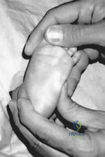
Question 30
A 2-year-old girl is brought in for a locked interphalangeal joint of her right thumb. Her mother reports a small nodule at the base of the thumb on the volar surface. Active extension is absent, but passive extension is painful and yields a palpable click. What is the most appropriate initial management?
Explanation
Pediatric trigger thumb presents with a locked IP joint in flexion and a palpable Notta's node at the A1 pulley. Unlike adult trigger digit, initial management in a young child (< 3 years) is observation, as up to 30-60% of cases will resolve spontaneously. Corticosteroid injections are generally not recommended in this age group. Surgical release of the A1 pulley is indicated if the condition persists beyond age 3 to 4 years.
Question 31
A 6-year-old boy sustains a severe Gartland Type III supracondylar humerus fracture. Upon arrival, his hand is pale and pulseless. After closed reduction and percutaneous pinning, the radial pulse remains unpalpable, but the hand becomes pink, warm, and has a capillary refill of less than 2 seconds. What is the next best step in management?
Explanation
The 'pink, pulseless hand' after reduction of a supracondylar humerus fracture is a well-known clinical entity. If the hand is well-perfused (pink, warm, strong capillary refill), the lack of a palpable radial pulse is likely due to arterial spasm rather than a complete irreversible occlusion or transection. The standard of care is admission, elevation, and close monitoring for 24-48 hours. Immediate surgical exploration is indicated only if the hand remains 'white and pulseless' after a proper reduction.
Question 32
A 4-year-old child with Osteogenesis Imperfecta (OI) type III is treated with intravenous pamidronate. What is the primary cellular mechanism of action by which this medication improves bone density and reduces fracture burden in this patient population?
Explanation
Bisphosphonates (such as pamidronate) are the medical treatment of choice for moderate to severe Osteogenesis Imperfecta. They function by inhibiting osteoclast activity and inducing osteoclast apoptosis, thereby decreasing bone resorption. This leaves osteoblast activity unopposed, increasing bone volume and density, though it does not correct the underlying genetic defect in Type I collagen synthesis.
Question 33
A 14-year-old boy presents with progressive mid-back pain. Lateral radiographs of his thoracic spine reveal rigid hyperkyphosis. According to the Sorensen criteria for classical Scheuermann's disease, what is the required radiographic threshold regarding vertebral body wedging?
Explanation
The diagnosis of classic Scheuermann's kyphosis using Sorensen criteria requires anterior wedging of greater than 5 degrees in at least three adjacent (contiguous) vertebral bodies. Other classic radiographic signs include Schmorl's nodes, endplate irregularities, and narrowed disc spaces, but the contiguous wedging is the definitive diagnostic criterion.
Question 34
Congenital Pseudarthrosis of the Tibia (CPT) is a notoriously difficult condition to treat and is characterized by bowing and spontaneous fractures that fail to heal. The condition has the strongest genetic and clinical association with a mutation on which chromosome?
Explanation
Congenital pseudarthrosis of the tibia (CPT) has a very strong association with Neurofibromatosis Type 1 (NF1), occurring in up to 50% of patients with CPT. NF1 is an autosomal dominant disorder caused by a mutation in the neurofibromin gene, which is located on Chromosome 17 (17q11.2).
Question 35
A 6-year-old girl complains of a painful 'clunk' and a snapping sensation on the lateral aspect of her right knee when moving from flexion to extension. MRI confirms a Watanabe Type III (Wrisberg variant) discoid lateral meniscus. What specific anatomical deficiency characterizes this specific variant?
Explanation
The Watanabe classification describes discoid menisci. Type I is complete, Type II is incomplete, and Type III is the Wrisberg variant. The hallmark of the Wrisberg variant is the absence of the normal posterior meniscotibial (coronary) ligament capsular attachments, leaving only the Wrisberg ligament attached to the posterior horn. This results in hypermobility and a symptomatic 'snapping' knee during extension as the meniscus subluxates.
Question 36
Pediatric hip fractures are rare and associated with a high rate of complications. According to the Delbet classification, which fracture type is associated with the highest rate of avascular necrosis (AVN) of the femoral head?
Explanation
The Delbet classification for pediatric hip fractures dictates that Type I (transepiphyseal) fractures have the highest rate of avascular necrosis, approaching 90-100% in some series, because it completely disrupts the precarious retinacular blood supply traversing the neck to the epiphysis. Type II (transcervical) is the most common pediatric hip fracture and also carries a very high AVN rate (~50%). AVN risk decreases moving distally down the neck to the intertrochanteric line.
Question 37
Proximal Focal Femoral Deficiency (PFFD), also known as Congenital Femoral Deficiency, presents with marked limb length discrepancy, external rotation, and flexion at the hip. It is most commonly associated with which of the following intra-articular knee anomalies?
Explanation
Proximal Focal Femoral Deficiency (PFFD) is heavily associated with fibular hemimelia, coxa vara, and knee instability. The most common intra-articular anomaly in these patients is agenesis or hypoplasia of the Anterior Cruciate Ligament (ACL). This must be carefully evaluated before attempting major limb lengthening procedures, as lengthening a knee with ACL deficiency can lead to posterior subluxation of the tibia.
Question 38
A 7-year-old girl presents with severe torticollis, neck pain, and a 'cock-robin' head position three weeks after undergoing a tonsillectomy. Radiographs show an increased atlantodens interval. In the pathogenesis of Grisel's syndrome, what is the primary mechanism leading to this atlantoaxial subluxation?
Explanation
Grisel's syndrome is a non-traumatic atlantoaxial rotatory subluxation associated with head and neck infections (or postoperative states like tonsillectomy/adenoidectomy). The robust venous plexus draining the retropharyngeal space communicates directly with the periodontoid vascular plexus. Inflammatory hyperemia spreads to the periodontoid tissues, causing decalcification of the anterior arch of C1 and subsequent laxity of the transverse ligament, leading to subluxation.
Question 39
A 12-year-old boy with Duchenne Muscular Dystrophy (DMD) has lost independent ambulation and presents with a progressive neuromuscular scoliosis of 28 degrees. When counseling the family regarding posterior spinal fusion to the pelvis, what are the standard criteria for proceeding with surgery to optimize outcomes and minimize prohibitive respiratory complications?
Explanation
In Duchenne Muscular Dystrophy, scoliosis progresses rapidly once ambulation is lost. To prevent severe deformity and restrictive lung disease, surgical intervention (posterior spinal fusion from the upper thoracic spine to the pelvis) is indicated early, typically when the Cobb angle reaches 20 to 30 degrees. The surgery must be performed while the patient's respiratory function is adequate; specifically, a Forced Vital Capacity (FVC) > 35% is generally required, as operating when FVC falls below 30-35% carries a prohibitively high risk of postoperative ventilator dependence and mortality.
Question 40
Madelung deformity is a congenital wrist deformity characterized by premature closure of the volar-ulnar aspect of the distal radial physis. It frequently presents bilaterally in adolescent females. This specific deformity is the hallmark skeletal manifestation of Léri-Weill dyschondrosteosis, which is caused by a mutation in which gene?
Explanation
Madelung deformity presents with a V-shaped radiocarpal joint and dorsal prominence of the distal ulna due to tethering by Vickers' ligament and premature distal radial physeal arrest. While it can occur idiopathically, when present as part of Léri-Weill dyschondrosteosis (mesomelic dwarfism), it is due to a mutation or deletion of the SHOX (Short Stature Homeobox) gene, which is located on the pseudoautosomal region of the X and Y chromosomes.
Question 41
A 6-week-old female is treated with a Pavlik harness for an ultrasonographically confirmed irreducible right hip dislocation. At the 2-week follow-up, the parents report the infant is no longer kicking her right leg. Examination reveals absent active knee extension on the right, but active ankle dorsiflexion and plantarflexion are preserved. Ultrasound confirms the hip remains dislocated. What is the most appropriate next step in management?
Explanation
The patient has developed a femoral nerve palsy, a known complication of the Pavlik harness, typically caused by excessive hip flexion pressing the nerve against the inguinal ligament. If the hip is still dislocated and a femoral nerve palsy develops, the harness must be discontinued (the so-called 'Pavlik holiday'). A period of rest allows for neurologic recovery before pursuing alternative treatments, such as closed reduction and spica casting. Continuing the harness or increasing flexion risks permanent nerve damage and vascular compromise.
Question 42
An 8.5-year-old boy presents with a 4-month history of a painless limp. Radiographs demonstrate fragmentation of the capital femoral epiphysis with >50% maintenance of the lateral pillar height. He has 15 degrees of abduction and a 10-degree flexion contracture. Based on his age and radiographic classification, which treatment provides the best long-term radiographic outcome?
Explanation
This patient has Legg-Calvé-Perthes disease with a Herring lateral pillar B classification (maintenance of >50% lateral pillar height). The Herring classification and age at onset dictate treatment. Studies have shown that for patients > 8 years old with Herring B or B/C border hips, surgical containment (such as a proximal femoral varus osteotomy or pelvic osteotomy) significantly improves radiographic outcomes (Stulberg classification) and reduces the risk of early osteoarthritis compared to nonoperative management.
Question 43
A 6-year-old boy sustains a Gartland type III extension supracondylar humerus fracture. On presentation, his hand is pink and warm, but the radial pulse is non-palpable. Urgent closed reduction and percutaneous pinning are performed. Postoperatively, the fracture is anatomically aligned on fluoroscopy, but the radial pulse remains absent. Capillary refill is 2 seconds and the hand remains warm. What is the most appropriate management?
Explanation
The management of a 'pulseless pink hand' following anatomical closed reduction and pinning of a pediatric supracondylar humerus fracture is close observation. The hand's pink color, warmth, and brisk capillary refill indicate that collateral circulation is sufficient to perfuse the extremity despite a presumed brachial artery vasospasm or non-occlusive intimal flap. Immediate vascular exploration is strictly indicated if the hand becomes dysvascular (white, cold, absent capillary refill) after reduction.
Question 44
A newborn male presents with bilateral radial longitudinal deficiency (absent radii and absent thumbs). A chromosomal breakage test using diepoxybutane is positive. If left untreated, what is the most common cause of mortality associated with this patient's underlying diagnosis?
Explanation
The patient's clinical presentation (radial clubhand) combined with a positive chromosomal breakage test using diepoxybutane (DEB) or mitomycin C definitively diagnoses Fanconi anemia. Fanconi anemia is an autosomal recessive condition that leads to progressive pancytopenia and bone marrow failure, which is the most common cause of mortality in these patients. It is critical to differentiate it from other causes of radial longitudinal deficiency, such as Holt-Oram (cardiac), TAR syndrome (thrombocytopenia but thumbs are present), and VACTERL.
Question 45
A 14-year-old boy presents with chronic, insidious-onset left hindfoot pain and recurrent ankle sprains. Examination shows a rigid, flat foot with severely restricted subtalar motion and peroneal spasticity. A lateral radiograph of the foot demonstrates the 'anteater nose' sign. Which anatomical structures are abnormally connected?
Explanation
The 'anteater nose' sign on a lateral foot radiograph represents a tubular elongation of the anterior process of the calcaneus, which is pathognomonic for a calcaneonavicular coalition. Tarsal coalitions commonly present in early adolescence with a rigid, painful flatfoot and peroneal spasticity. In contrast, a talocalcaneal coalition typically presents with the 'C-sign' on a lateral radiograph and talar beaking.
Question 46
A 2-year-old boy presents with a completely displaced, atraumatic fracture of the middle/distal third of the tibia. Radiographs show dysplastic, tapered bone ends at the fracture site. He has 7 café-au-lait spots measuring >5mm. Which of the following is the most important surgical principle in achieving primary osseous union for this condition?
Explanation
This child has congenital pseudarthrosis of the tibia (CPT), which is strongly associated with Neurofibromatosis Type 1 (indicated by the presence of multiple café-au-lait spots). The core pathology in CPT is a thickened, hamartomatous periosteum that impairs local blood supply and osteogenesis, leading to bone resorption and nonunion. The critical first step in surgical management is the aggressive, complete excision of this diseased periosteum and the pseudarthrotic tissue. This is followed by robust stabilization (often an intramedullary rod) and bone grafting to achieve union.
Question 47
A 14-year-old female gymnast presents with severe lower back pain radiating down the posterior aspect of both thighs. Radiographs demonstrate a Grade III L5-S1 isthmic spondylolisthesis. Her pelvic incidence is 75 degrees and her slip angle is 50 degrees. She has failed 6 months of conservative management. What is the most appropriate surgical treatment?
Explanation
This patient has a high-grade (Grade III) isthmic spondylolisthesis with high pelvic incidence and a high slip angle, all of which indicate severe sagittal imbalance and a high risk of progression. High-grade slips typically require posterior instrumented fusion extending to L4 (L4-S1), often with reduction and interbody support (ALIF or TLIF) to correct the severe slip angle, improve sagittal balance, and increase fusion rates. A simple pars repair is only appropriate for early Grade I slips without significant listhesis, and isolated in situ fusion for high-grade slips carries unacceptably high pseudoarthrosis rates.
Question 48
A 5-year-old child with a history of multiple low-energy fractures, easy bruising, and blue sclerae is diagnosed with Osteogenesis Imperfecta type I. Genetic testing is most likely to reveal a defect in the synthesis or structure of which of the following?
Explanation
Osteogenesis Imperfecta (OI) is a genetic disorder of connective tissue primarily caused by autosomal dominant mutations in the COL1A1 or COL1A2 genes. These genes are responsible for the production of Type I collagen, the major structural protein found in bone, skin, and sclerae. Type I OI is the most common and mildest form, resulting from a quantitative defect (decreased production) of structurally normal Type I collagen. Type II collagen defects are associated with spondyloepiphyseal dysplasia, COMP with pseudoachondroplasia, and FGFR3 with achondroplasia.
Question 49
A 9-year-old male presents with bilateral knee pain and a waddling gait. He is in the 10th percentile for weight and 5th percentile for height. Radiographs reveal bilateral slipped capital femoral epiphyses (SCFE). Given the patient's age and bilateral presentation, an endocrine workup is initiated. Which of the following conditions is most commonly associated with this presentation?
Explanation
Atypical Slipped Capital Femoral Epiphysis (SCFE) occurs in patients who fall outside the classic age range (boys <12 or >14), those who are underweight or short-statured, or those who present with synchronous bilateral involvement. These patients have a high likelihood of an underlying endocrine or metabolic disorder. Hypothyroidism is the single most common endocrine disorder associated with atypical SCFE. Other causes include panhypopituitarism, growth hormone deficiency, and renal osteodystrophy.
Question 50
A 9-year-old boy (Tanner stage I) sustains a complete mid-substance tear of the anterior cruciate ligament (ACL) while playing soccer. He has recurrent episodes of instability despite extensive physical therapy and bracing. To minimize the risk of growth arrest and severe angular deformity, which of the following ACL reconstruction techniques is most appropriate for this patient?
Explanation
In a Tanner stage I (skeletally immature) patient with significant remaining growth, transphyseal drilling carries a high risk of physeal arrest, which can lead to limb length discrepancy and angular deformity. The all-epiphyseal (physeal-sparing) technique using a hamstring autograft keeps the drill tunnels, graft, and fixation entirely within the epiphysis, completely avoiding the distal femoral and proximal tibial physes. This technique is considered the gold standard for prepubescent children with symptomatic ACL insufficiency who have failed conservative management.
Question 51
A 12-year-old girl with primary hypothyroidism presents with a stable left Slipped Capital Femoral Epiphysis (SCFE) and undergoes in situ pinning. Her right hip is currently asymptomatic and radiographically normal. What is the most appropriate management regarding her contralateral right hip?
Explanation
Patients with endocrine disorders (such as hypothyroidism) have a significantly higher risk (up to 80-100%) of developing a contralateral SCFE. Prophylactic in situ pinning of the contralateral hip is highly recommended in these patients.
Question 52
A 3-month-old infant is being treated with a Pavlik harness for developmental dysplasia of the hip (DDH). At the 2-week follow-up, the mother reports the infant is not kicking the left leg as much. On examination, the infant exhibits absent active knee extension on the left. What is the most appropriate next step in management?
Explanation
Decreased active knee extension in a Pavlik harness is the classic presentation of a femoral nerve palsy, typically caused by excessive hip flexion. The harness must be removed immediately to allow for neurologic recovery, which usually occurs within a few weeks.
Question 53
An 8-year-old boy is diagnosed with Legg-Calvé-Perthes disease. Radiographs demonstrate exactly 50% loss of lateral pillar height. According to the Herring lateral pillar classification and patient age, which treatment has been shown to provide the best long-term radiographic outcome (Stulberg I or II)?
Explanation
This patient has Herring Group B/C border disease. Multicenter prospective studies have shown that children over 8 years of age at the onset of symptoms with Herring Group B or B/C border hips have significantly better radiographic outcomes with surgical containment compared to nonoperative treatment.
Question 54
A 6-year-old girl sustains an extension-type Gartland III supracondylar humerus fracture. Examination reveals an inability to actively flex the interphalangeal joint of the thumb and the distal interphalangeal joint of the index finger. Which nerve is most likely injured?
Explanation
The anterior interosseous nerve (AIN) is the most commonly injured nerve in extension-type supracondylar humerus fractures. Injury presents with an inability to form an 'OK' sign due to weakness of the flexor pollicis longus and flexor digitorum profundus to the index finger.
Question 55
A 14-year-old boy presents with an ankle injury after an external rotation force.
Radiographs demonstrate a Salter-Harris III fracture of the anterolateral distal tibia (Tillaux fracture). Which of the following describes the normal sequence of distal tibial physeal closure that explains this fracture pattern?
Radiographs demonstrate a Salter-Harris III fracture of the anterolateral distal tibia (Tillaux fracture). Which of the following describes the normal sequence of distal tibial physeal closure that explains this fracture pattern?
Explanation
The distal tibial physis closes in a predictable pattern: Central, followed by Posteromedial, then Anteromedial, and finally Anterolateral. Because the anterolateral physis is the last to close, it is susceptible to avulsion (Tillaux fracture) from the anterior inferior tibiofibular ligament during external rotation.
Question 56
A 6-month-old infant is noted to have a left thoracic curve of 25 degrees on a supine radiograph. The rib-vertebral angle difference (RVAD) of Mehta is measured at 28 degrees. What is the most likely clinical course and appropriate management?
Explanation
In infantile idiopathic scoliosis, an RVAD (Mehta angle) greater than 20 degrees indicates a high risk of progressive deformity. Early serial elongation-derotation-flexion (EDF) casting is the gold standard treatment for progressive infantile curves.
Question 57
A 2-year-old boy with neurofibromatosis type 1 (NF1) presents with pronounced anterolateral bowing of the left tibia and a frank pseudarthrosis on radiographs. What is the most definitive surgical management strategy to achieve long-term union?
Explanation
Congenital pseudarthrosis of the tibia in NF1 is notoriously difficult to heal. The gold standard surgical management involves complete resection of the hamartomatous tissue, autogenous bone grafting, and intramedullary rodding (often combined with a cross-union to the fibula).
Question 58
A 4-year-old child with achondroplasia presents to the clinic with progressive weakness in the lower extremities, hyperreflexia, and new-onset central sleep apnea. What is the most critical anatomical region to evaluate urgently with an MRI?
Explanation
Achondroplasia causes impaired endochondral bone formation, leading to a narrowed foramen magnum. Severe stenosis at the cervicomedullary junction can cause profound upper motor neuron signs, central sleep apnea, and sudden death, requiring urgent decompression.
Question 59
A 4-year-old boy, previously treated successfully with the Ponseti method for idiopathic clubfoot, presents with dynamic supination of the foot during the swing phase of gait. Passive range of motion of the ankle and subtalar joints is normal. What is the most appropriate surgical treatment?
Explanation
Dynamic supination in a relapsed clubfoot that has maintained passive flexibility is treated with a full transfer of the anterior tibial tendon to the lateral cuneiform. A split transfer (SPLATT) does not provide enough eversion power in true clubfoot relapse.
Question 60
A 12-year-old boy complains of recurrent ankle sprains and rigid, painful flatfeet. A lateral radiograph demonstrates an elongated lateral process of the talus projecting toward the navicular. What is the most likely diagnosis?
Explanation
The elongation of the anterior process of the calcaneus (or sometimes described as a protrusion towards the navicular) is known as the 'anteater sign', which is pathognomonic for a calcaneonavicular coalition.
Question 61
A 5-year-old boy with Sillence Type III osteogenesis imperfecta sustains a recurrent mid-diaphyseal femur fracture. His previous fracture was treated non-operatively and healed with significant anterolateral bowing. What is the optimal surgical management for his current fracture to prevent future deformities?
Explanation
In severe osteogenesis imperfecta, telescoping intramedullary rods are the standard of care for long bone fractures and deformities. They accommodate the child's longitudinal growth while protecting the entire diaphysis from recurrent bowing and fractures.
Question 62
What is the primary cause of cubitus varus deformity following closed reduction and percutaneous pinning of a Gartland type III supracondylar humerus fracture?
Explanation
Cubitus varus after supracondylar humerus fractures is primarily a cosmetic deformity resulting from malunion. It specifically occurs due to the failure to adequately reduce internal rotation and correct coronal tilt in the presence of medial column comminution.
Question 63
A 4-year-old child with developmental dysplasia of the hip (DDH) is scheduled for an innominate osteotomy. Which of the following osteotomies hinges at the pubic symphysis to redirect the entire acetabulum, requiring a complete cut through the ilium from the sciatic notch to the anterior inferior iliac spine?
Explanation
The Salter osteotomy is a complete, redirectional transiliac cut that hinges at the pubic symphysis. In contrast, Pemberton and Dega are incomplete, volume-reducing osteotomies that hinge at the triradiate cartilage.
Question 64
A 6-week-old female is treated with a Pavlik harness for developmental dysplasia of the hip (DDH). At the 3-week follow-up, she exhibits decreased active extension of the knee on the affected side but cries when the leg is manipulated. What is the most likely cause of this clinical finding?
Explanation
Femoral nerve palsy is a known complication of Pavlik harness treatment, typically resulting from excessive hyperflexion of the hip. Treatment involves adjusting the anterior straps to decrease hip flexion, which usually leads to spontaneous resolution of the palsy.
Question 65
A 6-year-old boy presents with a displaced Gartland type III supracondylar humerus fracture. After closed reduction and percutaneous pinning, the hand is pink, warm, and has a capillary refill of 2 seconds, but the radial pulse remains absent on Doppler ultrasound. What is the most appropriate next step in management?
Explanation
A 'pulseless, pink' hand following successful reduction and pinning of a supracondylar fracture indicates adequate collateral perfusion. Observation is the standard of care, as the pulse typically returns over hours to days without the need for acute vascular exploration.
Question 66
An 8-year-old boy presents with a progressive limp and hip pain. Radiographs demonstrate sclerosis and fragmentation of the proximal femoral epiphysis consistent with Legg-Calve-Perthes disease. Which of the following radiographic findings in the lateral pillar classification is most predictive of a poor long-term outcome?
Explanation
The Lateral Pillar (Herring) classification is highly prognostic in Legg-Calve-Perthes disease. Group C, defined as greater than 50% collapse of the lateral pillar, is associated with the worst outcomes, often leading to early osteoarthritis, particularly in children over 8 years old.
Question 67
An 11-year-old girl with end-stage renal disease and renal osteodystrophy undergoes in situ percutaneous pinning for a left slipped capital femoral epiphysis (SCFE). Her right hip is completely asymptomatic and radiographically normal. What is the most appropriate management for the right hip?
Explanation
Patients with endocrinopathies, such as renal osteodystrophy or hypothyroidism, have an exceptionally high risk of developing bilateral SCFE. Prophylactic pinning of the contralateral asymptomatic hip is strongly recommended in these specific patient populations.
Question 68
A 9-year-old boy weighing 55 kg (121 lbs) sustains a midshaft femoral fracture. The surgeon considers intramedullary fixation using titanium elastic nails (TENs). Which complication is most highly associated with this choice of implant in this specific patient compared to alternative fixation methods?
Explanation
Titanium elastic nails have significantly higher complication rates, specifically loss of reduction and coronal/sagittal malunion, in patients weighing over 50 kg. For heavier children, alternative methods like submuscular plating or rigid lateral-entry intramedullary nails are preferred.
Question 69
A 14-year-old boy presents with rigid flat feet and recurrent ankle sprains. Examination shows restricted subtalar motion and peroneal spasticity. A CT scan confirms a talocalcaneal coalition. Which non-operative management is typically considered the most effective initial step?
Explanation
The initial non-operative management for a symptomatic tarsal coalition is immobilization in a short leg walking cast for 4 to 6 weeks. This rests the spastic peroneal muscles and inflamed joints; conservative measures like medial wedges can actually exacerbate pain by increasing stress on the rigid joint.
Question 70
A newborn is evaluated for a congenital right lower extremity deformity. Clinical examination reveals an anteromedial tibial bow, a deep skin dimple over the anterior tibia, an absent lateral ray of the foot, and significant limb length discrepancy. What is the most likely diagnosis?
Explanation
Fibular hemimelia classically presents with an anteromedial tibial bow, skin dimpling over the anterior tibia, absent lateral foot rays, and limb length discrepancy. It is the most common congenital longitudinal deficiency of the long bones.
Question 71
A 3-year-old child presents with progressive bilateral genu varum. Radiographs demonstrate a prominent medial metaphyseal beak of the proximal tibia with a metaphyseal-diaphyseal angle (Drennan's angle) of 20 degrees. What is the most appropriate initial management?
Explanation
A metaphyseal-diaphyseal angle greater than 16 degrees indicates a high risk for infantile Blount's disease. In a child under 4 years of age with Langenskiold stage I or II, bracing with KAFOs is the initial treatment of choice to unload the medial compartment.
Question 72
A 14-year-old girl sustains an ankle injury while playing soccer. Radiographs reveal a Salter-Harris III fracture of the anterolateral distal tibial epiphysis. What is the primary mechanism causing this specific fracture pattern?
Explanation
A juvenile Tillaux fracture is a Salter-Harris III fracture of the anterolateral distal tibia. It occurs because the distal tibial physis closes from medial to lateral, leaving the anterolateral portion vulnerable to avulsion by the AITFL during external rotation forces.
Question 73
A 2-month-old infant boy presents with his head tilted to the right and chin rotated to the left. A firm, painless, non-pulsatile mass is palpable in the right side of the neck. Ultrasound confirms a fibrotic mass within the sternocleidomastoid muscle. What is the most common associated musculoskeletal condition that must be screened for in this patient?
Explanation
Congenital muscular torticollis has a well-documented association with developmental dysplasia of the hip (DDH), with incidence rates reported up to 20%. Therefore, all infants presenting with congenital muscular torticollis should undergo rigorous screening for DDH via clinical exam and ultrasound.
None
Detailed Chapters & Topics
Dive deeper into specialized chapters regarding pediatric-orthopaedic-mcqs-online-bank-1
01
Chapter 1
52 min
Pediatric Orthopaedic Self Ass Review | Dr Hutaif Pedia -...
02
Chapter 2
35 min
Orthopedic Pediatrics Review | Dr Hutaif Pediatric Orth -...
03
Chapter 3
63 min
Ortho Peds Review | Dr Hutaif Pediatric Orthopedics Rev -...
04
Chapter 4
43 min
Orthopedic Pediatric Review | Dr Hutaif Pediatric Ortho -...
05
Chapter 5
147 min
Pediatric Orthopedic MCQs Online: Advanced Exam & Review
06
Chapter 6
61 min
Pediatric Orthopedic MCQs: Osteogenesis Imperfecta & SMA Comprehensive Review
07
Chapter 7
83 min
Pediatric Orthopaedic Scored And Re Review | Dr Hutaif - ...
08
Chapter 8
21 min
Pediatric Osteomyelitis: Epidemiology, Surgical Anatomy, and Clinical Approach
09
Chapter 9
46 min
Ellis-Van Creveld Syndrome MCQs | Orthopedic Board Review
10
Chapter 10
11 min
Congenital Anomalies of the Upper Extremity: Radial Head Dislocation and Pseudarthrosis
11
Chapter 11
17 min
Pediatric Hand and Wrist Fractures: A Masterclass in Surgical Management
12
Chapter 12
12 min
Congenital Elevation of the Scapula: Comprehensive Management of Sprengel Deformity
13
Chapter 13
11 min
Congenital Pseudarthrosis of the Ulna and Radioulnar Synostosis: A Masterclass in Surgical Management
14
Chapter 14
18 min
Osteochondrosis and Epiphysitis: Comprehensive Surgical Management
15
Chapter 15
85 min