Spondyloepiphyseal Dysplasia MCQs | Ortho Board Prep
Spondyloepiphyseal Dysplasia MCQs | Ortho Board Prep
Comprehensive 100-Question Exam
00:00
Start Quiz
Question 1
A 5-year-old boy presents with disproportionate short stature, a short trunk, and epiphyseal dysplasia of the femoral head. Genetic testing reveals a mutation in the COL2A1 gene. Which of the following proteins is primarily affected in this condition?
Explanation
Correct Answer: Type II procollagen
Congenital spondyloepiphyseal dysplasia (SEDc) is an inherited chondrodysplasia caused by a mutation in the COL2A1 gene, which encodes type II procollagen. This leads to defective cartilage formation, resulting in short stature, a short trunk, and epiphyseal dysplasia.
Question 2
A 10-year-old boy presents with a short neck, broad chest, and disproportionate short stature. Radiographs reveal characteristic vertebral anomalies. His mother is of normal stature and has no clinical symptoms, but her brother has a similar condition. What is the most likely inheritance pattern of this specific disorder?
Explanation
Correct Answer: X-linked recessive
Spondyloepiphyseal dysplasia tarda (SEDt) is an X-linked recessive progressive osteochondrodysplasia. It affects males only, while heterozygous carrier females are generally clinically and radiographically normal.
Question 3
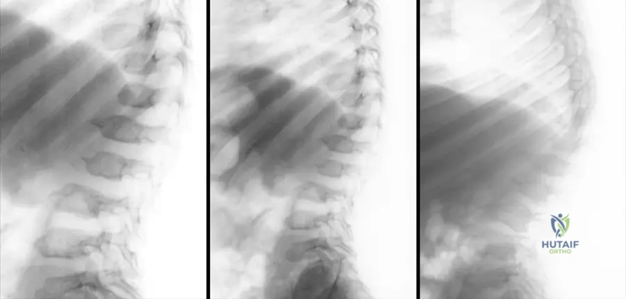
Based on the lateral spine radiograph of this 17-year-old male patient presenting with short trunk dwarfism, what is the characteristic vertebral deformity shown?
Explanation
Correct Answer: Champagne-bottle shaped vertebrae
The radiograph demonstrates the typical 'champagne-bottle' shaped vertebral bodies, which are a hallmark radiographic finding in Spondyloepiphyseal Dysplasia Tarda and Congenita. Progressive dorsolumbar kyphosis and platyspondyly are also commonly observed.
Question 4
A 6-year-old child with Spondyloepiphyseal Dysplasia Congenita is scheduled for elective lower extremity surgery. Which of the following preoperative evaluations is most critical to prevent a catastrophic neurological complication during anesthesia induction and intubation?
Explanation
Correct Answer: Flexion-extension radiographs of the cervical spine
Patients with Spondyloepiphyseal Dysplasia Congenita commonly have an os odontoideum with or without atlantoaxial instability. Preoperative flexion-extension cervical spine radiographs are critical to evaluate for instability before intubation to prevent iatrogenic spinal cord injury.
Question 5
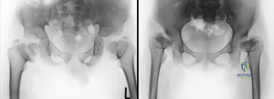
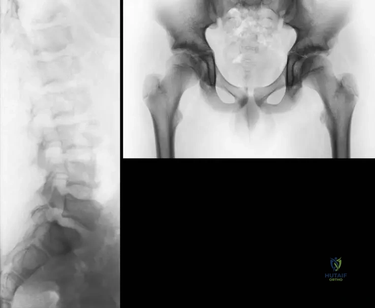
A 28-year-old female with a known history of an inherited chondrodysplasia presents with hip pain and a waddling gait. Review the provided radiograph. Which of the following proximal femoral deformities is most commonly associated with this condition in adulthood?
Explanation
Correct Answer: Coxa vara
The radiograph shows retarded ossification of the proximal femur, which is usually accompanied by coxa vara in the adult period for patients with Spondyloepiphyseal Dysplasia. This deformity contributes to the waddling gait and hip pain.
Question 6
A family seeks genetic counseling regarding Spondyloepiphyseal Dysplasia Tarda. The father is unaffected, and the mother is a known heterozygous carrier. What is the probability that their daughter will exhibit the clinical and radiographic manifestations of the disease?
Explanation
Correct Answer: 0%
Spondyloepiphyseal dysplasia tarda is an X-linked recessive disorder. Heterozygous carrier females are clinically and radiographically normal. The disease affects males only. Therefore, the probability of a daughter exhibiting clinical manifestations is 0% (though she has a 50% chance of being a carrier).
Question 7
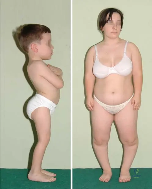
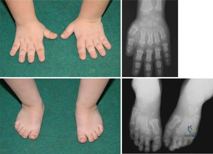
A young girl presents with disproportionate short stature. Clinical and radiographic evaluation of her extremities is shown below. Which of the following best describes the findings in the appendicular skeleton of patients with this condition?
Explanation
Correct Answer: Short, small tubular bones with broad feet
Patients with Spondyloepiphyseal Dysplasia typically present with short, small tubular bones in the hands and broad feet, as demonstrated in the provided clinical and radiographic images.
Question 8
A 5-year-old boy diagnosed with Spondyloepiphyseal Dysplasia Congenita is being monitored in the orthopedic clinic. Which of the following spinal deformities is most likely to progress as he ages, requiring close observation?
Explanation
Correct Answer: Dorsolumbar kyphosis
Progressive dorsolumbar kyphosis with platyspondyly and deformed vertebrae is a characteristic and progressive spinal deformity in patients with Spondyloepiphyseal Dysplasia Congenita, often requiring close clinical and radiographic monitoring.
Question 9
Review the anteroposterior and lateral thoracolumbar spine radiographs of this patient. The generalized flattening of the vertebral bodies observed is medically termed:
Explanation
Correct Answer: Platyspondyly
Platyspondyly refers to the generalized flattening of the vertebral bodies, which is clearly visible on the provided thoracolumbar spine radiographs along with narrow disc spaces. It is a hallmark of Spondyloepiphyseal Dysplasia.
Question 10
When differentiating Spondyloepiphyseal Dysplasia Congenita (SEDc) from Achondroplasia, which of the following clinical features is most characteristic of SEDc?
Explanation
Correct Answer: Disproportionate short stature with a markedly short trunk
SEDc is characterized by a short trunk due to a growth disorder of the spine (platyspondyly), whereas achondroplasia typically presents with a normal trunk length and rhizomelic (proximal) shortening of the limbs.
Question 11
A 5-year-old boy presents with disproportionate short stature, a short trunk, and the spinal radiographic findings shown below. Genetic testing is most likely to reveal a mutation affecting which of the following?
Explanation
Correct Answer: Type II procollagen
Spondyloepiphyseal dysplasia congenita (SEDc) is an inherited chondrodysplasia characterized by a short trunk, platyspondyly, and epiphyseal dysplasia. It is caused by a mutation in the COL2A1 gene, which encodes type II procollagen.
Question 12
A 10-year-old boy presents with a short neck, broad chest, and the characteristic vertebral shape seen in the radiograph below. His mother is clinically and radiographically normal. What is the inheritance pattern of this specific condition?
Explanation
Correct Answer: X-linked recessive
The radiograph shows 'champagne-bottle' shaped vertebrae, characteristic of Spondyloepiphyseal dysplasia tarda (SEDt). SEDt is an X-linked recessive progressive osteochondrodysplasia. It affects males only, while heterozygous carrier females are clinically and radiographically normal.
Question 13
A patient with congenital spondyloepiphyseal dysplasia is being evaluated prior to general anesthesia for a surgical procedure. Which of the following cervical spine abnormalities is most commonly associated with this condition and requires careful preoperative assessment?
Explanation
Correct Answer: Os odontoideum with atlantoaxial instability
Patients with Spondyloepiphyseal dysplasia congenita (SEDc) commonly present with os odontoideum, with or without atlantoaxial instability. This requires careful preoperative evaluation, especially before general anesthesia, to prevent catastrophic neurologic injury during intubation.
Question 14
A 28-year-old female with a history of a skeletal dysplasia presents with hip pain. Her pelvic radiograph is shown below. What is the primary deformity seen in the proximal femur that typically develops in this condition?
Explanation
Correct Answer: Coxa vara
The radiograph demonstrates retarded ossification of the proximal femur, which is usually accompanied by coxa vara in older patients with spondyloepiphyseal dysplasia.
Question 15
A 12-year-old male presents with progressive dorsolumbar kyphosis. The lateral radiograph of his thoracolumbar spine is shown below. The characteristic shape of the vertebral bodies in this condition is classically described as:
Explanation
Correct Answer: Champagne-bottle vertebrae
Spondyloepiphyseal dysplasia tarda (SEDt) is characterized by defective growth and typical 'champagne-bottle' shaped vertebrae, as seen in the provided radiograph.
Question 16
A young girl with a known chondrodysplasia presents for evaluation. Clinical and radiographic images of her hands and feet are shown below. Which of the following best describes the extremity manifestations typical of this condition?
Explanation
Correct Answer: Short small tubular bones with broad feet
The images demonstrate short small tubular bones in the hands and broad feet, which are characteristic extremity findings in patients with spondyloepiphyseal dysplasia.
Question 17
A family presents for genetic counseling. The father has a history of spondyloepiphyseal dysplasia tarda (SEDt). Which of the following statements regarding the transmission and manifestation of this specific disorder is correct?
Explanation
Correct Answer: The disease affects males only and manifests in childhood
Spondyloepiphyseal dysplasia tarda is an X-linked recessive disorder. It manifests in childhood with disproportionate short stature. Heterozygous carrier females are generally clinically and radiographically normal, and the disease affects males only.
Question 18
A 17-year-old boy with spondyloepiphyseal dysplasia congenita presents with worsening back deformity. Based on the typical progression of this disease and the provided radiograph, what is the most likely spinal deformity?
Explanation
Correct Answer: Progressive dorsolumbar kyphosis with platyspondyly
Patients with spondyloepiphyseal dysplasia congenita typically develop progressive dorsolumbar kyphosis with platyspondyly and deformed vertebrae, as demonstrated in the radiograph.
Question 19
A 35-year-old male with a history of X-linked recessive spondyloepiphyseal dysplasia tarda presents with worsening joint pain and stiffness in his hips and knees. Which of the following is a known long-term complication associated with this condition?
Explanation
Correct Answer: Progressive arthropathy
Spondyloepiphyseal dysplasia tarda (SEDt) is an osteochondrodysplasia that can be associated with progressive arthropathy, leading to significant joint pain and stiffness in adulthood.
Question 20
A 4-year-old child is being evaluated for short stature. Radiographs reveal platyspondyly and epiphyseal dysplasia of the femoral head. The child has a disproportionately short trunk. This presentation is most consistent with spondyloepiphyseal dysplasia congenita. Which of the following features most reliably distinguishes this condition from achondroplasia?
Explanation
Correct Answer: Presence of a short trunk
Spondyloepiphyseal dysplasia congenita is associated with a short trunk due to a growth disorder of the spine (platyspondyly). In contrast, achondroplasia is characterized by a relatively normal trunk length and rhizomelic (proximal) shortening of the limbs.
Question 21
A 5-year-old boy presents with disproportionate short stature, a short trunk, and platyspondyly. Radiographs show epiphyseal dysplasia of the femoral head. Genetic testing is most likely to reveal a mutation in which of the following genes?
Explanation
Correct Answer: C
Spondyloepiphyseal dysplasia congenita (SEDc) is an inherited chondrodysplasia caused by a mutation in the COL2A1 gene, which encodes type II procollagen. This leads to a growth disorder of the spine and epiphyses, resulting in a short trunk, platyspondyly, and epiphyseal dysplasia. FGFR3 mutations are associated with achondroplasia, COL1A1 with osteogenesis imperfecta, COMP with pseudoachondroplasia, and RUNX2 with cleidocranial dysplasia.
Question 22
A 10-year-old boy presents with disproportionate short stature, a broad chest, and a short neck. Radiographs reveal 'champagne bottle' shaped vertebrae. His parents are unaffected, but his maternal uncle has a similar condition. What is the inheritance pattern of this specific disorder?
Explanation
Correct Answer: D
Spondyloepiphyseal dysplasia tarda (SEDt) is an X-linked recessive progressive osteochondrodysplasia. It manifests in childhood with disproportionate short stature, a short neck and trunk, and a broad chest. Because it is X-linked recessive, heterozygous carrier females are generally clinically and radiographically normal, and the disease exclusively affects males.
Question 23
A 12-year-old male presents with short stature and back pain. The lateral radiograph of the thoracolumbar spine is shown below. What is the characteristic radiographic sign seen in the vertebral bodies?
Explanation
Correct Answer: C
The radiograph demonstrates platyspondyly and narrow disc spaces with the characteristic 'champagne-bottle' shaped vertebrae of the lower thoracic spine, which is a hallmark finding in Spondyloepiphyseal dysplasia tarda. 'Picture frame' vertebrae are seen in Paget's disease, 'Rugger jersey' spine in renal osteodystrophy, and 'Bullet-shaped' vertebrae in achondroplasia or mucopolysaccharidoses.
Question 24
A 6-year-old child with known spondyloepiphyseal dysplasia congenita is scheduled for elective lower extremity surgery. Which of the following preoperative evaluations is most critical to prevent a catastrophic neurological complication?
Explanation
Correct Answer: B
Patients with spondyloepiphyseal dysplasia congenita commonly have an os odontoideum with or without atlantoaxial instability. Prior to any surgery requiring general anesthesia and intubation, it is critical to obtain flexion-extension radiographs of the cervical spine to rule out instability, which could lead to catastrophic spinal cord injury during neck manipulation.
Question 25
A 28-year-old female with a history of spondyloepiphyseal dysplasia presents with hip pain and a waddling gait. Her pelvic radiograph is shown below. Which of the following proximal femoral deformities is most commonly associated with this condition in adulthood?
Explanation
Correct Answer: B
The radiograph shows retarded ossification of the proximal femur, which is usually accompanied by coxa vara in the elderly period or adulthood in patients with spondyloepiphyseal dysplasia. This deformity contributes to the waddling gait and progressive arthropathy seen in these patients.
Question 26
Which of the following clinical features best distinguishes spondyloepiphyseal dysplasia from achondroplasia?
Explanation
Correct Answer: B
Spondyloepiphyseal dysplasia is characterized by a short trunk due to a growth disorder of the spine, leading to disproportionate short stature where the trunk is short relative to the limbs. In contrast, achondroplasia is characterized by rhizomelic (proximal) limb shortening with a relatively normal trunk length.
Question 27
A young female patient presents with the clinical and radiographic findings of the hands and feet shown below. In the context of spondyloepiphyseal dysplasia, what is the primary underlying defect causing these skeletal manifestations?
Explanation
Correct Answer: C
The images show short, small tubular bones and broad feet. Spondyloepiphyseal dysplasia is an inherited chondrodysplasia characterized by a growth disorder of the spine and the epiphyses of the limbs, leading to these characteristic changes in the hands and feet. It is not a defect of mineralization (rickets/osteomalacia) or osteoclast function (osteopetrosis).
Question 28
A couple seeks genetic counseling. The father has spondyloepiphyseal dysplasia tarda, and the mother is unaffected and is not a carrier. What is the probability that their sons will be affected by the disease?
Explanation
Correct Answer: A
Spondyloepiphyseal dysplasia tarda is an X-linked recessive disorder. An affected father will pass his Y chromosome to all of his sons, meaning none of his sons (0%) will inherit the defective X chromosome or the disease. He will pass his defective X chromosome to all of his daughters, making them obligate carriers, but they will be clinically normal.
Question 29
A 5-year-old boy presents with the progressive spinal deformity shown in the radiograph below. Which of the following terms best describes the generalized flattening of the vertebral bodies seen in this condition?
Explanation
Correct Answer: B
The radiograph demonstrates progressive dorsolumbar kyphosis with platyspondyly. Platyspondyly refers to the generalized flattening of the vertebral bodies, which is a hallmark radiographic finding in spondyloepiphyseal dysplasia congenita. Platybasia refers to flattening of the skull base.
Question 30
Spondyloepiphyseal dysplasia congenita is primarily caused by a defect in which of the following structural components of cartilage?
Explanation
Correct Answer: B
Spondyloepiphyseal dysplasia congenita occurs through a mutation in the COL2A1 gene, which encodes type II procollagen. Type II collagen is the primary structural collagen found in articular and hyaline cartilage, explaining the epiphyseal and spinal growth disturbances seen in this condition.
Question 31
A 5-year-old boy presents with disproportionate short stature, a short trunk, and progressive dorsolumbar kyphosis. Radiographs reveal platyspondyly and epiphyseal dysplasia of the femoral head. Genetic testing is most likely to reveal a mutation in which of the following genes?
Explanation
Correct Answer: COL2A1
Congenital spondyloepiphyseal dysplasia (SEDC) is an inherited chondrodysplasia characterized by short stature and a short trunk due to a growth disorder of the spine and epiphyses. It is caused by a mutation in the COL2A1 gene, which encodes type II procollagen. FGFR3 mutations are associated with achondroplasia, COL1A1 with osteogenesis imperfecta, COMP with pseudoachondroplasia, and RUNX2 with cleidocranial dysplasia.
Question 32
A 10-year-old boy presents with progressive arthropathy, a short neck, and a broad chest. Radiographs show characteristic vertebral anomalies. His mother is clinically and radiographically normal, but his maternal uncle has a similar condition. What is the inheritance pattern of this disorder?
Explanation
Correct Answer: X-linked recessive
The clinical picture describes Spondyloepiphyseal Dysplasia Tarda (SEDT). SEDT is an X-linked recessive progressive osteochondrodysplasia. It manifests in childhood and affects males only, while heterozygous carrier females are generally clinically and radiographically normal.
Question 33
A 17-year-old male presents with a short trunk and progressive back deformity. The lateral radiograph of his spine is shown below. What is the characteristic radiographic finding seen in the vertebral bodies of this condition?
Explanation
Correct Answer: Champagne-bottle shaped vertebral bodies
The radiograph demonstrates the typical 'champagne-bottle' shaped vertebral bodies characteristic of Spondyloepiphyseal Dysplasia. This is accompanied by progressive dorsolumbar kyphosis and platyspondyly. 'Picture frame' vertebrae are seen in Paget's disease, 'Rugger jersey' in renal osteodystrophy, 'Bamboo spine' in ankylosing spondylitis, and 'Bullet-shaped' vertebrae in achondroplasia or mucopolysaccharidoses.
Question 34
A 28-year-old female with a known history of an inherited chondrodysplasia presents with hip pain and a waddling gait. Her pelvic radiograph is shown below. Which of the following deformities is most commonly associated with the delayed ossification of the proximal femur seen in this condition?
Explanation
Correct Answer: Coxa vara
In Spondyloepiphyseal Dysplasia, there is retarded ossification of the proximal femur in young patients, which is usually accompanied by the development of coxa vara in the older period, as seen on the radiograph of this 28-year-old female. This contributes to the progressive arthropathy and gait abnormalities.
Question 35
Patients with congenital spondyloepiphyseal dysplasia (SEDC) are at high risk for a specific cervical spine anomaly that requires careful preoperative evaluation before any surgery requiring intubation. Which of the following is the most common cervical spine manifestation in these patients?
Explanation
Correct Answer: Os odontoideum with atlantoaxial instability
In SEDC, os odontoideum with or without atlantoaxial instability is a common and critical finding. It poses a significant risk for spinal cord compression, especially during neck extension for endotracheal intubation, making preoperative cervical spine flexion-extension radiographs mandatory.
Question 36
Which of the following statements accurately describes the epidemiology and clinical manifestation of Spondyloepiphyseal Dysplasia Tarda (SEDT)?
Explanation
Correct Answer: It affects males only, manifests in childhood, and is associated with progressive arthropathy.
SEDT is an X-linked recessive disorder, meaning it affects males only, while heterozygous carrier females are clinically and radiographically normal. It manifests in childhood with disproportionate short stature, a short neck and trunk, and a broad chest, and is commonly associated with progressive arthropathy.
Question 37
A young girl with disproportionate short stature presents for evaluation. Clinical and radiographic images of her hands and feet are shown below. Which of the following best describes the findings in the appendicular skeleton of patients with this condition?
Explanation
Correct Answer: Short, small tubular bones with broad feet
The images demonstrate short, small tubular bones in the hands and broad feet, which are characteristic appendicular findings in patients with Spondyloepiphyseal Dysplasia. Arachnodactyly is seen in Marfan syndrome, and Madelung deformity is associated with Leri-Weill dyschondrosteosis.
Question 38
Congenital spondyloepiphyseal dysplasia is caused by a mutation in the COL2A1 gene. This mutation primarily affects the synthesis of which of the following structural proteins?
Explanation
Correct Answer: Type II procollagen
The COL2A1 gene encodes type II procollagen, which is the major collagenous component of articular cartilage and the nucleus pulposus of intervertebral discs. Mutations in this gene lead to the defective cartilage matrix seen in SEDC.
Question 39
A patient with a known osteochondrodysplasia presents with back pain. Anteroposterior and lateral radiographs of the thoracolumbar spine are shown below. Which of the following terms best describes the generalized flattening of the vertebral bodies seen in this imaging?
Explanation
Correct Answer: Platyspondyly
Platyspondyly refers to the generalized flattening of the vertebral bodies, which is a hallmark radiographic feature of Spondyloepiphyseal Dysplasia. The radiographs also show narrow disc spaces and the characteristic 'champagne-bottle' shaped vertebrae of the lower thoracic spine.
Question 40
A key differentiating factor between Spondyloepiphyseal Dysplasia Congenita (SEDC) and Spondyloepiphyseal Dysplasia Tarda (SEDT) is their genetic inheritance and onset. Which of the following is true regarding SEDT?
Explanation
Correct Answer: It is an X-linked recessive disorder that manifests in childhood.
SEDT is an X-linked recessive disorder that manifests later in childhood, unlike SEDC, which is congenital (present at birth) and typically caused by an autosomal dominant mutation in the COL2A1 gene. SEDT affects males exclusively and leads to progressive osteochondrodysplasia.
Question 41
A 4-year-old child presents with disproportionate short stature, a short trunk, and epiphyseal dysplasia of the femoral heads. Genetic testing reveals a mutation in the COL2A1 gene. Which of the following proteins is primarily affected in this condition?
Explanation
Correct Answer: Type II procollagen
Spondyloepiphyseal dysplasia (SED) congenita is caused by a mutation in the COL2A1 gene, which encodes for type II procollagen. This leads to defective cartilage formation, resulting in the characteristic short trunk, platyspondyly, and epiphyseal dysplasia.
Question 42
Review the lateral radiograph of the thoracolumbar spine of a 17-year-old boy presenting with progressive dorsolumbar kyphosis.
Based on the characteristic vertebral morphology shown, what is the most likely diagnosis?
Explanation
Correct Answer: Spondyloepiphyseal dysplasia
The radiograph demonstrates platyspondyly and the typical 'champagne-bottle' shaped vertebral bodies, which are hallmark radiographic features of Spondyloepiphyseal dysplasia (SED). Achondroplasia typically presents with posterior scalloping and narrowing of interpedicular distances, while Morquio syndrome features anterior central beaking.
Question 43
A 10-year-old boy presents with a short neck, broad chest, and disproportionate short stature. Radiographs reveal 'champagne bottle' shaped vertebrae. His mother is clinically and radiographically normal, but his maternal uncle has a similar condition. What is the inheritance pattern of this specific disorder?
Explanation
Correct Answer: X-linked recessive
The clinical scenario describes Spondyloepiphyseal dysplasia tarda, which is an X-linked recessive progressive osteochondrodysplasia. It affects males only, while heterozygous carrier females are generally clinically and radiographically normal.
Question 44
Patients with Spondyloepiphyseal Dysplasia Congenita are at high risk for a specific cervical spine anomaly that requires careful preoperative evaluation before any general anesthesia. Which of the following is the most common cervical spine manifestation in these patients?
Explanation
Correct Answer: Atlantoaxial instability due to os odontoideum
In Spondyloepiphyseal dysplasia congenita, os odontoideum with or without atlantoaxial instability is a common and critical finding. It must be evaluated prior to any surgical procedure requiring intubation to prevent catastrophic spinal cord injury.
Question 45
A 28-year-old female with a known skeletal dysplasia presents with hip pain. Her pelvic radiograph is shown below.
Which of the following best describes the characteristic proximal femoral deformity seen in this condition during adulthood?
Explanation
Correct Answer: Coxa vara with retarded ossification of the proximal femur
In Spondyloepiphyseal dysplasia, there is retarded ossification of the proximal femur in youth, which typically progresses to coxa vara in adulthood, as demonstrated in the provided radiograph.
Question 46
Which of the following clinical profiles is most characteristic of Spondyloepiphyseal Dysplasia Tarda?
Explanation
Correct Answer: Disproportionate short stature with a short trunk, short neck, and broad chest manifesting in childhood
SED tarda manifests in childhood with disproportionate short stature, a short neck and trunk, and a broad chest. The short stature is primarily due to a growth disorder of the spine (short trunk), unlike achondroplasia which features rhizomelic limb shortening with a relatively normal trunk.
Question 47
A young female patient with a short trunk and short stature presents for evaluation. Clinical and radiographic images of her hands and feet are provided.
What is the predominant finding in the appendicular skeleton shown in these images?
Explanation
Correct Answer: Short, small tubular bones and broad feet
The images demonstrate short, small tubular bones in the hands and broad feet, which are characteristic appendicular findings in patients with Spondyloepiphyseal dysplasia.
Question 48
In Spondyloepiphyseal Dysplasia Tarda, the vertebral bodies develop a pathognomonic radiographic appearance due to defective growth. Which of the following terms best describes this characteristic vertebral shape?
Explanation
Correct Answer: Champagne-bottle vertebrae
The 'champagne-bottle' shaped vertebra is a classic radiographic hallmark of Spondyloepiphyseal dysplasia tarda, resulting from defective growth of the vertebral ring apophyses. 'Picture-frame' is seen in Paget's disease, 'Rugger-jersey' in renal osteodystrophy, and 'Bullet-shaped' in achondroplasia or Morquio syndrome.
Question 49
Anteroposterior and lateral thoracolumbar spine radiographs of a patient with a genetic skeletal dysplasia are shown.
In addition to the characteristic vertebral body shape, what other spinal radiographic feature is prominently demonstrated?
Explanation
Correct Answer: Platyspondyly and narrow disc spaces
The radiographs show platyspondyly (flattened vertebral bodies) and narrow disc spaces, alongside the characteristic 'champagne-bottle' shaped vertebrae of the lower thoracic spine, typical of Spondyloepiphyseal dysplasia.
Question 50
A 35-year-old male with a history of X-linked recessive Spondyloepiphyseal Dysplasia Tarda presents to the orthopedic clinic. Which of the following long-term musculoskeletal complications is most strongly associated with this condition as the patient ages?
Explanation
Correct Answer: Progressive arthropathy
Spondyloepiphyseal dysplasia tarda is frequently associated with progressive arthropathy, particularly affecting the hips and spine, leading to early-onset osteoarthritis and significant morbidity in adulthood.
Question 51
A 4-year-old child with spondyloepiphyseal dysplasia congenita (SEDC) is scheduled for a valgus osteotomy for coxa vara. Prior to intubation, what is the most critical screening study required?
Explanation
Question 52
Spondyloepiphyseal dysplasia congenita (SEDC) is associated with mutations in the COL2A1 gene. Which of the following tissues is primarily affected by this genetic defect?
Explanation
Question 53
A 14-year-old male presents with worsening hip pain and early-onset hip osteoarthritis. He has short stature with a disproportionately short trunk. Radiographs reveal a "heaped up" appearance of the posterior portion of the vertebral endplates. What is the most likely diagnosis?
Explanation
Question 54
A patient with spondyloepiphyseal dysplasia congenita is noted to have severe, progressive coxa vara. If surgical intervention is planned, what is the primary goal of the recommended procedure?
Explanation
Question 55
Spondyloepiphyseal dysplasia (SED) can present similarly to Morquio syndrome. Which of the following clinical features most reliably distinguishes SED congenita from Morquio syndrome?
Explanation
Question 56
A biopsy of the epiphyseal cartilage from a patient with spondyloepiphyseal dysplasia congenita (SEDC) is examined histologically. Which of the following findings is most characteristic of this disorder?
Explanation
Question 57
Spondyloepiphyseal dysplasia tarda is typically inherited in which of the following patterns?
Explanation
Question 58
Which of the following non-orthopedic specialists is most essential in the routine multidisciplinary surveillance of a child with spondyloepiphyseal dysplasia congenita (SEDC)?
Explanation
Question 59
A 25-year-old male with spondyloepiphyseal dysplasia tarda presents for evaluation of progressive back pain. Lateral radiographs of the thoracolumbar spine are likely to demonstrate:
Explanation
Question 60
A child with a known diagnosis of spondyloepiphyseal dysplasia congenita develops upper motor neuron signs, including hyperreflexia and a positive Babinski sign. What is the most appropriate next step in management?
Explanation
Question 61
You are evaluating a newborn with a disproportionately short trunk, flat face, and cleft palate. Radiographs show absent ossification centers for the pubic bones, distal femoral epiphyses, and proximal tibial epiphyses. What is the most likely diagnosis?
Explanation
Question 62
A 12-year-old boy presents with progressive hip pain. Radiographs show flattened femoral heads and early degenerative changes, but his spine radiographs are entirely normal. Which of the following is the most likely diagnosis?
Explanation
Question 63
In addition to the spine and large joints, which of the following appendicular skeletal deformities is frequently observed at birth in patients with spondyloepiphyseal dysplasia congenita?
Explanation
Question 64
What distinguishes the clinical presentation of Pseudoachondroplasia from Spondyloepiphyseal Dysplasia (SED)?
Explanation
Question 65
What is the genetic mutation associated with Spondyloepiphyseal Dysplasia (SED) tarda?
Explanation
Question 66
A 7-year-old child with spondyloepiphyseal dysplasia congenita presents with a waddling gait. Radiographs demonstrate an abnormal neck-shaft angle of 90 degrees with a vertical physeal orientation. Which complication is most likely if this is left untreated?
Explanation
Question 67
A patient with spondyloepiphyseal dysplasia congenita requires an atlantoaxial fusion due to symptomatic instability. Which of the following factors makes cervical spine surgery particularly challenging in this population?
Explanation
Question 68
Spondyloepiphyseal dysplasia congenita is characterized by a defect in Type II collagen. Which of the following conditions shares the same underlying defective protein?
Explanation
Question 69
A 6-year-old girl with SEDC is evaluated for progressive spinal deformity. Which spinal deformity is most classically associated with the natural history of this condition?
Explanation
Question 70
Which of the following best describes the classical radiographic appearance of the pelvis and hips in an infant with Spondyloepiphyseal Dysplasia Congenita (SEDC)?
Explanation
Question 71
A 3-year-old child presents with disproportionate short trunk dwarfism, a cleft palate, and a waddling gait. Radiographs reveal coxa vara and delayed ossification of the femoral heads. Genetic testing confirms a mutation in the COL2A1 gene. Which type of collagen is primarily defective in this patient?
Explanation
Question 72
A 12-year-old male with a short trunk and barrel chest is diagnosed with Spondyloepiphyseal Dysplasia Tarda (SEDT). He has normal vision and intelligence. Which of the following best describes the inheritance pattern of the most common form of this specific condition?
Explanation
Question 73
A 5-year-old girl with Spondyloepiphyseal Dysplasia Congenita is scheduled to undergo strabismus surgery under general anesthesia. Which of the following pre-operative orthopedic evaluations is absolutely mandatory?
Explanation
Question 74
You are examining a newborn diagnosed with Spondyloepiphyseal Dysplasia Congenita. In addition to monitoring the skeletal system and cervical spine, what other specialized screening must be routinely performed throughout this patient's childhood?
Explanation
Question 75
Which of the following genes is responsible for X-linked Spondyloepiphyseal Dysplasia Tarda?
Explanation
Question 76
A 4-year-old child presents with short trunk dwarfism and a waddling gait. Radiographs show universal platyspondyly. The patient has a cleft palate and clear corneas on eye examination. Unlike Morquio syndrome, which can present similarly, this patient's condition is primarily caused by a mutation in:
Explanation
Question 77
A 6-year-old boy with Spondyloepiphyseal Dysplasia Congenita is noted to have a progressively worsening waddling gait. Pelvic radiographs reveal bilateral coxa vara with a Hilgenreiner-epiphyseal angle (HEA) of 65 degrees. What is the most appropriate management?
Explanation
Question 78
Which of the following histologic findings is classically characteristic of the chondrocytes in a patient with Spondyloepiphyseal Dysplasia Congenita?
Explanation
Question 79
A newborn presents with short trunk disproportionate dwarfism. Radiographic evaluation reveals marked delay in ossification of several centers. Which of the following ossification centers is characteristically delayed or completely absent at birth in Spondyloepiphyseal Dysplasia Congenita?
Explanation
Question 80
In a 14-year-old male with Spondyloepiphyseal Dysplasia Tarda, what is the most characteristic pathognomonic finding on a lateral radiograph of the lumbar spine?
Explanation
Question 81
Review the clinical image.
A 2-year-old child presents with delayed walking and short stature. The pelvic radiograph reveals coxa vara and distinctly absent ossification of the femoral heads and severely delayed ossification of the pubic symphysis. What is the most likely diagnosis?
Explanation
Question 82
An adult male with a known history of Spondyloepiphyseal Dysplasia Tarda presents to the orthopedic clinic with progressively worsening, debilitating groin pain. Given the natural history of this disease, which surgical procedure is he most likely to require in his 30s or 40s?
Explanation
Question 83
The mutation in Spondyloepiphyseal Dysplasia Congenita impairs the normal structural integrity of hyaline cartilage. Which of the following intervertebral disc components is also primarily composed of this defective protein?
Explanation
Question 84
A 7-year-old girl with Spondyloepiphyseal Dysplasia Congenita is noted to have progressive coxa vara. Radiographs show a neck-shaft angle of 85 degrees. What is the primary biomechanical rationale for performing a subtrochanteric valgus osteotomy in this patient?
Explanation
Question 85
Morquio syndrome and Spondyloepiphyseal Dysplasia Congenita both present with short trunk dwarfism and atlantoaxial instability. Which of the following clinical features reliably distinguishes Morquio syndrome from SED Congenita on physical examination?
Explanation
Question 86
Which of the following represents the most immediately life-threatening complication of Spondyloepiphyseal Dysplasia Congenita in early childhood?
Explanation
Question 87
What spinal deformity is most frequently associated with Spondyloepiphyseal Dysplasia Congenita and typically progresses during childhood, often requiring surgical fusion?
Explanation
Question 88
Based on the lateral spine radiograph showing characteristic heaped-up bone on the posterior vertebral endplates, a 14-year-old boy is diagnosed with SED Tarda.
What is the inheritance pattern for the most common form of this disease?
Explanation
Question 89
While performing a physical examination on a newborn later diagnosed with Spondyloepiphyseal Dysplasia Congenita, you note a bilateral structural foot deformity. What is the most common foot anomaly associated with this skeletal dysplasia?
Explanation
Question 90
A pregnant woman with a known family history of X-linked Spondyloepiphyseal Dysplasia Tarda undergoes genetic counseling. Genetic testing confirms she is a carrier of the TRAPPC2 mutation. Her husband is unaffected. What is the probability that their son will be affected by the condition?
Explanation
Question 91
A 13-year-old male presents with bilateral hip pain and stiffness. Examination reveals a short trunk with normal limb proportions. Radiographs demonstrate premature osteoarthritis of the hips and characteristic humped vertebral endplates in the lumbar spine. His maternal uncle required bilateral total hip arthroplasties at age 35. A mutation in which of the following genes is most likely responsible?
Explanation
Question 92
A 5-year-old child with spondyloepiphyseal dysplasia congenita (SEDC) is scheduled to undergo a proximal femoral valgus osteotomy for severe coxa vara. Which of the following preoperative evaluations is absolutely critical before proceeding with general anesthesia?
Explanation
Question 93
A newborn is evaluated for severe disproportionate short stature. Radiographic survey reveals absent ossification of the pubic bones, calcaneus, talus, and distal femoral epiphyses. Which of the following histological findings is most characteristic of the cartilage in this patient's underlying condition?
Explanation
Question 94
Which of the following clinical features most reliably differentiates Spondyloepiphyseal Dysplasia Congenita (SEDC) from Morquio syndrome (Mucopolysaccharidosis Type IV) in a 6-year-old patient with short-trunk dwarfism and atlantoaxial instability?
Explanation
Question 95
A 4-year-old boy with a known COL2A1 mutation is being evaluated in the orthopedic clinic for a waddling gait.
Based on the typical natural history of his skeletal dysplasia, which of the following hip deformities is most likely present?
Explanation
Question 96
Because of the widespread distribution of the defective protein in Spondyloepiphyseal Dysplasia Congenita, patients are at high risk for extraskeletal complications. Routine screening by which of the following specialists is most imperative?
Explanation
Question 97
A 10-year-old girl with Spondyloepiphyseal Dysplasia Congenita presents with increasing lower extremity weakness, hyperreflexia, and a positive Babinski sign. Cervical radiographs reveal an atlantodens interval (ADI) of 8 mm. What is the primary pathoanatomic etiology of her cervical instability?
Explanation
Question 98
A newborn presents with a short trunk, cleft palate, and a barrel chest. Radiographs show flattened vertebral bodies (platyspondyly) and the absence of ossification in the knees and feet. The parents are of normal height and have no significant medical history. Which type of collagen is quantitatively or qualitatively abnormal in this infant?
Explanation
Question 99
In a patient with Spondyloepiphyseal Dysplasia Tarda (SEDT), which of the following radiographic spinal abnormalities is considered the hallmark pathognomonic finding?
Explanation
Question 100
A 28-year-old male with an established diagnosis of Spondyloepiphyseal Dysplasia Tarda (SEDT) presents with severe, debilitating groin pain bilaterally. Conservative management has failed.
Based on the natural history of this disorder, which of the following surgical interventions is he most likely to require?
Explanation
None
