Dysplasia Epiphysealis Hemimelica (DEH) MCQs - Ortho Board
Dysplasia Epiphysealis Hemimelica (DEH) MCQs - Ortho Board
Comprehensive 100-Question Exam
00:00
Start Quiz
Question 1
Which of the following demographic profiles is most characteristic for the onset of Dysplasia Epiphysealis Hemimelica (DEH)?
Explanation
Correct Answer: A 6-year-old male
Dysplasia epiphysealis hemimelica (DEH) is a rare skeletal developmental disorder that typically presents in young children. The age of onset is usually between 2 and 14 years, and males are affected twice as frequently as females.
Question 2
A biopsy of a lesion suspected to be Dysplasia Epiphysealis Hemimelica (DEH) is sent for histological examination. The pathologist notes that the tissue is histologically indistinguishable from which of the following benign bone tumors?
Explanation
Correct Answer: Osteochondroma
Histologically, DEH is similar to an osteochondroma. However, they are distinguished by their site of origin: an osteochondroma arises from the metaphysis or diaphysis, whereas DEH arises from the epiphysis.
Question 3
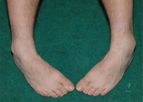
A 7-year-old boy presents with the clinical finding shown in the image below. He reports mild aching pain and limited range of motion. Given the most likely diagnosis of Dysplasia Epiphysealis Hemimelica, which anatomical structure is most commonly affected in this specific region?
Explanation
Correct Answer: Talus
The image demonstrates a bone-hard swelling at the lateral side of the ankle. DEH usually occurs in the lower limb, with the distal femur, distal tibia, and talus being the most commonly affected sites.
Question 4
Which of the following radiographic findings is the hallmark of Dysplasia Epiphysealis Hemimelica?
Explanation
Correct Answer: Asymmetric epiphyseal enlargement with multiple ossification centers
Characteristically, DEH lesions show on radiographs as asymmetric epiphyseal enlargement with multiple ossification centers, reflecting the hemimelic (one-sided) nature of the epiphyseal involvement.
Question 5
Review the MRI and CT images of a 9-year-old patient presenting with a hard, asymmetric ankle mass. The lesion originates from the epiphysis. Based on the typical distribution of this disease, which of the following statements is true regarding its anatomical predilection?
Explanation
Correct Answer: It characteristically involves either the medial or lateral epiphyseal side
The term 'hemimelica' refers to the characteristic involvement of only half of the epiphysis (either the medial or lateral side). Upper limb involvement is extremely rare, and the lesion arises from the epiphysis, not the diaphysis.
Question 6
While Dysplasia Epiphysealis Hemimelica (DEH) shares histological similarities with osteochondroma, it is primarily distinguished by its site of origin. From which specific anatomical region of the bone does DEH arise?
Explanation
Correct Answer: Epiphysis
DEH is a developmental disorder affecting the epiphyses in young children. This is a key distinguishing factor from osteochondromas, which arise from the metaphysis or diaphysis.
Question 7
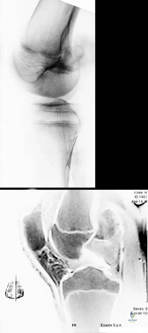
A 10-year-old male presents with limited knee flexion and a palpable mass in the popliteal fossa. Radiograph and MRI are shown below. The mass is seen protruding from the distal femoral epiphysis. What is the estimated incidence of this condition in the general population?
Explanation
Correct Answer: 1 in 1,000,000
Dysplasia epiphysealis hemimelica (DEH) is an extremely rare skeletal developmental disorder with an estimated incidence of 1 in 1,000,000.
Question 8
A 5-year-old boy is brought to the orthopedic clinic by his parents due to a noticeable deformity around his right knee. Which of the following symptom clusters is most characteristic of Dysplasia Epiphysealis Hemimelica (DEH) at presentation?
Explanation
Correct Answer: Bone-hard mass, deformity, aching pains, and limited range of motion
The most common presenting symptoms of DEH include the presence of a mass with the consistency of bone, visible deformity, aching pains, and a limited range of motion in the affected joint.
Question 9
Dysplasia Epiphysealis Hemimelica (DEH) predominantly affects the lower extremities. Which of the following groups of bones represents the most common sites of involvement?
Explanation
Correct Answer: Distal femur, distal tibia, and talus
DEH usually occurs in the lower limb. The distal femur, distal tibia, and talus are the most commonly affected sites. Upper limb involvement is extremely rare.
Question 10
The term 'hemimelica' in Dysplasia Epiphysealis Hemimelica refers to a specific pattern of involvement. Which of the following best describes this characteristic pattern?
Explanation
Correct Answer: Involvement restricted to either the medial or lateral side of the epiphysis
Characteristically, the involvement in DEH is hemimelic, meaning that only one half (either the medial or the lateral side) of the epiphysis is involved, leading to asymmetric growth and deformity.
Question 11
A 6-year-old boy presents with the clinical appearance shown below. Palpation reveals a bone-hard mass. Based on the most likely diagnosis, what is the expected histological appearance of this lesion?
Explanation
Correct Answer: Identical to an osteochondroma
The clinical image demonstrates a moderate, painless, bone-hard swelling at the lateral side of the ankle, characteristic of Dysplasia Epiphysealis Hemimelica (DEH). Histologically, DEH lesions are similar to osteochondromas. However, they are distinguished by their origin: DEH arises from the epiphysis, whereas osteochondromas arise from the metaphysis or diaphysis.
Question 12
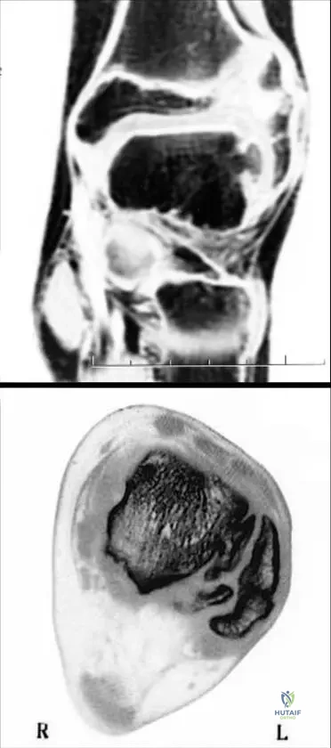
Review the MRI and CT images of a 9-year-old male's ankle below. The lesion is localized to the lateral side of the talus. Which of the following best describes the characteristic radiographic feature of this condition?
Explanation
Correct Answer: Asymmetric epiphyseal enlargement with multiple ossification centers
The images show Dysplasia Epiphysealis Hemimelica (DEH) of the talus. Characteristically, radiographs of DEH show asymmetric epiphyseal enlargement with multiple ossification centers. The involvement is typically hemimelic, affecting either the medial or lateral side of the epiphysis.
Question 13
A 12-year-old boy presents with limited range of motion in his knee. Imaging is provided below. The mass protrudes from the distal femoral epiphysis into the popliteal fossa. How does this condition primarily differ from an osteochondroma?
Explanation
Correct Answer: It arises from the epiphysis rather than the metaphysis or diaphysis
The imaging demonstrates Dysplasia Epiphysealis Hemimelica (DEH) of the distal femur. While histologically similar to an osteochondroma, DEH is uniquely characterized by its origin from the epiphysis. Osteochondromas classically arise from the metaphysis or diaphysis.
Question 14
What is the typical age of onset for Dysplasia Epiphysealis Hemimelica (DEH)?
Explanation
Correct Answer: 2 to 14 years
Dysplasia epiphysealis hemimelica (DEH) is a rare skeletal developmental disorder affecting the epiphyses in young children. The age of onset is usually between 2 and 14 years.
Question 15
Regarding the epidemiology of Dysplasia Epiphysealis Hemimelica (DEH), which of the following statements is correct?
Explanation
Correct Answer: It affects males twice as frequently as females
DEH is a very rare condition with an incidence of 1 in 1,000,000. Males are affected twice as frequently as females. The etiology remains unknown, and it is not described as having a strong autosomal dominant inheritance or specific ethnic exclusivity in standard texts.
Question 16
Which of the following anatomical locations is LEAST likely to be affected by Dysplasia Epiphysealis Hemimelica (DEH)?
Explanation
Correct Answer: Proximal humerus
DEH usually occurs in the lower limb, with the distal femur, distal tibia, and talus being the most commonly affected sites. Upper limb involvement is extremely rare.
Question 17
The term 'hemimelic' in Dysplasia Epiphysealis Hemimelica refers to which of the following characteristics?
Explanation
Correct Answer: Involvement of either the medial or lateral side of the epiphysis
Characteristically, the involvement in DEH is hemimelic, meaning that only one half (either the medial or the lateral side) of the affected epiphysis is involved.
Question 18
A 7-year-old boy presents with aching pain and a bone-hard mass on the medial aspect of his knee. Radiographs show asymmetric epiphyseal enlargement with multiple ossification centers. A biopsy is taken. The pathologist is most likely to report findings consistent with which of the following?
Explanation
Correct Answer: Osteochondroma
The clinical and radiographic presentation is classic for Dysplasia Epiphysealis Hemimelica (DEH). Histologically, the lesion is similar to an osteochondroma, consisting of a bony outgrowth covered by a cartilage cap, but it is distinguished by its epiphyseal origin.
Question 19
Which of the following is the most common presenting symptom complex for a patient with Dysplasia Epiphysealis Hemimelica?
Explanation
Correct Answer: Bone-hard mass, deformity, aching pains, and limited range of motion
The most common presenting symptoms of DEH include the presence of a mass with the consistency of bone, deformity, aching pains, and limited range of motion in the affected joint.
Question 20
A 10-year-old male presents with a bony prominence on the medial side of his ankle. Radiographs reveal an irregular, multi-lobulated ossified mass arising from the medial epiphysis of the distal tibia. What is the estimated incidence of this specific developmental disorder?
Explanation
Correct Answer: 1 in 1,000,000
The patient's presentation is consistent with Dysplasia Epiphysealis Hemimelica (DEH) of the distal tibia. DEH is a very rare skeletal developmental disorder with an estimated incidence of 1 in 1,000,000.
Question 21
A 6-year-old boy presents with the clinical appearance shown below. Palpation reveals a bone-hard, painless mass. Given the most likely diagnosis of Dysplasia Epiphysealis Hemimelica, which of the following is the most common histological finding of this lesion?
Explanation
Correct Answer: Histologically identical to an osteochondroma
According to the text, Dysplasia Epiphysealis Hemimelica (DEH) is histologically similar/identical to an osteochondroma. The key differentiating factor is anatomical location: osteochondromas arise from the metaphysis or diaphysis, whereas DEH arises from the epiphysis.
Question 22
A 10-year-old male presents with ankle stiffness and a palpable mass. The MRI and CT scans are shown below. Based on the provided text, which of the following statements regarding this condition is true?
Explanation
Correct Answer: It is characterized by asymmetric epiphyseal enlargement with multiple ossification centers.
The imaging shows DEH localized on the lateral side of the talus. Radiographically, DEH is characterized by asymmetric epiphyseal enlargement with multiple ossification centers. It primarily affects the lower limbs, arises from the epiphysis, and males are affected twice as frequently as females.
Question 23
A 12-year-old boy presents with limited knee range of motion and aching pain. Imaging is shown below, demonstrating a protruding bone mass extending into the popliteal fossa. The mass originates from the epiphysis. What is the estimated incidence of this specific developmental disorder?
Explanation
Correct Answer: 1 in 1,000,000
The images demonstrate DEH of the distal femur. Dysplasia epiphysealis hemimelica is a very rare skeletal developmental disorder with an estimated incidence of 1 in 1,000,000.
Question 24
A pediatric orthopedic surgeon is reviewing a case series of Dysplasia Epiphysealis Hemimelica (DEH). According to established epidemiological data, which demographic profile is most typical for the initial presentation of this disorder?
Explanation
Correct Answer: Males between 2 and 14 years of age
DEH is a rare skeletal developmental disorder affecting young children. The age of onset is usually between 2 and 14 years, and males are affected twice as frequently as females.
Question 25
The term 'hemimelica' in Dysplasia Epiphysealis Hemimelica refers to which characteristic pattern of involvement?
Explanation
Correct Answer: Involvement of either the medial or lateral side of the affected epiphysis
Characteristically, the involvement in DEH is hemimelic, meaning that only one half (either the medial or the lateral side) of the epiphysis is involved.
Question 26
A 9-year-old boy presents with a bony mass around the knee. A biopsy is performed, and the pathologist notes that the histology is identical to an osteochondroma. Which of the following radiographic features would definitively differentiate Dysplasia Epiphysealis Hemimelica (DEH) from a standard osteochondroma in this patient?
Explanation
Correct Answer: The lesion arising directly from the epiphysis
While DEH and osteochondroma share identical histological features, they are differentiated by their site of origin. Osteochondromas arise from the metaphysis or diaphysis, whereas DEH arises exclusively from the epiphysis.
Question 27
Which of the following constellations of symptoms is most characteristic for a child presenting with Dysplasia Epiphysealis Hemimelica (DEH)?
Explanation
Correct Answer: Bone-hard mass, deformity, aching pains, and limited range of motion
The most common presenting symptoms of DEH include the presence of a mass with the consistency of bone, deformity, aching pains, and a limited range of motion in the affected joint.
Question 28
A resident is evaluating a 7-year-old male with suspected Dysplasia Epiphysealis Hemimelica (DEH). Which of the following anatomical locations is considered extremely rare for this pathology?
Explanation
Correct Answer: Distal radius
DEH usually occurs in the lower limb, with the distal femur, distal tibia, and talus being the most commonly affected sites. Upper limb involvement (such as the distal radius) is extremely rare.
Question 29
When evaluating plain radiographs of a patient with Dysplasia Epiphysealis Hemimelica (DEH), what is the classic radiographic hallmark of the lesion?
Explanation
Correct Answer: Asymmetric epiphyseal enlargement with multiple ossification centers
On radiographs, DEH lesions characteristically show asymmetric epiphyseal enlargement (due to the hemimelic nature of the disease) accompanied by multiple ossification centers.
Question 30
Despite its distinct clinical and radiographic presentation, the exact cause of Dysplasia Epiphysealis Hemimelica (DEH) remains a topic of research. According to current literature, what is the established etiology of DEH?
Explanation
Correct Answer: The etiology is currently unknown
According to the provided text, Dysplasia epiphysealis hemimelica is a rare skeletal developmental disorder, and its etiology is still unknown.
Question 31
A 7-year-old boy presents with the clinical finding shown below. If a biopsy of the underlying bony mass is performed, the histological appearance will most closely resemble which of the following benign bone tumors?
Explanation
Correct Answer: Osteochondroma
The image demonstrates a moderate, painless, bone-hard swelling at the lateral side of the ankle, characteristic of Dysplasia Epiphysealis Hemimelica (DEH). Histologically, DEH lesions are similar to osteochondromas (featuring a cartilage cap over trabecular bone). The key distinguishing factor is their anatomical origin: osteochondromas arise from the metaphysis or diaphysis, whereas DEH arises from the epiphysis.
Question 32
Review the advanced imaging of the hindfoot in an 8-year-old child presenting with a stiff, painful ankle. Based on the typical behavior of this pathology, which radiographic feature is most characteristic?
Explanation
Correct Answer: Asymmetric epiphyseal enlargement with multiple ossification centers
The MRI and CT slides show DEH localized on the lateral side of the talus. Characteristically, DEH presents on radiographs as asymmetric epiphyseal enlargement with multiple ossification centers. The involvement is typically hemimelic, meaning it is confined to either the medial or lateral side of the epiphysis.
Question 33
The imaging below demonstrates a protruding bone mass in a 10-year-old patient. Unlike a standard exostosis, this specific lesion originates from which anatomical region?
Explanation
Correct Answer: Epiphysis
The provided radiograph and MRI show a protruding bone mass extending into the popliteal fossa. This is a classic presentation of Dysplasia Epiphysealis Hemimelica (DEH). While histologically identical to an osteochondroma, DEH is defined by its origin from the epiphysis, whereas true osteochondromas arise from the metaphysis or diaphysis.
Question 34
Regarding the epidemiology of Dysplasia Epiphysealis Hemimelica (DEH), which of the following statements is most accurate?
Explanation
Correct Answer: The typical age of onset is between 2 and 14 years
Dysplasia Epiphysealis Hemimelica is a rare developmental disorder with an onset usually between 2 and 14 years of age. It affects males twice as frequently as females, has an incidence of 1 in 1,000,000, and upper limb involvement is extremely rare.
Question 35
A 5-year-old boy undergoes excision of a bony mass causing a deformity at the medial aspect of the distal tibia. Histological examination reveals a cartilage-capped bony outgrowth. Which of the following features definitively distinguishes Dysplasia Epiphysealis Hemimelica from a solitary osteochondroma in this patient?
Explanation
Correct Answer: The origin of the lesion from the epiphysis
Histologically, DEH is indistinguishable from an osteochondroma. The definitive distinguishing feature is the anatomical site of origin: DEH arises from the epiphysis, whereas osteochondromas arise from the metaphysis or diaphysis.
Question 36
Dysplasia Epiphysealis Hemimelica (DEH) most commonly affects the lower extremities. Which of the following bones is considered one of the most frequently involved sites?
Explanation
Correct Answer: Talus
DEH usually occurs in the lower limb. The most commonly affected sites are the distal femur, distal tibia, and the talus. Upper limb involvement is extremely rare.
Question 37
The term 'hemimelica' in Dysplasia Epiphysealis Hemimelica describes which specific characteristic of the disease?
Explanation
Correct Answer: It is confined to either the medial or lateral side of the affected epiphysis
Characteristically, the involvement in DEH is hemimelic, meaning that the lesion is localized to either the medial or the lateral epiphyseal side of the affected bone.
Question 38
A 9-year-old male presents with the swelling shown below. He reports aching pain and limited range of motion. What is the estimated incidence of the underlying condition in the general population?
Explanation
Correct Answer: 1 in 1,000,000
The image shows a clinical presentation of Dysplasia Epiphysealis Hemimelica (DEH). DEH is an extremely rare skeletal developmental disorder with an estimated incidence of 1 in 1,000,000.
Question 39
A 12-year-old boy presents with knee pain and the imaging findings shown below. The mass is protruding into the popliteal fossa. Based on the diagnosis of Dysplasia Epiphysealis Hemimelica, what is the most likely gender distribution for this condition?
Explanation
Correct Answer: Males are affected twice as frequently as females
The imaging demonstrates DEH of the distal femoral epiphysis. Epidemiologically, males are affected twice as frequently as females by this rare developmental disorder.
Question 40
A 6-year-old child is evaluated for a suspected skeletal developmental disorder affecting the epiphyses. Which of the following constellations of symptoms is most classically associated with the presentation of Dysplasia Epiphysealis Hemimelica?
Explanation
Correct Answer: Bone-hard mass, deformity, aching pains, and limited range of motion
The most common presenting symptoms of Dysplasia Epiphysealis Hemimelica (DEH) include the presence of a mass with the consistency of bone, visible deformity, aching pains, and a limited range of motion in the affected joint.
Question 41
A 6-year-old boy presents with the clinical finding shown below. The mass is bone-hard and painless. Given the most likely diagnosis, which of the following bones is most frequently affected by this condition?
Explanation
Correct Answer: Talus
The clinical image demonstrates a bone-hard swelling at the lateral side of the ankle, characteristic of Dysplasia Epiphysealis Hemimelica (DEH) or Trevor's disease. DEH usually occurs in the lower limb, with the distal femur, distal tibia, and talus being the most commonly affected sites. Upper limb involvement is extremely rare.
Question 42
A 10-year-old child presents with limited range of motion in the ankle. The imaging studies are shown below. Which of the following radiographic features is most characteristic of this pathology?
Explanation
Correct Answer: Asymmetric epiphyseal enlargement with multiple ossification centers
The imaging shows DEH localized on the lateral side of the talus. Characteristically, DEH lesions show on radiographs as asymmetric epiphyseal enlargement with multiple ossification centers. The involvement is typically hemimelic (affecting either the medial or lateral side of the epiphysis).
Question 43
A 12-year-old boy presents with a posterior knee mass and limited flexion. Imaging is shown below. If a biopsy is taken from the protruding mass, the histological appearance will most closely resemble which of the following lesions?
Explanation
Correct Answer: Osteochondroma
The imaging demonstrates a protruding bone mass from the distal femoral epiphysis into the popliteal fossa, typical of Dysplasia Epiphysealis Hemimelica (DEH). Histologically, the lesion is identical to an osteochondroma. The key differentiating factor is location: osteochondromas arise from the metaphysis or diaphysis, whereas DEH arises from the epiphysis.
Question 44
Dysplasia epiphysealis hemimelica (DEH) is a rare developmental disorder. Which of the following statements regarding the epidemiology of DEH is correct?
Explanation
Correct Answer: Males are affected twice as frequently as females.
DEH is a rare skeletal developmental disorder with an incidence of 1 in 1,000,000. Males are affected twice as frequently as females. The age of onset is usually between 2 and 14 years, and it predominantly affects the lower limbs.
Question 45
The term 'hemimelica' in Dysplasia Epiphysealis Hemimelica refers to which of the following characteristic patterns of involvement?
Explanation
Correct Answer: Involvement restricted to either the medial or lateral side of the epiphysis.
Characteristically, the involvement in DEH is hemimelic, meaning that only one half (either the medial or the lateral side) of the affected epiphysis is involved. This leads to asymmetric growth and subsequent joint deformity.
Question 46
A 5-year-old boy is evaluated for a bony mass around the knee. Which of the following is NOT a typical presenting symptom of Dysplasia Epiphysealis Hemimelica (DEH)?
Explanation
Correct Answer: Pathological fracture
The most common presenting symptoms of DEH include the presence of a mass with the consistency of bone, joint deformity, aching pains, and limited range of motion. Pathological fractures are not a typical presenting feature of this condition.
Question 47
Both Dysplasia Epiphysealis Hemimelica (DEH) and osteochondroma share similar histological features. Which of the following characteristics best differentiates DEH from a typical osteochondroma?
Explanation
Correct Answer: DEH arises from the epiphysis, whereas osteochondroma arises from the metaphysis or diaphysis.
Histologically, DEH is similar to an osteochondroma (both have a cartilage cap and underlying trabecular bone). However, osteochondromas arise from the metaphysis or diaphysis, whereas DEH uniquely arises from the epiphysis, leading to intra-articular pathology and joint deformity.
Question 48
Based on the imaging provided of a patient with Dysplasia Epiphysealis Hemimelica, the mass is protruding from the distal femoral epiphysis into the popliteal fossa. Which of the following structures is at highest risk of compression from this specific lesion?
Explanation
Correct Answer: Popliteal artery
The MRI slide in the sagittal plane shows a protruding bone mass from the distal femoral epiphysis extending directly into the popliteal fossa. The popliteal fossa contains the popliteal artery, popliteal vein, and tibial nerve. A large space-occupying lesion in this area places these neurovascular structures, particularly the popliteal artery, at risk of compression.
Question 49
A pediatrician refers a patient with an asymmetric, bone-hard swelling of the medial ankle. You suspect Dysplasia Epiphysealis Hemimelica (DEH). What is the most common age range for the onset of this condition?
Explanation
Correct Answer: 2 to 14 years
Dysplasia epiphysealis hemimelica (DEH) is a rare skeletal developmental disorder affecting the epiphyses in young children. The age of onset is usually between 2 and 14 years.
Question 50
The imaging below demonstrates a lesion characteristic of Dysplasia Epiphysealis Hemimelica. Which of the following best describes the typical anatomical distribution of this disease?
Explanation
Correct Answer: It usually occurs in the lower limb, with the distal femur, distal tibia, and talus being most commonly affected.
The provided MRI and CT images show DEH localized on the lateral side of the talus. DEH usually occurs in the lower limb, with the distal femur, distal tibia, and talus being the most commonly affected sites. Upper limb involvement is extremely rare, and the disease is characteristically asymmetric (hemimelic).
Question 51
Which of the following is true regarding the genetic etiology of Dysplasia Epiphysealis Hemimelica (DEH)?
Explanation
Question 52
DEH most frequently affects which anatomical aspect of the involved extremity?
Explanation
Question 53
According to the Azouz classification for Dysplasia Epiphysealis Hemimelica, which of the following defines a "Classic" (Type 2) presentation?
Explanation
Question 54
An 8-year-old male presents with asymmetric medial swelling of his left knee and a progressive varus deformity. Radiographs show a mass arising from the medial distal femoral epiphysis. If aggressive surgical excision is performed, what is the most significant long-term risk?
Explanation
Question 55
Which of the following best describes the natural history of the lesions in Dysplasia Epiphysealis Hemimelica?
Explanation
Question 56
A 4-year-old child presents with irregular, multi-centric radiopaque foci adjacent to the medial aspect of the distal tibial epiphysis on plain radiographs. Over the next two years, these foci are expected to:
Explanation
Question 57
You evaluate a 6-year-old boy whose parents report a painless, hard swelling on the medial aspect of his ankle.
He has full range of motion and no deformity. What is the most appropriate management?
Explanation
Question 58
In a patient with Dysplasia Epiphysealis Hemimelica of the distal femur, what advanced imaging modality is considered the gold standard for evaluating the extent of the unossified cartilaginous cap and its relationship to the articular surface prior to surgery?
Explanation
Question 59
The original term "tarso-epiphyseal aclasis" was used by Trevor in 1950 to describe this condition because it most frequently involves which tarsal bone?
Explanation
Question 60
Which of the following best describes the fundamental pathogenesis of Dysplasia Epiphysealis Hemimelica (Trevor disease)?
Explanation
Question 61
In a patient diagnosed with Dysplasia Epiphysealis Hemimelica, which of the following anatomic locations is most frequently affected?
Explanation
Question 62
According to the Azouz classification for Dysplasia Epiphysealis Hemimelica, which of the following defines the "classic" (or regional) form of the disease?
Explanation
Question 63
A 4-year-old child presents with an enlarging mass on the medial aspect of the right knee. Imaging confirms Dysplasia Epiphysealis Hemimelica of the medial distal femoral epiphysis. If left untreated, which of the following deformities is most likely to develop?
Explanation
Question 64
Which of the following MRI findings is most characteristic of Dysplasia Epiphysealis Hemimelica and helps confirm the diagnosis preoperatively?
Explanation
Question 65
A 5-year-old girl is diagnosed with DEH of the ankle. What is the primary indication for surgical excision in this patient?
Explanation
Question 66
Which of the following is the most common anatomic location for the development of Dysplasia Epiphysealis Hemimelica (Trevor disease)?
Explanation
Question 67
Dysplasia Epiphysealis Hemimelica (DEH) is characterized by asymmetric cartilage overgrowth. Which aspect of the joint is most frequently involved, and what is the typical inheritance pattern?
Explanation
Question 68
According to the Azouz classification for Dysplasia Epiphysealis Hemimelica, which of the following accurately describes the 'classic' form of the disease?
Explanation
Question 69
A 5-year-old boy presents with an asymmetric valgus deformity of the right knee. MRI is performed to evaluate an irregular ossific mass adjacent to the medial condyle. What is the most characteristic MRI finding of this lesion?
Explanation
Question 70
A parent of an 8-year-old child recently diagnosed with Dysplasia Epiphysealis Hemimelica asks about the risk of the lesion undergoing malignant transformation. What is the correct evidence-based counseling?
Explanation
Question 71
Which of the following represents the most appropriate primary indication for surgical excision of a lesion in a patient with Dysplasia Epiphysealis Hemimelica?
Explanation
Question 72
A 4-year-old boy presents with a painless mass on the medial aspect of his ankle. Radiographs show an irregular, ossified mass arising from the talar epiphysis.
For an asymptomatic patient with this confirmed condition, what is the most appropriate initial management?
Explanation
Question 73
When performing surgical excision of an epiphyseal mass in a growing child with Dysplasia Epiphysealis Hemimelica, what is the most significant potential local complication related directly to the surgical technique?
Explanation
Question 74
A 6-year-old child presents with limited knee flexion and an asymmetric palpable mass on the medial knee.
Radiographs show multicentric ossification centers adjacent to the medial femoral condyle. What is the standard surgical approach when excising such a symptomatic lesion?
Explanation
Question 75
In a patient with untreated Dysplasia Epiphysealis Hemimelica (Trevor disease) involving the ankle, which of the following secondary changes is most likely to develop due to chronic mechanical joint incongruity?
Explanation
Question 76
A 4-year-old male presents with painless swelling over the medial aspect of the ankle and a progressive varus deformity. Radiographs demonstrate irregular, multicentric ossification centers arising from the medial aspect of the talar epiphysis. What is the most likely diagnosis?
Explanation
Question 77
A 5-year-old boy is diagnosed with symptomatic Dysplasia Epiphysealis Hemimelica of the distal femur. Surgical intervention is planned. Which imaging modality is most critical for preoperative evaluation of the unossified cartilaginous extent of the lesion and joint congruity?
Explanation
Question 78
According to the Azouz classification for Dysplasia Epiphysealis Hemimelica, a patient presenting with multiple lesions confined to a single lower extremity has which type of the disease?
Explanation
Question 79
Which of the following anatomical characteristics reliably differentiates Dysplasia Epiphysealis Hemimelica (DEH) from a typical solitary osteochondroma?
Explanation
Question 80
A 25-year-old patient presents with severe knee pain and restricted range of motion. He was diagnosed with untreated Dysplasia Epiphysealis Hemimelica of the proximal tibia during childhood. What is the most likely long-term complication explaining his current symptoms?
Explanation
Question 81
Parents of a 3-year-old boy diagnosed with Trevor disease ask about the likelihood of their future children inheriting the condition. What is the correct genetic counseling to provide?
Explanation
Question 82
Dysplasia Epiphysealis Hemimelica is characterized by asymmetric epiphyseal overgrowth. Which portion of the affected epiphysis is most frequently involved?
Explanation
Question 83
A biopsy from a localized lesion in a 6-year-old child with Trevor disease is analyzed. Which of the following histological descriptions best matches the expected findings?
Explanation
Question 84
A 4-year-old girl is incidentally found to have a small, asymptomatic DEH lesion on her medial talus. She has full range of motion and no clinically evident deformity. What is the most appropriate management plan?
Explanation
Question 85
When performing an intra-articular surgical excision of a symptomatic DEH lesion in a growing child, the surgeon must exercise extreme caution to prevent which of the following severe iatrogenic complications?
Explanation
Question 86
Overgrowth of the medial aspect of the distal femoral epiphysis due to Trevor disease is most likely to result in which of the following clinical deformities?
Explanation
Question 87
While Dysplasia Epiphysealis Hemimelica primarily affects long bone epiphyses, it can occasionally present in the wrist or foot. Why are the carpal and tarsal bones susceptible to this condition?
Explanation
Question 88
A 4-year-old boy presents with ankle pain and the radiographic finding shown below.
Which bone/joint complex is most commonly affected by this condition overall?
Explanation
Question 89
Which of the following is a key distinguishing feature of Multiple Hereditary Exostoses (MHE) compared to Dysplasia Epiphysealis Hemimelica (DEH)?
Explanation
Question 90
During the surgical excision of an intra-articular DEH lesion of the knee, removing too much tissue can lead to poor outcomes. To preserve joint function, the surgeon must be particularly careful not to resect which structure?
Explanation
Question 91
An 8-year-old patient has DEH involving the ipsilateral proximal femur, distal femur, talus, and navicular bone. According to the Azouz classification, this wide distribution is termed:
Explanation
Question 92
Which of the following demographic profiles is most consistent with the typical presentation of Dysplasia Epiphysealis Hemimelica?
Explanation
Question 93
A 6-year-old boy undergoes surgical excision of a symptomatic DEH lesion from the medial talus. Two years later, the swelling returns and joint motion becomes restricted again. What is the most common reason for this recurrence?
Explanation
Question 94
What is the primary pathophysiologic mechanism by which DEH causes joint pain and restricted range of motion?
Explanation
Question 95
A 3-year-old child presents with an asymmetrical mass on the medial aspect of the knee and early signs of deformity.
Which of the following best describes the fundamental pathogenesis of this condition?
Explanation
Question 96
A 6-year-old boy presents with an asymmetric cartilaginous overgrowth affecting the medial aspect of the distal femur, proximal tibia, and talus of the right lower extremity. Clinical and radiographic evaluation confirms the absence of lesions in the left lower extremity and upper limbs. According to the Azouz classification for Dysplasia Epiphysealis Hemimelica (DEH), how is this specific pattern categorized?
Explanation
Question 97
A 5-year-old girl is diagnosed with Dysplasia Epiphysealis Hemimelica (DEH) of the medial distal tibia after a painless, hard bump was noted by her parents. Clinical examination reveals a mild varus deformity of 5 degrees, but she is completely asymptomatic with full, unrestricted range of motion. What is the most appropriate initial management for this patient?
Explanation
Question 98
An 8-year-old boy presents with a bony prominence over the medial aspect of the knee. Radiographs reveal an irregular, multi-lobulated ossified mass arising from the medial epiphysis of the distal femur.
If surgical intervention is planned, which advanced imaging modality is most critical for accurately evaluating the unossified cartilaginous cap and joint congruity prior to excision?
Explanation
Question 99
A 9-year-old male undergoes excision of a large Dysplasia Epiphysealis Hemimelica (Trevor disease) lesion on the medial aspect of his distal femoral epiphysis due to a severe mechanical block to knee flexion. The surgeon aggressively resects the mass deep into the epiphyseal base. Which of the following is the most significant long-term risk of this aggressive surgical approach?
Explanation
Question 100
A 4-year-old boy presents with asymmetric enlargement of the right ankle joint. Radiographs confirm a diagnosis of Dysplasia Epiphysealis Hemimelica of the distal tibia.
Regarding the characteristic anatomical distribution of this pathology within a single affected long bone epiphysis, which pattern is overwhelmingly the most common?
Explanation
None
