
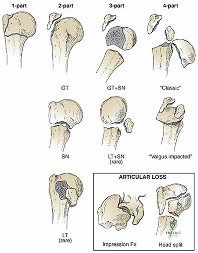
Please interpret this imaging (Figure 2) and describe your immediate clinical assessment approach for this patient.
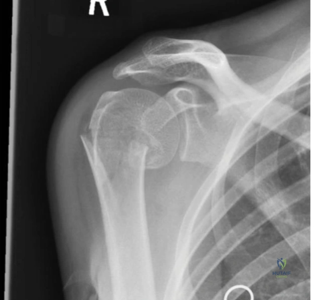
Candidate: The radiograph shows a two-part proximal humerus fracture involving the surgical neck, with significant varus displacement. My immediate priority is to assess the neurovascular status of the limb—specifically the axillary nerve function—and obtain a full trauma series, including an axillary view, to ensure no fracture-dislocation or glenoid involvement.
Candidates often jump straight into describing the "Neer classification" or asking about surgery before completing the primary survey. Failing to explicitly mention the neurovascular exam or the necessity of an axillary view—to rule out dislocation—is a frequent oversight.
A high-scoring answer starts with the "True AP/Axillary/Scapular Y" triage, identifies the injury (Neer 2-part, varus-displaced surgical neck), and emphasizes the critical neurovascular status (Axillary nerve). It mentions the "varus" nature as a specific concern for medial cortical instability.
Given the varus displacement shown in the radiograph, why is this an important prognostic feature, and how does it change your management plan?
Candidate: Varus displacement implies a disruption of the medial cortical hinge. This is prognostic for instability and a higher risk of secondary varus collapse or malunion if treated conservatively. Management must prioritize restoring the medial column support, either through anatomic reduction or use of medial-column screws/struts to prevent further displacement.
Stating only that "varus is bad" without explaining the biomechanics of the medial hinge. Candidates often fail to mention the importance of the "calcar" support, which is the most critical factor for internal fixation success.
The candidate highlights that the medial column is the primary load-bearing pillar. Mentioning that the "head-shaft angle" is key and that restoration of the calcar (medial cortical contact) is the single most important factor to prevent "varus collapse" after plating demonstrates elite-level clinical reasoning.
Consider the patient is an 80-year-old with osteoporosis. How do the risks change, and what is your specific strategy for fixation?
Candidate: In an elderly patient, bone quality is the limiting factor. I would use a locking plate construct to enhance pull-out strength in osteopenic bone. Key intra-operative strategies include medial cortical reduction, calcar screw placement, and perhaps the use of cement augmentation or an allograft strut to support the medial hinge.
Ignoring the "Reverse Shoulder Arthroplasty (RSA)" option. In the elderly, especially with severe comminution or high risk of avascular necrosis, ORIF may fail. Failing to offer the patient an arthroplasty option for complex fractures in advanced age is a clinical blind spot.
The candidate balances ORIF vs. Arthroplasty. They articulate the "locking plate" technique while referencing the increased reliance on "reverse shoulder arthroplasty" for older, low-demand patients or cases where tuberosity healing is deemed unlikely due to severe osteoporosis or fracture pattern.
Detailed Chapters & Topics
Dive deeper into specialized chapters regarding orthopedic-md-oral-examinationmanagement-of-proximal-humerus-fracture