Proximal Humerus Fracture: Orthopedic MD Explains Treatment & Management
Key Takeaway
Here are the crucial details you must know about Proximal Humerus Fracture: Orthopedic MD Explains Treatment & Management. For a proximal humerus fracture orthopedic intervention, surgery typically involves fixing the displaced fracture with a proximal humerus locking plate, often through a deltopectoral approach. This procedure aims to achieve the best functional outcome, preventing mal- or non-union, despite potential risks like fixation failure, neurovascular injury, or stiffness.
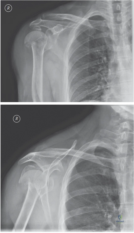
A 62-year-old female presents to the Emergency Department following a mechanical fall onto her right shoulder. She is holding her arm in internal rotation. Please interpret these radiographs and provide your classification and assessment of the fracture geometry.
Candidate: These are AP and axillary views of the proximal humerus showing a displaced fracture. Based on Neer’s classification, I would classify this as a 3-part fracture involving the surgical neck and the greater tuberosity. There is significant varus angulation of the humeral head and medial displacement of the shaft.
Failing to define what makes a fracture "displaced" (missing the Neer criteria of >1cm or >45°). Candidates often guess the classification without describing the anatomical landmarks (bicipital groove, tuberosity status) or assessing the critical blood supply risks (e.g., medial hinge disruption).
A systematic description: 1. Patient demographics. 2. Radiological interpretation (describe the segments). 3. Classification (Neer 3-part). 4. Assessment of "High-Risk" features: specifically checking the integrity of the medial hinge (as disruption carries a high risk of AVN) and the neck-shaft angle.
Given this patient is an active 62-year-old, how would you counsel her regarding the management options, specifically referencing the current evidence base?
Candidate: I would discuss both non-operative and operative management. The PROFHER trial showed no significant difference in functional outcomes between surgical and non-surgical treatment for displaced proximal humerus fractures at 2 years. However, surgery is indicated if there is significant deformity that would cause chronic impingement or functional deficit.
Suggesting surgery as the default without acknowledging the PROFHER trial or the UK-based consensus. Failing to mention patient-specific factors such as bone quality (DEXA scan/osteoporosis) and hand dominance.
Use a structured approach: "Shared Decision Making." 1. Acknowledge the PROFHER trial findings (equivalence in PROMs). 2. Discuss contra-indications for surgery (e.g., severe osteoporosis, medical comorbidities). 3. Discuss the specific goals for this patient (active 62-year-old) and the potential for malunion (varus) leading to long-term subacromial pain.
You have decided to proceed with ORIF. Describe your surgical approach and the specific complications you would highlight during the consent process.
Candidate: I would use a deltopectoral approach. Key complications include axillary nerve injury, AVN of the humeral head due to compromise of the arcuate artery, screw cutout, and secondary varus collapse.
Neglecting to mention the "danger zone" for the axillary nerve or failing to discuss the importance of medial column support. Simply listing "infection/nerve damage" is insufficient for an FRCS candidate; you must demonstrate an understanding of fracture biology.
Answer with anatomical precision: "I utilize the deltopectoral interval, protecting the cephalic vein. I would specifically mention the risk to the axillary nerve, which is at risk inferior to the glenoid. I emphasize the importance of medial column restoration (using calcar screws or bone grafting) to prevent the most common failure mode: secondary varus collapse and screw cutout."
