Operative Management of Hand Tumors and Ganglion Cysts
Key Takeaway
Ganglion cysts are the most common focal hand masses, typically arising from the scapholunate interosseous ligament. While conservative management includes aspiration, surgical excision remains the gold standard for definitive treatment. This guide details the step-by-step surgical technique for dorsal wrist ganglion excision, emphasizing capsular margin removal and preservation of the scapholunate ligament to minimize recurrence and optimize postoperative wrist biomechanics.
Comprehensive Introduction and Patho-Epidemiology
The operative management of hand tumors and tumor-like conditions encompasses a vast spectrum of pathology, ranging from ubiquitous benign cystic lesions to exceedingly rare, life-threatening soft tissue and bone sarcomas. Primary malignant tumors of the hand and upper extremity are exceptionally uncommon, yet they demand a high index of suspicion, rigorous diagnostic workup, and aggressive multidisciplinary management. Unlike benign tumorous conditions, malignancies in the upper extremity carry a significant risk of morbidity, limb loss, and mortality. The hand is a complex, densely packed anatomic region where critical neurovascular, tendinous, and skeletal structures exist in close proximity, making oncologic resection with negative margins a formidable reconstructive challenge.
Rhabdomyosarcoma is a highly aggressive malignant mesenchymal tumor exhibiting skeletal muscle differentiation. While it is the most common soft tissue sarcoma in childhood, its presentation in the hand and upper extremity is exceptionally rare. Historically, most reported cases of rhabdomyosarcoma in the hand have been fatal, standing in stark contrast to the relatively better prognoses of other primary bone or soft tissue tumors in the upper extremity. The tumor typically presents as a rapidly enlarging, firm, and often painless mass. Early hematogenous spread to the lungs and lymphatic dissemination are common. Pathologically, the alveolar subtype is frequently associated with t(2;13) or t(1;13) chromosomal translocations, resulting in PAX3-FOXO1 or PAX7-FOXO1 fusion oncogenes, which portend a particularly poor prognosis and necessitate aggressive neoadjuvant chemotherapy, wide oncologic surgical resection, and adjuvant radiation therapy.
Ewing sarcoma of the hand is another rare but highly aggressive malignancy, typically affecting children and young adults. It is a primitive neuroectodermal tumor driven primarily by the t(11;22)(q24;q12) translocation, yielding the EWSR1-FLI1 fusion protein. Radiographically, it is characterized by a permeative pattern of bone destruction accompanied by an aggressive periosteal reaction, often described as an "onion-skin" or "sunburst" appearance. Any permeative osteolytic lesion in the tubular bones of the hand in a young patient must be considered Ewing sarcoma until proven otherwise, making a prompt incisional biopsy mandatory. With the advent of modern multidisciplinary treatment protocols—combining multi-agent systemic chemotherapy with localized radiation therapy and wide surgical excision—current 5-year survival rates have improved significantly, ranging from 50% to 75% for localized disease.
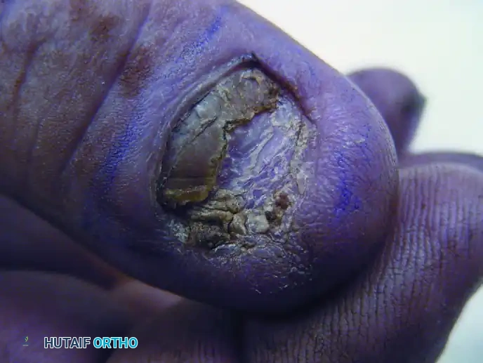
Squamous cell carcinoma (SCC) is the most common primary malignancy of the nail bed and the hand overall. It is frequently misdiagnosed as a chronic paronychia, verruca vulgaris, or pyogenic granuloma, leading to a dangerous delay in definitive treatment. Patients typically present with chronic ulceration, nail plate deformity, or a non-healing subungual mass. The pathogenesis is strongly linked to high-risk human papillomavirus (HPV) strains, particularly HPV 16 and 18, as well as chronic ultraviolet radiation exposure. While local tissue destruction can be extensive, the metastatic potential of subungual SCC is relatively low compared to SCC of other cutaneous sites, provided that wide local excision is achieved before deep osseous or lymphatic invasion occurs.
Conversely, ganglion cysts represent the most common cause of focal hand masses, accounting for 50% to 70% of all soft tissue tumors of the hand and wrist. They characteristically arise from the synovium of joints, tendon sheaths, or directly from tendons. While the exact etiology remains debated, the prevailing theory suggests that a history of acute trauma or recurrent chronic microtrauma leads to mucoid degeneration of connective tissue and the formation of a one-way valve mechanism. This allows synovial fluid to escape the joint capsule but prevents its return, leading to the concentration of hyaluronic acid and the formation of a thick, highly viscous, mucin-filled cyst. Understanding the precise topographic origins of these cysts is paramount for successful surgical extirpation.
Detailed Surgical Anatomy and Biomechanics
A profound understanding of the complex functional anatomy and biomechanics of the hand and wrist is the foundation of successful tumor extirpation and ganglion excision. The dorsal wrist anatomy is defined by the extensor retinaculum, which is divided into six distinct fibro-osseous compartments. Dorsal wrist ganglia, which comprise 60% to 70% of upper extremity ganglia, almost exclusively originate from the distal dorsal portion of the scapholunate (SL) interosseous ligament. The SL ligament is a C-shaped structure with dorsal, membranous, and volar components. The dorsal SL ligament is the thickest and biomechanically most critical component for maintaining carpal stability, resisting volar and ulnar translation of the scaphoid. Surgical excision of a dorsal ganglion requires resection of the capsular root without violating this intrinsic dorsal SL ligament, lest the patient develop a dorsal intercalated segment instability (DISI) deformity.
The volar wrist presents a more perilous anatomic landscape due to the intimate proximity of critical neurovascular structures. Volar wrist ganglia typically present just radial to the flexor carpi radialis (FCR) tendon and usually arise from the radioscaphocapitate (RSC) or radiolunate (RL) ligaments. The RSC ligament is a primary stabilizer of the palmar midcarpal joint. The radial artery courses directly over or is intimately adherent to the pseudocapsule of the volar ganglion. The palmar cutaneous branch of the median nerve, which arises approximately 5 cm proximal to the wrist crease and travels between the FCR and palmaris longus (PL), is highly susceptible to iatrogenic injury during volar approaches. Meticulous adventitial dissection is required to peel the radial artery off the cyst wall while preserving the deep ligamentous stabilizers.
The digital anatomy is equally critical, particularly when addressing subungual squamous cell carcinoma or flexor tendon sheath ganglia. The perionychium consists of the nail bed (sterile and germinal matrices), the nail fold (eponychium), and the hyponychium. The germinal matrix is responsible for the vast majority of nail plate generation. The distal phalanx tuft lies mere millimeters beneath the nail bed, making it highly susceptible to direct invasion by subungual malignancies. The vascular supply to the digit is provided by the proper digital arteries, which form a rich anastomotic network in the pulp space and the dorsal nail fold. Flexor tendon sheath ganglia (retinacular cysts) typically arise from the A1 or A2 pulleys. The biomechanical function of these pulleys is to prevent bowstringing of the flexor tendons during digital flexion; therefore, excision of a retinacular cyst must involve only a minimal window of the pulley to maintain its mechanical integrity.
Neuroanatomy plays a dual role in tumor surgery: structures to be protected and structures to be targeted for pain relief. The terminal branch of the posterior interosseous nerve (PIN) lies on the radial floor of the fourth extensor compartment, providing deep nociceptive innervation to the dorsal wrist capsule. Hypertrophy or stretching of the capsule by a dorsal ganglion can cause severe, poorly localized wrist pain mediated by the PIN. Prophylactic neurectomy of this terminal sensory branch is a powerful adjunct during dorsal ganglion excision. Conversely, the superficial sensory branch of the radial nerve (SRN) and the dorsal ulnar sensory nerve (DUSN) must be rigorously protected during superficial dissection, as iatrogenic neuromas of these nerves are notoriously recalcitrant to treatment and can cause debilitating neuropathic pain.
Exhaustive Indications and Contraindications
The decision-making algorithm for operative intervention in hand tumors is dictated by the biologic behavior of the lesion, the degree of functional impairment, and the patient's systemic health. For suspected malignant lesions, the primary indication for surgery is the necessity for definitive oncologic extirpation with wide negative margins. Any rapidly growing mass, deep-seated lesion larger than 3 cm, or lesion exhibiting aggressive radiographic features (e.g., permeative bone destruction, cortical breakthrough) mandates an initial biopsy. Once a diagnosis of sarcoma or invasive carcinoma is confirmed, definitive surgery is indicated, often following neoadjuvant chemotherapy or radiation.
For benign conditions such as ganglion cysts, the indications for surgical intervention are largely relative and driven by patient symptomatology. Absolute indications include secondary neurologic compression, such as a Guyon canal ganglion causing profound, painless atrophy of the ulnar-innervated intrinsic muscles, or a massive volar cyst causing impending radial artery occlusion. Relative indications include persistent pain refractory to conservative measures, significant restriction of wrist or digital range of motion, and unacceptable cosmetic deformity. The size of a dorsal ganglion is not proportional to the pain experienced by the patient; occult, small cysts hidden beneath the extensor retinaculum can cause incapacitating pain due to mass effect on the PIN during extreme wrist extension, serving as a valid indication for surgical exploration and excision.
Contraindications to surgical intervention must be carefully weighed against the natural history of the disease. For malignant tumors, absolute contraindications are rare but may include a medically unstable patient unable to tolerate general anesthesia, or a tumor that is so locally advanced and systemically disseminated that surgery would offer no palliative or oncologic benefit. In such cases, palliative radiation or systemic therapy may be the only viable options. For benign ganglion cysts, absolute contraindications include active local skin infection (cellulitis) over the operative site, which could lead to deep space seeding or septic arthritis upon capsular violation.
Relative contraindications for elective ganglion excision include poor patient compliance, unrealistic cosmetic expectations, and complex regional pain syndrome (CRPS) in the affected extremity. Operating on a patient with active CRPS for a benign condition can lead to a catastrophic exacerbation of neuropathic pain and stiffness. Furthermore, in the setting of suspected sarcomas, an unplanned or poorly executed excisional biopsy by an inexperienced surgeon is a strict contraindication. Biopsies must be meticulously planned so that the entire biopsy tract can be excised en bloc during the definitive oncologic procedure without compromising future reconstructive options or limb salvage.
| Condition Category | Primary Indications for Operative Intervention | Absolute and Relative Contraindications |
|---|---|---|
| Malignant Tumors (Sarcoma/SCC) | Confirmed malignancy requiring wide local excision; Limb salvage procedures; Palliative resection for fungating/bleeding masses; En bloc resection of biopsy tracts. | Unstable medical condition; Disseminated metastasis where local control offers no palliative benefit; Unplanned excision without prior staging. |
| Dorsal/Volar Ganglion Cysts | Refractory pain limiting activities of daily living; Neurologic compression (e.g., median/ulnar nerve); Mechanical restriction of joint motion; Failure of aspiration/injection. | Active local infection; Asymptomatic cysts (relative); Active Complex Regional Pain Syndrome (CRPS); Unrealistic cosmetic expectations. |
| Retinacular Cysts (A1/A2) | Painful snapping or mechanical triggering; Exquisite tenderness to palpation interfering with grip; Failure of needle puncture/corticosteroid injection. | Active local infection; Bleeding diatheses (relative); Concomitant untreated severe osteoarthritis of the adjacent joint. |
Pre-Operative Planning, Templating, and Patient Positioning
Thorough pre-operative planning is the cornerstone of successful hand tumor surgery, relying heavily on advanced imaging modalities and multidisciplinary collaboration. For suspected malignancies, plain radiographs are the initial step, evaluating for cortical erosion, periosteal reactions (such as the onion-skin appearance of Ewing sarcoma), or matrix calcifications. Magnetic Resonance Imaging (MRI) with and without intravenous gadolinium contrast is the gold standard for evaluating soft tissue extent, neurovascular involvement, and intramedullary marrow infiltration. MRI is critical for defining the exact anatomic stalk of complex, multilocular ganglion cysts, particularly those extending volarly or into the carpal tunnel, ensuring that the surgeon can plan an approach that completely extirpates the capsular origin.
In the context of oncologic surgery, pre-operative templating involves determining the necessary margins for wide local excision. For subungual squamous cell carcinoma, the depth of invasion dictates the surgical plan. If MRI or radiographs demonstrate intact cortical bone with a sufficient soft tissue envelope, a wide local excision of the nail bed with full-thickness skin grafting may be planned. However, if there is gross osseous invasion or the margins are perilously close, pre-operative templating will include planning for a partial ostectomy of the distal phalanx or a distal interphalangeal (DIP) joint amputation. The reconstructive ladder must be considered pre-operatively, ensuring that appropriate donor sites for skin grafts, local advancement flaps (e.g., V-Y flaps), or regional flaps are prepped and draped.
Patient positioning and anesthesia are optimized to provide a motionless, bloodless surgical field. The procedure is optimally performed under a regional block (axillary or supraclavicular) or general anesthesia. Regional anesthesia not only provides excellent intraoperative conditions but also facilitates prolonged postoperative analgesia, reducing the need for systemic opioids. The patient is placed supine with the operative arm extended on a radiolucent hand table, allowing for intraoperative fluoroscopy if required for margin assessment or joint localization.
Exsanguination and tourniquet application require specific modifications depending on the pathology. For benign ganglion cysts, the limb is tightly exsanguinated with an Esmarch bandage, and an upper arm tourniquet is inflated to 250 mmHg. However, for malignant tumors such as rhabdomyosarcoma or Ewing sarcoma, exsanguination with an Esmarch bandage is strictly contraindicated due to the theoretical risk of mechanically forcing tumor cells into the systemic circulation. Instead, the arm is simply elevated for 3 to 5 minutes to allow for venous drainage before tourniquet inflation. This subtle but critical modification in pre-operative preparation is a hallmark of meticulous oncologic surgical technique.
Step-by-Step Surgical Approach and Fixation Technique
Oncologic Resection of Subungual Squamous Cell Carcinoma
Surgical management of subungual SCC dictates wide local excision to prevent local recurrence and distal metastasis. The procedure begins with the complete removal of the nail plate to fully expose the tumor bed. Depending on the depth of invasion and bone involvement, treatment ranges from wide local excision of the nail bed with full-thickness skin grafting to distal interphalangeal (DIP) joint amputation.
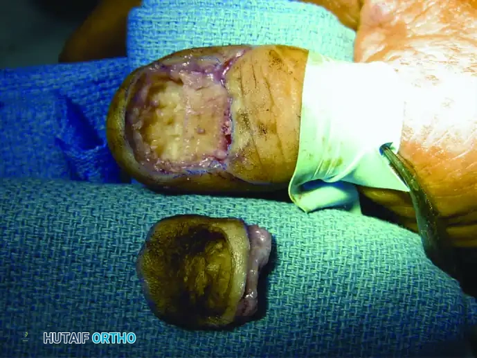
Figure 1: Clinical presentation of a subungual squamous cell carcinoma of the nail bed, demonstrating chronic ulceration and destruction of the nail plate.
Using a #15 blade, a wide margin (typically 4-5 mm) is marked around the visible extent of the tumor. The incision is carried down to the periosteum of the distal phalanx. If the tumor is strictly confined to the soft tissue, the entire complex is elevated off the bone. However, when bone is not grossly invaded but close margins are suspected, a partial ostectomy of the distal phalanx must be performed.
Figure 2: Wide surgical excision of the squamous cell carcinoma, including a portion of the underlying distal phalanx, to ensure negative oncologic margins while allowing for adequate soft tissue closure.
Using a sharp osteotome or a sagittal saw, the dorsal cortex or the distal tuft of the phalanx is resected en bloc with the tumor. Intraoperative frozen sections may be utilized to confirm negative margins. Once oncologic clearance is achieved, reconstruction focuses on providing a durable, sensate tip. If a partial ostectomy was performed, the remaining volar pad can often be advanced dorsally. Alternatively, a full-thickness skin graft harvested from the hypothenar eminence or the groin is secured over the defect.
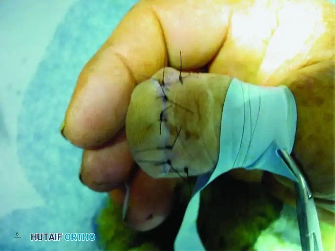
Figure 3: Final closure after wide excision. Meticulous soft tissue handling is required to provide a durable, sensate tip.
Excision of Dorsal Wrist Ganglion
The cornerstone of successful ganglion excision is the complete removal of the cyst along with a generous margin of the adjacent joint capsule at its base. Make a 2.0- to 3.0-cm transverse incision in one of the dorsal wrist creases, centered directly over the scapholunate interval. Carry the incision through the dermis only, then spread the underlying subcutaneous tissues longitudinally to avoid transecting the superficial sensory branches of the radial nerve (SRN) or the dorsal ulnar sensory nerve (DUSN). Open the ulnar border of the third extensor compartment and retract the extensor pollicis longus (EPL) and radial wrist extensor tendons radially. Retract the extensor digitorum communis (EDC) ulnarly to expose the dorsal wrist capsule.
Using a combination of sharp and blunt dissection, isolate the ganglion in its entirety down to its stalk. Excise the cyst along with a 1 cm x 1 cm portion of its capsular origin at the dorsal scapholunate ligament. It is critical not to injure the intrinsic scapholunate interosseous ligament; dissect the capsular tissue cleanly to expose, but not violate, the SL ligament. To reduce postoperative pain, perform a posterior interosseous nerve (PIN) neurectomy by resecting a 1.0-cm segment of the terminal branch on the radial floor of the fourth extensor compartment. Deflate the tourniquet, achieve meticulous hemostasis, and close the skin without attempting to close the joint capsule, which prevents postoperative wrist stiffness.
Excision of Volar Wrist Ganglion
Volar ganglion excision demands immense respect for the radial artery. A longitudinal or curvilinear incision is made over the flexor carpi radialis (FCR) tendon. The palmar cutaneous branch of the median nerve must be identified and protected ulnarly. The radial artery is frequently draped over the cyst and must be meticulously dissected off the pseudocapsule using tenotomy scissors in the adventitial plane.
Once the artery is safely mobilized and retracted, the stalk of the ganglion is traced deep between the FCR and the radial artery down to the radioscaphocapitate (RSC) or radiolunate (RL) ligaments. A window of the volar capsule encompassing the stalk is excised. Similar to the dorsal approach, the intrinsic carpal ligaments must be preserved to prevent catastrophic midcarpal instability. The wound is irrigated, and hemostasis is confirmed prior to layered closure.
Complications, Incidence Rates, and Salvage Management
Surgical intervention for hand tumors and ganglion cysts, while often curative, is fraught with potential complications that can severely compromise hand function. The most common complication following ganglion excision is recurrence. Historically, recurrence rates following simple aspiration were as high as 60-70%. With meticulous open surgical excision that includes the capsular root, recurrence rates drop to 5-15%. Recurrence is almost universally due to failure to identify and excise the true capsular stalk, leaving behind the one-way valve mechanism. Salvage management for a recurrent ganglion involves a more extensive revision surgery, often requiring a wider capsular resection and occasionally local tissue interposition to obliterate the defect.
Neurovascular complications are particularly devastating. In volar ganglion excisions, injury to the radial artery can result in pseudoaneurysm formation, thrombosis, or distal ischemia, occurring in approximately 1-3% of cases. If the radial artery is inadvertently transected, it must be primarily repaired using microvascular techniques, or if the palmar arch is incomplete (as determined by a pre-operative Allen's test), an interposition vein graft may be required. Injury to the superficial sensory nerves (SRN, DUSN, or palmar cutaneous branch of the median nerve) leads to painful neuroma formation. Salvage for a symptomatic neuroma involves resection of the neuroma and burying the proximal nerve stump deep into adjacent muscle bellies or bone to prevent mechanical irritation.
In the realm of oncologic surgery, complications are inherently more severe. Positive margins following the resection of a sarcoma or subungual SCC dramatically increase the risk of local recurrence and metastasis. The incidence of local recurrence for subungual SCC treated with inadequate margins can approach 20-30%. Salvage management in these scenarios inevitably involves more proximal amputation (e.g., ray resection or transmetacarpal amputation) and adjuvant radiation therapy. Furthermore, aggressive resection of bone and soft tissue can lead to profound functional deficits, stiffness, and delayed wound healing, particularly if the patient has received neoadjuvant radiation.
Carpal instability is a rare but catastrophic complication following overzealous capsular resection during ganglion surgery. If the intrinsic dorsal scapholunate interosseous ligament is violated during a dorsal ganglion excision, the patient may develop scapholunate dissociation and a subsequent DISI deformity. This presents as chronic, activity-related wrist pain and a clunking sensation. Salvage management for iatrogenic SL instability requires complex ligament reconstruction (e.g., modified Brunelli procedure) or, in chronic cases with secondary arthritic changes, a partial wrist fusion (e.g., scaphoid excision and four-corner fusion).
| Complication | Estimated Incidence | Etiology / Risk Factors | Salvage Management / Treatment |
|---|---|---|---|
| Ganglion Recurrence | 5% - 15% (Open Excision) | Inadequate excision of the capsular stalk; Failure to resect the one-way valve. | Revision open excision with extended capsular resection; MRI to confirm stalk anatomy. |
| Neuroma Formation (SRN/DUSN) | 2% - 5% | Iatrogenic transection or aggressive traction during superficial dissection. | Neuroma excision and proximal transposition into muscle/bone; targeted muscle reinnervation (TMR). |
| Radial Artery Injury | 1% - 3% (Volar approaches) | Inadequate adventitial dissection; Blind clamping of bleeding vessels. | Microvascular primary repair; Interposition vein grafting if Allen's test indicates incomplete arch. |
| Iatrogenic Carpal Instability | < 1% | Violation of the intrinsic Scapholunate (SL) or Radioscaphocapitate (RSC) ligaments. | Ligament reconstruction (e.g., Brunelli); Partial wrist arthrodesis for chronic/arthritic cases. |
| Local Oncologic Recurrence | Variable (Tumor dependent) | Positive surgical margins; Inadequate pre-operative staging; Aggressive tumor biology. | Wider re-excision; Proximal amputation (Ray resection); Adjuvant radiation/chemotherapy. |
Phased Post-Operative Rehabilitation Protocols
The postoperative rehabilitation following hand tumor and ganglion surgery is as critical as the surgical execution itself. The hand is uniquely prone to stiffness, tendon adhesions, and capsular contractures; therefore, a meticulously phased rehabilitation protocol is mandatory. The immediate postoperative phase (Days 0 to 14) focuses on wound healing, edema control, and the prevention of digital stiffness. Following a dorsal or volar wrist ganglion excision, a soft, bulky compressive dressing is applied with a volar orthoplast splint holding the wrist in slight extension (20 degrees). Patients are instructed to keep the extremity elevated above the level of the heart. Crucially, active range-of-motion (ROM) of the fingers, thumb, and elbow must begin on postoperative day one to prevent extensor tendon adhesions and promote venous return.
The intermediate phase (Weeks 2 to 6) commences with the first formal postoperative clinical appointment at 10 to 14 days, during which sutures are removed. The bulky dressing and rigid splint are discontinued, and the patient is transitioned to a removable wrist orthosis to be worn during heavy activities and at night. A formal, therapist-directed hand therapy program is initiated. Modalities such as scar massage with silicone gel sheets are employed to soften the incision and prevent tethering of the underlying tendons. Active and active-assisted ROM exercises for the wrist are aggressively pursued to restore terminal flexion and extension. The joint capsule, which was intentionally left open during surgery, is allowed to heal via secondary intention, but early motion ensures it heals with sufficient laxity to permit full physiological movement.
The advanced phase (Weeks 6 to 12) focuses on strengthening, proprioception, and work hardening. Heavy lifting, extreme weight-bearing across the wrist (such as push-ups), and forceful gripping are restricted for the first 4 to 6 weeks. As the capsular defect consolidates, progressive resistance exercises utilizing putty, hand grippers, and weighted wrist curls are introduced. Full recovery and the complete resolution of the deep aching pain associated with the initial pathology may take up to 3 months. Patients must be counseled pre-operatively that transient wrist stiffness and mild incisional site discomfort are expected parts of the recovery trajectory.
For patients undergoing major oncologic resections, the rehabilitation protocol is significantly more complex and highly individualized. If a distal amputation or partial ostectomy was performed for subungual SCC, desensitization of the residual stump is a primary focus. Techniques including tapping, fluidotherapy, and varied texture immersion are utilized to downregulate hypersensitive nerve endings. If the patient requires adjuvant radiation therapy for a sarcoma, the therapist must proactively manage radiation-induced fibrosis with aggressive stretching and custom dynamic splinting. In cases of limb salvage requiring complex flap reconstruction, motion protocols are strictly dictated by the viability and healing timeline of the soft tissue envelope, often delaying aggressive mobilization to ensure graft or flap survival.
Summary of Landmark Literature and Clinical Guidelines
The evolution of operative management for hand tumors and ganglion cysts is heavily rooted in landmark anatomical studies and rigorous oncologic protocols. The surgical principles for ganglion excision were fundamentally shaped by Angelides and Wallace in their seminal 1976 paper. They definitively demonstrated that dorsal wrist ganglia arise from a tortuous duct connecting the cyst to the radiocarpal joint, specifically at the dorsal scapholunate ligament. Their work established the modern gold standard: that simple cyst enucleation is insufficient, and excision must include the capsular root and the one-way valve mechanism to minimize recurrence. Subsequent comparative studies by Dias et al. validated that meticulous open excision yields statistically significant lower recurrence rates (less than 10%) compared to simple aspiration or rupture, solidifying surgical excision as the definitive treatment for symptomatic cysts.
In the realm of hand malignancies, clinical guidelines are driven by large, multi-institutional cooperative groups due to the rarity of the diseases. For Ewing sarcoma, the Euro-E.W.I.N.G. (European Ewing Tumour Working Initiative of National Groups) protocols and the Children's Oncology Group (COG) have established the absolute necessity
