Malignant Tumors of the Hand: Principles of Surgical Oncology and Resection
Key Takeaway
Malignant tumors of the hand are rare but require aggressive, multidisciplinary management. Chondrosarcoma is the most common primary bone malignancy, while soft tissue sarcomas include undifferentiated pleomorphic sarcoma and epithelioid sarcoma. Successful surgical intervention relies on achieving negative oncologic margins through wide local excision, ray resection, or amputation. Limb-sparing surgery combined with adjuvant therapy is increasingly utilized, though survival heavily depends on tumor stage and size.
Comprehensive Introduction and Patho-Epidemiology
Malignant tumors of the hand represent an exceedingly rare and highly complex subset of musculoskeletal oncology. Because the vast majority of masses presenting in the hand and wrist are benign—such as ganglion cysts, giant cell tumors of the tendon sheath, and epidermoid inclusion cysts—the index of suspicion for malignancy is often inappropriately low among primary care providers and even general orthopedic surgeons. A landmark 65-year retrospective review (1920–1985) from the Mayo Clinic by Frassica and Amadia highlighted the sheer infrequency of these lesions, noting that their rarity frequently leads to delayed diagnosis, inadvertent marginal excisions (the dreaded "whoops" surgery), and ultimately, inadequate initial oncologic management. When a malignancy is finally identified, the surgeon is faced with the daunting task of achieving wide oncologic margins in an anatomical region where neurovascular bundles, tendons, and bone exist in intimate, unforgiving proximity.
The patho-epidemiology of hand malignancies can be broadly categorized into primary bone tumors, soft tissue sarcomas, and cutaneous malignancies. Cutaneous malignancies, specifically squamous cell carcinoma (SCC) and malignant melanoma, are by far the most common cancers of the hand overall. Squamous cell carcinoma predominantly arises on the sun-exposed dorsal surfaces and generally carries an excellent prognosis following wide local excision. Malignant melanoma, particularly the acral lentiginous subtype presenting as subungual melanoma (Hutchinson’s sign), requires aggressive surgical management, often necessitating digital amputation and sentinel lymph node biopsy. Conversely, primary deep soft tissue and bone sarcomas are exceptionally rare. Among soft tissue sarcomas, undifferentiated pleomorphic sarcoma (formerly malignant fibrous histiocytoma) is the most common in older adults, whereas epithelioid sarcoma and synovial sarcoma frequently present in young adults. Epithelioid sarcoma is particularly notorious for its superficial, nodular presentation—often mimicking a benign granuloma or chronic infection—and its unusually high propensity for lymphatic metastasis, a feature uncommon in most other soft tissue sarcomas.
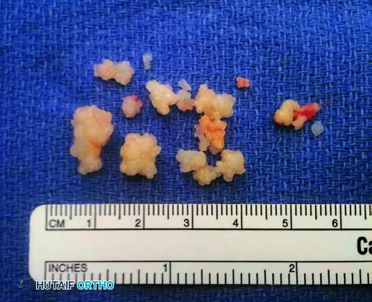
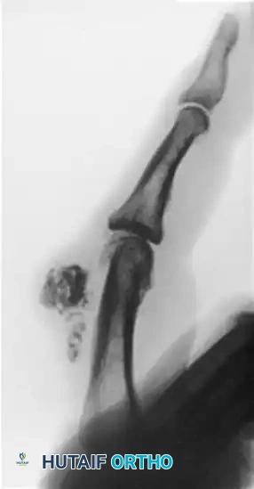
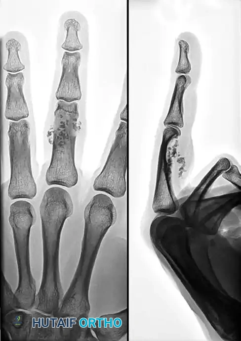
Primary bone malignancies of the hand are dominated by chondrosarcoma, which typically arises in the metacarpals and phalanges. Differentiating a low-grade chondrosarcoma from a benign, active enchondroma remains one of the most formidable diagnostic dilemmas in orthopedic oncology. Pain in the absence of a pathologic fracture, cortical breakthrough, or rapid recurrence following curettage must immediately raise the suspicion of chondrosarcoma. Osteogenic sarcoma (osteosarcoma) of the hand is even rarer, presenting at an average age of 49 years—significantly older than the adolescent demographic typical of long-bone osteosarcomas. A distinct subset of these hand osteosarcomas has been historically linked to prior irradiation or exposure to radium salts. Interestingly, while early literature suggested that malignant bone tumors of the hand rarely metastasize, contemporary evidence confirms that distant metastasis can and does occur, particularly following local recurrence. However, the overall prognosis and long-term survival for primary bone sarcomas located in the hand generally remain superior to identical histological lesions located in the axial skeleton or proximal appendicular skeleton.
The ultimate success of surgical intervention is strictly dependent on the surgical margins achieved, classified as R0 (microscopically negative), R1 (microscopically positive), or R2 (macroscopically positive). Historically, proximal amputation was considered the gold standard for any hand sarcoma to guarantee an R0 resection. Today, the paradigm has shifted toward a multidisciplinary limb-salvage approach. Combining meticulous limb-sparing surgery with adjuvant brachytherapy, external beam radiation, and systemic chemotherapy has yielded 5-year survival rates exceeding 70% for high-grade soft tissue sarcomas of the extremity. Nevertheless, limb salvage is not universally applicable. Local recurrence of a malignant hand tumor, while not definitively shown to independently affect overall survival in all histological subtypes, is a catastrophic functional event. Furthermore, larger tumor size (greater than 5 cm), deep fascial involvement, and advanced histological grade have a profound negative impact on patient prognosis, often dictating the necessity of amputation to preserve life over limb.
Detailed Surgical Anatomy and Biomechanics
The surgical anatomy of the hand is characterized by a high density of critical structures confined within a minimal cross-sectional area. Unlike the thigh or the arm, where large muscle bellies can be sacrificed to achieve wide oncologic margins without catastrophic functional loss, the hand lacks expendable muscular compartments. The soft tissues of the hand are divided into distinct fascial compartments: the thenar, hypothenar, central palmar, and interosseous compartments. Sarcomas typically respect these fascial boundaries in their early stages, growing longitudinally along the path of least resistance rather than transversely through robust fascial septa. However, the complex synovial sheaths of the flexor tendons and the rigid osteofascial tunnels (such as the carpal tunnel and Guyon’s canal) can act as conduits for rapid proximal or distal tumor extension, complicating the definition of safe oncologic margins.
Vascular anatomy plays a pivotal role in the surgical planning of malignant hand tumors. The blood supply to the hand is derived from the superficial and deep palmar arches, which are formed by the anastomoses of the radial and ulnar arteries. Tumors located in the central palmar space frequently encase these vital vascular structures. Attempting to dissect a high-grade sarcoma off a major artery invariably violates the tumor pseudocapsule—a reactive zone composed of compressed normal tissue, neovascularity, and microscopic satellite tumor cells. Violation of this pseudocapsule results in an R1 or R2 resection, drastically increasing the risk of local recurrence. Therefore, if a tumor intimately involves the superficial or deep arch, the surgeon must be prepared to resect the vascular structures en bloc with the tumor, necessitating complex microsurgical vascular reconstruction with reversed interpositional vein grafts to salvage the distal digits.
Lymphatic drainage of the hand is another critical anatomical consideration, particularly when dealing with epithelioid sarcoma, clear cell sarcoma, or malignant melanoma. The superficial lymphatic vessels of the digits and palm drain primarily into the dorsal lymphatic network, which ascends the forearm to the epitrochlear and axillary lymph nodes. The deep lymphatic system follows the major neurovascular bundles. Understanding this drainage pattern is essential for staging and for performing sentinel lymph node biopsies or formal regional lymphadenectomies. Furthermore, the venous drainage of the hand, which primarily relies on the dorsal venous network, must be carefully managed during surgery. Aggressive manipulation of a tumor can lead to venous tumor embolization, highlighting the importance of meticulous, gentle handling of the mass and the avoidance of exsanguinating Esmarch bandages during tourniquet preparation.
The biomechanics of the hand heavily influence the reconstructive strategy following oncologic resection, particularly in the context of ray resections for bone sarcomas. The hand functions as a dynamic, adaptable tool relying on the stability of the fixed central unit (the second and third metacarpals articulating with the trapezoid and capitate) and the mobility of the border digits (the first, fourth, and fifth rays). Resection of a central ray (long or ring finger) creates a biomechanically disadvantageous cleft, leading to the dropping of small objects and a significant reduction in grip strength. To restore biomechanical integrity, the adjacent ray is often transposed (e.g., transposing the index ray to the base of the long metacarpal) to close the cleft and restore a functional cascade. Conversely, resection of a border ray (index or small finger) requires an oblique osteotomy of the metacarpal base to smooth the contour of the hand, preventing a prominent bony stump that could cause pain or skin breakdown during power grip.
Exhaustive Indications and Contraindications
The decision-making process for the surgical management of malignant hand tumors must be governed by a multidisciplinary musculoskeletal oncology tumor board, incorporating orthopedic oncologists, hand surgeons, medical oncologists, radiation oncologists, and musculoskeletal radiologists. The primary indication for surgical intervention is the presence of a biopsy-proven malignant primary bone or soft tissue tumor where complete extirpation is anatomically feasible. Limb-sparing surgery (wide local excision or ray resection) is indicated when an R0 margin can be achieved while preserving a functional, sensate, and vascularized hand. For low-grade lesions, such as low-grade chondrosarcoma of the phalanx, a wide en bloc resection is curative and represents the absolute standard of care.
Contraindications to limb-sparing surgery are defined by the inability to achieve negative oncologic margins without rendering the hand completely non-functional. Absolute contraindications to limb salvage include massive tumors that diffusely involve multiple anatomical compartments, encasement of both the median and ulnar nerves precluding sensory and motor preservation, and involvement of the major proximal vascular inflow where reconstruction is deemed impossible or carries an unacceptable risk of failure. In these scenarios, a below-elbow or transradial amputation is unequivocally indicated to prioritize patient survival and prevent the horrific morbidity of a fungating, locally recurrent tumor. Furthermore, a limb-sparing procedure is contraindicated if the resulting appendage would be painful, insensate, and mechanically useless, as a well-fitted upper extremity prosthesis often provides superior functional outcomes compared to a severely compromised, salvaged hand.
Adjuvant therapies carry their own set of indications and contraindications that directly impact surgical planning. Neoadjuvant or adjuvant external beam radiation therapy (EBRT) is strongly indicated for high-grade soft tissue sarcomas (>5 cm) and for cases where surgical margins are marginally close (e.g., adjacent to a preserved major nerve). However, radiation in the hand is fraught with complications, including severe joint stiffness, tendon adhesions, radiation-induced fractures, and delayed wound healing. Therefore, a history of prior irradiation to the same extremity is an absolute contraindication for further EBRT and often tips the scale toward amputation. Systemic chemotherapy is indicated for highly chemosensitive tumors (such as Ewing sarcoma or osteosarcoma, though rare in the hand) and for high-grade soft tissue sarcomas in young, healthy patients to mitigate the risk of distant micrometastasis.
| Clinical Scenario | Indicated Procedure / Management | Contraindications / Relative Contraindications |
|---|---|---|
| Low-Grade Chondrosarcoma (Phalanx/Metacarpal) | En bloc wide local excision or Ray Resection. | Intralesional curettage is STRICTLY contraindicated (leads to recurrence). |
| High-Grade Soft Tissue Sarcoma (Superficial, <5cm) | Wide local excision with 1-2 cm margins + Adjuvant Radiation. | Marginal excision ("shelling out") is absolutely contraindicated. |
| Sarcoma Encasing Median/Ulnar Nerves & Arches | Below-elbow amputation or transradial amputation. | Limb salvage is contraindicated if R0 margins leave a useless, insensate hand. |
| Epithelioid Sarcoma of the Digit | Ray amputation + Sentinel Lymph Node Biopsy / Regional Node evaluation. | Observation of lymph nodes without staging is contraindicated due to high lymphatic spread rate. |
| Locally Recurrent High-Grade Sarcoma | Proximal amputation (e.g., wrist disarticulation or below-elbow). | Repeat limb salvage is generally contraindicated unless margins are widely clearable. |
| Subungual Melanoma (Breslow Depth > 2mm) | Distal interphalangeal (DIP) or metacarpophalangeal (MCP) amputation. | Skin grafting over positive deep margins is contraindicated. |
Pre-Operative Planning, Templating, and Patient Positioning
Thorough pre-operative planning is the cornerstone of successful oncologic surgery in the hand. The diagnostic workup begins with advanced cross-sectional imaging. Magnetic Resonance Imaging (MRI), both with and without intravenous gadolinium contrast, is mandatory for all suspected soft tissue sarcomas. T1-weighted images provide excellent anatomical detail and define the tumor's relationship to surrounding fat and neurovascular structures. T2-weighted and Short Tau Inversion Recovery (STIR) sequences highlight the extent of peritumoral edema and the reactive zone, which must be encompassed within the resection margins. For primary bone tumors, a fine-cut Computed Tomography (CT) scan is preferred to evaluate the exact degree of cortical destruction, endosteal scalloping (a hallmark of chondrosarcoma when >2/3 of cortical thickness is involved), and subtle periosteal reactions. A complete systemic staging workup, including a high-resolution CT of the chest to rule out pulmonary metastasis, must be completed prior to any surgical intervention.
During the pre-operative evaluation, the surgeon must also consider benign but locally aggressive mimickers that can present similarly to malignancies. Osteochondromas, while common in the appendicular skeleton, can present in the hand as dense, exophytic masses causing mechanical block or sudden growth, which may raise suspicion for secondary malignant transformation.
Similarly, synovial chondromatosis—a rare metaplastic condition of the synovial membrane—presents with insidious swelling and multiple spotty calcifications clustered around a joint, which can radiographically mimic a synovial sarcoma or chondrosarcoma.
Accurate biopsy and histopathological differentiation of these entities are paramount to avoid unnecessary radical resections.
The biopsy is arguably the most critical step in the pre-operative sequence and must be meticulously planned. It is a fundamental rule of orthopedic oncology that the biopsy should be performed by the exact surgeon who will perform the definitive resection. Poorly planned biopsies can contaminate adjacent compartments, turning a salvageable limb into an obligate amputation. Longitudinal incisions must always be used, completely avoiding transverse incisions that contaminate multiple neurovascular planes. The biopsy tract must be strategically placed so that it can be excised en bloc with the tumor during the definitive surgery. Meticulous hemostasis is required before closure to prevent a post-biopsy hematoma, which acts as a vehicle to disseminate microscopic tumor cells far beyond the primary tumor bed.
Patient positioning and the management of the pneumatic tourniquet require strict adherence to oncologic principles. The patient is positioned supine with the operative arm extended on a radiolucent hand table, allowing for unobstructed intraoperative fluoroscopy.
Clinical Pearl: When operating on a suspected or confirmed malignancy, the surgeon must never exsanguinate the limb using an Esmarch bandage. Mechanical compression of the tumor mass can force malignant cells into the systemic venous circulation, causing iatrogenic tumor embolization.
Instead, the arm should be elevated at 60 degrees for 3 to 5 minutes to allow for gravity-assisted venous drainage before inflating the pneumatic tourniquet to 250 mmHg. This technique provides a bloodless surgical field necessary for meticulous dissection without compromising oncologic safety.
Step-by-Step Surgical Approach and Fixation Technique
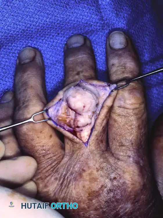
The definitive surgical approach begins with an incision designed to incorporate the previous biopsy tract, ensuring a 1 to 2 cm margin of normal skin surrounding the scar, depending on the tumor's histological grade and type. Flaps are raised full-thickness, maintaining the subcutaneous fat on the skin to preserve vascularity, unless the tumor superficially invades the dermis, in which case the skin must be sacrificed en bloc. The fundamental principle of sarcoma resection is the management of the "pseudocapsule." Sarcomas invariably form a compressed layer of normal tissue and tumor cells at their periphery. The surgeon must never visualize the tumor itself during the dissection; seeing the tumor implies that the pseudocapsule has been breached and the margin is contaminated. Dissection must proceed through normal, unreactive tissue entirely outside the reactive zone defined on the pre-operative MRI.
For primary bone sarcomas of the metacarpals or proximal phalanges, such as a confirmed chondrosarcoma, a ray resection is frequently the procedure of choice. The incision is typically a dorsal or volar V-shaped or racquet-shaped incision encompassing the base of the involved digit. The extensor and flexor tendons are identified proximally and transected cleanly. The digital nerves are isolated, drawn distally under tension, and transected sharply so they retract deep into the intrinsic musculature, minimizing the risk of symptomatic neuroma formation. The deep transverse metacarpal ligaments are divided. The metacarpal is then osteotomized at its base using an oscillating saw, or disarticulated at the carpometacarpal (CMC) joint, depending on proximal tumor extent. The entire ray is then delivered en bloc, ensuring no violation of the cortical bone or tumor mass.
Reconstruction and fixation following a ray resection are dictated by the specific ray removed. If the central ray (long finger) is resected, a transposition osteotomy of the index ray is performed to close the central defect. The index metacarpal is osteotomized at its metadiaphyseal junction, translated ulnarly to articulate with the base of the third metacarpal or the capitate, and fixed rigidly. Fixation techniques vary but typically involve the use of multiple parallel or crossed Kirschner wires (K-wires), or more robustly, a low-profile titanium locking compression plate (LCP) and screws. Rigid fixation is paramount to allow for early mobilization and to ensure primary bone healing of the osteotomy site. The deep transverse metacarpal ligament must be reconstructed between the transposed index ray and the ring finger to prevent scissoring and rotational deformity during active flexion.
Closure following an R0 resection often presents a significant reconstructive challenge, as primary closure is rarely possible without undue tension. If a healthy, vascularized bed of muscle or fascia remains, a split-thickness or full-thickness skin graft may be applied and secured with a tie-over bolster dressing. However, if the resection exposes bare bone, tendons devoid of paratenon, or major neurovascular structures, vascularized tissue coverage is mandatory. Local rotational flaps (e.g., a cross-finger flap or reverse homodigital island flap) may suffice for small digital defects. For larger defects on the dorsum of the hand or palm, regional flaps such as the radial forearm pedicled flap, or microvascular free tissue transfer (e.g., an anterolateral thigh flap or lateral arm flap) must be executed by a skilled reconstructive microsurgeon to provide durable, pliable soft tissue coverage that can withstand potential adjuvant radiation therapy.
Complications, Incidence Rates, and Salvage Management
The surgical management of malignant hand tumors is fraught with both oncologic and reconstructive complications. The most devastating oncologic complication is local recurrence, which occurs in approximately 10-15% of patients treated with limb-sparing surgery for high-grade soft tissue sarcomas, even with negative margins and adjuvant radiation. Local recurrence in the hand is particularly disastrous because the virgin anatomical planes have been obliterated by the primary surgery and subsequent scarring. Distant metastasis, most commonly to the lungs, occurs in 20-30% of patients with high-grade lesions. The risk of metastasis is directly correlated with tumor size, depth, and histological grade, rather than the choice between limb-salvage and amputation, provided R0 margins were initially achieved.
Surgical and functional complications are highly prevalent due to the delicate anatomy of the hand and the necessity of radical resection. Wound dehiscence and skin graft or flap necrosis occur in up to 20% of cases, particularly when adjuvant radiation is administered pre-operatively or early post-operatively. Joint stiffness, tendon adhesions, and severe contractures are almost universal to some degree, resulting from extensive soft tissue stripping, prolonged immobilization, and radiation-induced fibrosis. Furthermore, the sacrifice of digital nerves or major mixed nerves results in sensory deficits and intrinsic muscle wasting, leading to a profound loss of fine motor dexterity, pinch strength, and an increased susceptibility to unrecognized thermal or mechanical trauma in the insensate digits.
| Complication | Estimated Incidence | Salvage Management / Mitigation Strategy |
|---|---|---|
| Local Tumor Recurrence | 10% - 15% (High-grade STS) | Proximal amputation (wrist disarticulation or below-elbow). Re-resection is rarely feasible. |
| Pulmonary Metastasis | 20% - 30% (High-grade STS/Bone) | Systemic chemotherapy; Pulmonary metastasectomy (wedge resection) if lesions are isolated and resectable. |
| Flap Necrosis / Wound Dehiscence | 15% - 20% | Debridement and secondary coverage (free tissue transfer). Delay adjuvant radiation until fully healed. |
| Severe Joint Stiffness / Contracture | 40% - 60% | Aggressive occupational therapy; surgical tenolysis or capsulotomy (only after oncologic clearance, usually >1 year). |
| Symptomatic Neuroma | 5% - 10% | Surgical excision of neuroma and burying the nerve stump deep into muscle, or targeted muscle reinnervation (TMR). |
| Radiation-Induced Fracture | 2% - 5% | Internal fixation with robust plating and bone grafting; often requires vascularized bone graft (e.g., free fibula) due to poor biology. |
Salvage management of these complications requires a highly individualized approach. For local oncologic recurrence, salvage limb-sparing surgery is almost universally contraindicated due to the high risk of a second recurrence and the extensive morbidity involved. In these scenarios, a definitive proximal amputation is the standard of care to achieve local disease control. For reconstructive failures, such as a necrotic free flap, immediate debridement and application of a temporizing negative pressure wound therapy (NPWT) device is indicated, followed by a secondary free tissue transfer once the wound bed is optimized. Management of functional complications, such as profound stiffness or tendon adhesions, should be delayed until the patient has completed all adjuvant therapies and has demonstrated a disease-free interval of at least 12 to 18 months, at which point palliative procedures like tenolysis or capsulotomy may be cautiously considered.
Phased Post-Operative Rehabilitation Protocols
Post-operative rehabilitation following the resection of a malignant hand tumor is a delicate balancing act between protecting the complex soft tissue and bony reconstruction and preventing the devastating complications of hand stiffness and contracture. The rehabilitation protocol is strictly phased and must be customized based on the extent of the resection, the type of reconstruction (e.g., skin graft vs. free flap, rigid internal fixation vs. K-wires), and the timing of adjuvant therapies. A specialized certified hand therapist (CHT) must be integrated into the care team immediately post-operatively.
Phase I: Protection and Healing (Weeks 0 to 3)
Immediately following surgery, the hand is immobilized in a bulky, non-compressive dressing reinforced with a custom-molded volar orthosis. The hand is placed in the intrinsic-plus position—the wrist extended 20 to 30 degrees, the metacarpophalangeal (MCP) joints flexed 70 to 90 degrees, and the interphalangeal (IP) joints fully extended. This position places the collateral ligaments of the MCP joints at maximal stretch, preventing severe extension contractures. Strict elevation is mandated to control edema, which is invariably exacerbated by the disruption of venous and lymphatic drainage during tumor resection. Wound care is paramount; flaps and grafts are monitored continuously for signs of venous congestion or arterial insufficiency. Active and passive range of motion of the uninvolved digits, elbow, and shoulder is initiated immediately to prevent proximal stiffness.
Phase II: Early Mobilization and Tendon Gliding (Weeks 3 to 6)
Once the surgical incisions have healed and the reconstructive flaps are deemed stable, the bulky dressing is removed, and a removable thermoplastic splint is fabricated. If rigid internal fixation was utilized for an osteotomy or ray transposition, early active and active-assisted range of motion of the involved digits can commence. Tendon gliding exercises are introduced to prevent adhesions between the flexor/extensor tendons and the surgical bed, a complication frequently seen after wide fascial excisions. If adjuvant external beam radiation therapy is planned, it is typically initiated during this phase (usually 3 to 4 weeks post-operatively) to allow sufficient time for primary wound healing while minimizing the delay in oncologic treatment. The therapist must closely monitor the skin for radiation dermatitis and adjust the use of splints to prevent friction and skin breakdown.
Phase III: Strengthening and Functional Restoration (Weeks 6 to 12+)
As bone healing consolidates and soft tissues mature, the focus shifts to functional restoration. Progressive resistive exercises are introduced to rebuild grip and pinch strength, which are often profoundly diminished following ray resections or extensive muscle excision. Sensory re-education protocols are initiated for patients who have undergone nerve repairs or grafting, utilizing textures and vibration to stimulate cortical plasticity and improve tactile discrimination. Desensitization techniques are employed to manage hypersensitivity over surgical scars and transposed skin flaps. The ultimate goal of this phase is to maximize the patient's independence in activities of daily living (ADLs) and facilitate a return to work, utilizing adaptive equipment if permanent biomechanical deficits remain.
Concurrent with the physical rehabilitation, the patient enters a rigorous, long-term oncologic surveillance program. The surveillance schedule is dictated by the tumor histology and grade but typically involves clinical examinations of the surgical site and regional lymph nodes, local MRI of the hand to monitor for subclinical local recurrence, and serial high-resolution Chest CTs to detect early pulmonary metastasis. A standard surveillance protocol dictates imaging every 3 months for the first 2 years, every 6 months for years 3 through 5, and annually thereafter for a minimum of 10 years. The orthopedic oncologist must remain vigilant, as late recurrences, particularly with tumors like chondrosarcoma and epithelioid sarcoma, are well-documented in the literature.
Summary of Landmark Literature and Clinical Guidelines
The foundation of modern surgical oncology in the upper extremity is built upon several landmark retrospective studies and evolving consensus guidelines. The seminal 65-year retrospective review by Frassica and Amadia from the Mayo Clinic remains a cornerstone in understanding the epidemiology of hand malignancies. This study definitively established the extreme rarity of these tumors and highlighted the critical pitfall of delayed diagnosis. It demonstrated that because general practitioners and surgeons rarely encounter malignant hand masses, initial re
