
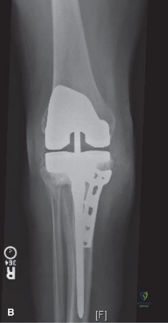
We have a 58-year-old male 4 years post-primary TKA presenting with chronic pain and instability. Looking at this patient's initial presentation, how would you systematically evaluate his knee to determine the cause of his failure, and what are the specific clinical signs you are looking for in the physical exam?
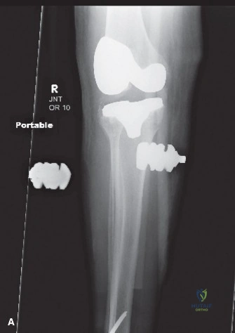
Candidate: I would start by ruling out infection with serum markers (ESR/CRP). Clinically, I would assess the scar, range of motion, and stability. I am looking for signs of aseptic loosening such as pain on weight-bearing and mechanical symptoms like clunking. I would perform varus/valgus stress tests to assess ligamentous laxity and check for an extensor mechanism deficit.
The candidate fails to provide a structured approach. They often jump to "revision surgery" without discussing the "unexplained pain" caveat. They also miss the importance of characterizing the instability (e.g., flexion vs. extension vs. global) and fail to mention that every painful TKA is infected until proven otherwise.
Structure the response: 1. Infection exclusion: Mandatory ESR/CRP, if abnormal, proceed to joint aspiration for cell count, differential, and cultures (ICM criteria). 2. Mechanical evaluation: Differentiate instability types (extension, flexion, mid-flexion, global) and assess for aseptic loosening (radiographic lucencies, component subsidence). 3. Exam focus: Document ROM (assess for extension contracture), patellofemoral tracking, and perform stress testing in both full extension and 30 degrees of flexion to evaluate MCL/LCL integrity. Acknowledge that revision for "unexplained pain" is a contraindication.
Suppose you have determined the patient has significant medial collateral ligament (MCL) attenuation and bone loss consistent with AORI Type 2B. You are in the OR; how do you manage the exposure, and what is your decision-making process regarding the constraint ladder?
Candidate: I would start with a medial parapatellar arthrotomy. If that’s tight, I’d perform a quadriceps snip or a tibial tubercle osteotomy for better exposure. Regarding the constraint, I would use a Constrained Condylar Knee (CCK) since the MCL is compromised. I would use augments to manage the AORI 2B bone loss.
Candidates often forget to mention joint line restoration. Simply "using augments" is insufficient; the examiner wants to know that distal femoral augments are used to prevent joint line elevation, which causes patella baja and mid-flexion instability.
Structure the answer: 1. Exposure: Adhere to the 'least invasive' principle—start with a medial parapatellar approach, then progress to quadriceps snip, and finally a TTO if necessary for exposure. 2. Joint Line: Emphasize restoring the joint line to 25-30mm distal to the medial epicondyle using distal femoral augments. 3. Constraint: Utilize the 'Constraint Ladder'. Since the MCL is incompetent, move to a CCK. Only escalate to a Rotating Hinge if the epicondylar attachments are deficient or global instability persists. 4. Fixation: Use diaphyseal engaging stems for alignment/offloading and metaphyseal cones/sleeves for AORI 2B bone defects to provide immediate rigid fixation.
Detailed Chapters & Topics
Dive deeper into specialized chapters regarding painful-total-knee-replacement