Periprosthetic Joint Infection in Total Knee Arthroplasty: Epidemiology, Risk Factors, and Foundational Principles
Key Takeaway
Periprosthetic joint infection (PJI) following total knee arthroplasty (TKA) is a devastating complication characterized by microorganisms in periprosthetic tissue. Its incidence ranges from 0.5-2%, escalating with revision. PJI is influenced by patient, intraoperative, and postoperative risk factors, necessitating a deep understanding of knee anatomy and biomechanics for accurate diagnosis and management.
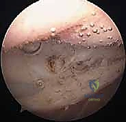
A 68-year-old patient presents with a painful total knee arthroplasty performed 5 years ago. Radiographs show no evidence of loosening, but synovial aspiration is performed due to clinical suspicion of periprosthetic joint infection (PJI). You are presented with the following intraoperative arthroscopic image taken during a diagnostic procedure. What does this image show, and how does it influence your management?
Candidate: The image shows evidence of synovitis, likely villonodular or inflammatory changes, with some fibrin or debris. In a PJI context, this represents synovial hypertrophy and reactive inflammation. It confirms the inflammatory process, and I would proceed by taking multiple tissue biopsies for histopathology and cultures to confirm the diagnosis of PJI.
Candidates often focus solely on the visual appearance without linking it to the pathophysiology of biofilm or the diagnostic criteria. Failing to mention that biopsies must be taken from multiple distinct anatomical sites (at least 5 samples) is a major oversight. Additionally, some candidates suggest using arthroscopy for "debridement," which is generally contraindicated in chronic PJI as it does not allow for radical synovectomy or implant-bone interface clearance.
The image demonstrates synovial hypertrophy and inflammatory proliferative changes typical of a chronic periprosthetic infection. My management would be as follows:
1. Diagnostic Confirmation: Ensure I have fulfilled the MSIS or ICM major/minor criteria.
2. Tissue Sampling: Perform at least 5 distinct synovial and periprosthetic tissue biopsies using separate instruments for each to avoid cross-contamination.
3. Histology: Send for frozen section; >5 PMNs per high-power field in 5 separate fields is highly suggestive of infection.
4. Decision Making: If confirmed, this is a Type IV (Tsukayama) chronic infection, necessitating a two-stage exchange arthroplasty. Arthroscopic debridement is insufficient due to the biofilm-protected sessile bacteria; a radical open synovectomy and implant removal are mandatory.
You have decided to proceed with a two-stage exchange for a chronic PJI. During the first stage, you perform an extensive debridement. The patient has significant bone loss. How do you classify the bone loss, and what are your options for reconstruction during the second stage?
Candidate: I would use the AORI (Anderson Orthopaedic Research Institute) classification to grade the bone loss. For reconstruction, I would use metal augmentation, bone graft, or cones, and likely use a constrained implant or a rotating hinge if the ligaments are incompetent.
The candidate fails to explain the AORI classification levels (Type 1: intact metaphyseal; Type 2: damaged metaphysis; Type 3: deficient metaphysis). They also fail to emphasize the importance of diaphyseal stem fixation in revision surgery to bypass the metaphyseal bone loss and provide stability.
I classify bone loss using the AORI system:
• Type 1: Intact metaphyseal bone.
• Type 2: Damaged metaphysis (2A: one condyle; 2B: both condyles).
• Type 3: Deficient metaphysis, involving the epicondyles/tibial plateau, requiring structural support.
Reconstruction Strategy:
1. Use metaphyseal-filling components like porous tantalum cones or sleeves to restore structural integrity.
2. Always utilize diaphyseal-engaging stems to offload the metaphysis and ensure rotational stability.
3. Determine constraint based on collateral ligament status: CCK (Condylar Constrained Knee) for manageable laxity; Rotating Hinge for total ligamentous deficiency or severe bone loss where tensioning is impossible.
