Revision TKA: Safely Removing Well-Fixed Components by Targeting the Interface
Key Takeaway
This topic focuses on Revision TKA: Safely Removing Well-Fixed Components by Targeting the Interface, Revision Total Knee Arthroplasty with removal of well-fixed components addresses issues like infection, malalignment, instability, or periprosthetic fracture, even when the implant itself isn't loose. Meticulous surgical technique is crucial to safely separate components, where the implant-bone interface is targeted to preserve bone stock and prevent iatrogenic fracture, ensuring a better patient outcome.
Introduction and Epidemiology
The burden of revision total knee arthroplasty is expanding at an unprecedented rate, driven by the exponential growth in primary arthroplasty volume and the expanding indications for joint replacement in younger, more active demographics. Current epidemiological estimates indicate that by the year 2030, the volume of primary total knee arthroplasty cases in the United States will have increased to approximately 3,480,000 annually. Consequently, the number of revision procedures is expected to rise proportionally, reaching an estimated 268,200 cases per year.
While many revisions are performed for aseptic loosening where the implant-bone interface has already failed, a significant and technically demanding subset of revisions requires the extraction of well-fixed components. Indications for removing well-fixed total knee components include deep periprosthetic joint infection, severe malalignment, malpositioning, catastrophic polyethylene wear with osteolysis, instability, periprosthetic fracture, arthrofibrosis, or the necessity to upsize constraint due to aseptic loosening of the contralateral component.
Achieving the goal of safe removal of well-fixed components depends entirely on meticulous surgical technique, a comprehensive understanding of the implant-cement-bone interfaces, and the availability of appropriate extraction instrumentation. In many respects, component extraction is the most critical phase of the revision procedure. Careless or impatient technique invariably leads to catastrophic degradation of the remaining bone stock, iatrogenic fracture, and disruption of the extensor mechanism. These preventable complications ultimately dictate the necessity for highly constrained implants, massive structural allografts, or highly porous metaphyseal cones and sleeves, thereby compromising the quality of the final revision construct and the long-term survivorship of the joint.
Surgical Anatomy and Biomechanics
Extensor Mechanism Anatomy
Removal of well-fixed components necessitates extensile exposure, which is fundamentally dictated by the anatomy of the extensor mechanism. The quadriceps tendon, patella, and patellar tendon form a continuous tensile structure that must be mobilized laterally to access the tibiofemoral articulation. In the setting of a revision, the suprapatellar pouch is often obliterated by dense fibrous scar tissue, effectively tethering the extensor mechanism to the anterior distal femur.
Failure to respect the tension on the patellar tendon insertion at the tibial tubercle during exposure and extraction is the leading cause of iatrogenic patellar tendon avulsion—a devastating complication with limited salvage options. Understanding the vascular supply to the patella, predominantly derived from the superior and inferior genicular anastomoses, is also critical. Extensive lateral release combined with medial arthrotomy can devascularize the patella, leading to necrosis and fragmentation during component extraction.

Osseous and Ligamentous Architecture
The osseous anatomy of the distal femur and proximal tibia in a revision setting is often distorted by osteolysis, stress shielding, and prior bone resections. The metaphyseal bone is particularly vulnerable during the extraction of well-fixed cemented or porous-coated components. The Anderson Orthopaedic Research Institute classification is utilized to quantify this bone loss, categorizing defects into contained metaphyseal defects or uncontained defects extending to the diaphysis.
The collateral ligaments dictate the coronal stability of the knee and must be protected during the aggressive use of osteotomes and saws. The medial collateral ligament originates on the medial femoral epicondyle and inserts broadly on the proximal medial tibia. The lateral collateral ligament originates on the lateral epicondyle and inserts on the fibular head. During the removal of a well-fixed tibial tray, the posterior neurovascular structures (popliteal artery and vein) are at extreme risk if an osteotome or saw blade plunges posterior to the tibial cortex.

Indications and Contraindications
The decision to revise a total knee arthroplasty and remove well-fixed components must be based on a definitive diagnosis. Failure to identify a specific mechanical, infectious, or biological cause for the patient’s pain before performing the revision portends a remarkably poor prognosis.
Diagnostic Evaluation
The history and physical examination should be meticulously directed to determine whether the patient’s pain is extrinsic or intrinsic to the articulation. Extrinsic sources of pain, such as lumbar radiculopathy, ipsilateral hip osteoarthritis, or complex regional pain syndrome, must be excluded.
Physical examination must include visual inspection of previous incisions to select the most appropriate, lateral-most incision to avoid wound necrosis. Passive and active range of motion are assessed, noting that postoperative range of motion predominantly depends on the preoperative state. Extensor lag must be documented as it indicates a deficient extensor mechanism. Coronal and sagittal plane instability are evaluated to determine the required constraint of the revision implants.
Operative Versus Non Operative Management
| Indication Category | Operative Management (Revision TKA) | Non Operative Management |
|---|---|---|
| Infection | Two-stage exchange, DAIR (if acute), or Single-stage exchange | Chronic antibiotic suppression (if medically unfit for surgery) |
| Instability | Component removal and revision to higher constraint (e.g., CCK or Hinge) | Hinged knee brace, physical therapy (limited efficacy) |
| Malalignment | Removal of well-fixed components, axis correction | Shoe lifts, offloading braces, observation |
| Periprosthetic Fracture | Distal femoral replacement or revision stem bypass | Casting/Bracing (only for strictly non-displaced, stable fractures) |
| Arthrofibrosis | Component removal, extensive lysis of adhesions | Aggressive physical therapy, manipulation under anesthesia |
| Extrinsic Pain | Absolute Contraindication | Treat primary source (e.g., lumbar spine decompression, THA) |
Pre Operative Planning and Patient Positioning
Radiographic Evaluation
Comprehensive imaging is mandatory for preoperative templating and strategy formulation. Standing anteroposterior, lateral, and patellofemoral (Merchant) radiographs are essential to evaluate component fixation, sizing, and gross osteolysis. Full-length standing anteroposterior radiographs are critical to determine the overall mechanical alignment of the lower limb and to identify extra-articular deformities that may influence the revision axis.

Advanced imaging, specifically computed tomography with metal artifact reduction sequences, is highly valuable for assessing component rotation, specifically the epicondylar axis relationship of the femoral component and the rotational alignment of the tibial tray. CT is also the gold standard for quantifying metaphyseal bone loss and planning for metaphyseal cones or sleeves. Laboratory evaluation must include erythrocyte sedimentation rate and C-reactive protein. Any elevation mandates a preoperative joint aspiration for cell count, differential, and extended cultures to rule out periprosthetic joint infection.
Patient Positioning and Setup
The patient is positioned supine on the operating table. A radiolucent table is preferred if intraoperative fluoroscopy is anticipated for stem trajectory or fracture management. A tourniquet is placed proximally on the thigh, though its inflation may be delayed or avoided entirely in patients with severe peripheral vascular disease. A lateral post or a specialized leg holder is utilized to allow dynamic positioning of the knee from full extension to 120 degrees of flexion.

Detailed Surgical Approach and Technique
Incision and Extensile Exposure
The previous surgical incision is utilized whenever possible. If multiple parallel incisions are present, the most lateral viable incision is selected to preserve the vascular supply to the anterior skin flap, which relies on medially based perforators. A standard medial parapatellar arthrotomy is initially performed.
In the setting of a stiff knee or robust well-fixed components, standard exposure is frequently inadequate. The surgeon must seamlessly transition to extensile exposure techniques to prevent patellar tendon avulsion.
1. Quadriceps Snip: An oblique incision is made extending superiorly and laterally from the apex of the standard medial parapatellar arthrotomy across the rectus femoris tendon. This provides excellent exposure and requires no modification to postoperative rehabilitation.
2. Tibial Tubercle Osteotomy: Indicated for severe stiffness, patellar baja, or when profound distal femoral and proximal tibial exposure is required. An 8 to 10 cm osteotomy of the anterior tibial crest is performed, leaving the lateral muscular hinge intact. It is later repaired with cerclage wires.
3. V-Y Quadricepsplasty: Reserved for extreme cases of extensor mechanism contracture, though largely supplanted by the tibial tubercle osteotomy due to extensor lag complications.
Principles of Targeting the Interface
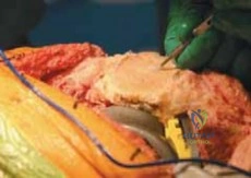
The fundamental principle of removing well-fixed components is to direct all mechanical force strictly at the implant-cement interface or the cement-bone interface, depending on the extraction strategy. Prying, levering, or applying asymmetric extraction forces before the interface is completely disrupted will result in massive loss of metaphyseal bone.

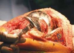
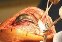
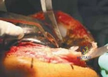
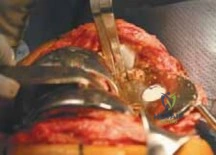
Femoral Component Extraction
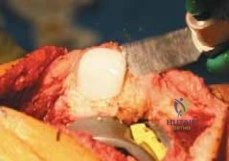
- Anterior Flange Disruption: Thin, flexible osteotomes are driven between the anterior flange of the femoral component and the anterior femoral cortex. The osteotome must be kept perfectly flush against the metal to avoid notching the anterior femur.
- Distal Condylar Interface: A reciprocating saw with a thin blade or flexible osteotomes are utilized to disrupt the cement mantle under the distal medial and lateral condyles.
- Chamfer Disruption via Gigli Saw: The anterior and posterior chamfers are notoriously difficult to access with straight osteotomes. A Gigli saw is passed behind the femoral component, often utilizing a right-angle clamp. By employing a reciprocating motion, the Gigli saw efficiently cuts through the cement mantle or the porous ingrowth at the chamfer interfaces.
- Posterior Condylar Release: Offset osteotomes are carefully directed behind the posterior condyles. Extreme caution must be exercised here to avoid penetrating the posterior capsule and injuring the popliteal artery.
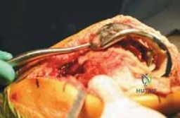
- Extraction: Once the interface is universally disrupted, a femoral extraction device or a slap hammer is attached to the component. The extraction force must be applied strictly in the axial plane corresponding to the distal femoral geometry. If the component does not easily disengage after three strikes, the interface has not been adequately disrupted, and further osteotome work is required.
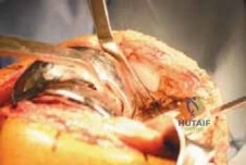
Tibial Component Extraction
- Polyethylene Removal: The modular polyethylene insert is removed first to increase the joint space and provide direct visualization of the tibial tray.
- Peripheral Interface Disruption: A thin osteotome or reciprocating saw is passed around the periphery of the tibial tray, targeting the prosthesis-cement interface. This preserves the cement mantle temporarily, which acts as a protective shield for the underlying trabecular bone.
- Keel or Stem Disruption: If the tibial component features a central keel or cemented stem, a narrow, flexible osteotome or a specialized ultrasonic cement removal tool is directed down the central axis alongside the keel.
- Extraction: A specialized tibial extraction tool is secured to the tray. Gentle, symmetric upward impaction is applied. Asymmetric levering will inevitably cause a fracture of the medial or lateral tibial plateau.
Patellar Component Extraction
If the patellar component is well-fixed, appropriately positioned, and compatible with the new femoral geometry, it should be retained to preserve patellar bone stock. If removal is mandatory (e.g., infection, severe wear, incompatibility), a thin oscillating saw is passed precisely at the prosthesis-cement interface. The remaining cement is then carefully removed with a high-speed burr or small curettes, preserving the often-thin residual patellar shell.
Cement and Debris Removal
Once the metallic components are extracted, the residual cement mantle must be removed. This is achieved using a combination of hand instruments (curettes, gouges), ultrasonic cement removal devices, and high-speed burrs. The goal is to remove all acrylic debris and fibrous tissue to expose bleeding, viable bone for the subsequent revision reconstruction, while minimizing the expansion of existing cavitary defects.

Complications and Management
The extraction of well-fixed components is fraught with potential intraoperative and postoperative complications. Anticipation, recognition, and immediate management of these events are hallmarks of an experienced revision arthroplasty surgeon.
Extensor Mechanism Disruption
Avulsion of the patellar tendon from the tibial tubercle is a catastrophic event. If it occurs intraoperatively, it must be repaired immediately using heavy non-absorbable sutures passed through transosseous tunnels, often augmented with a wire cerclage or a synthetic mesh tape. Postoperatively, the patient must be immobilized in extension for 6 to 8 weeks, significantly compromising final range of motion. Prevention through the early use of extensile exposures is paramount.
Iatrogenic Bone Loss and Fracture
Despite meticulous technique, extraction often results in the loss of metaphyseal cancellous bone. Cavitary defects must be managed with morcellized allograft, whereas uncontained segmental defects require structural allografts or highly porous titanium cones and metaphyseal sleeves.
Iatrogenic fractures of the femoral condyles or tibial plateau during extraction require immediate internal fixation or bypass. Epicondylar fractures may destabilize the collateral ligaments, necessitating the use of a constrained condylar knee or a rotating hinge construct.
Table of Complications and Salvage Strategies
| Complication | Estimated Incidence | Salvage Strategy / Management |
|---|---|---|
| Iatrogenic Bone Loss (Severe) | 15 - 30% | Highly porous metaphyseal cones/sleeves, structural bulk allograft, transition to diaphyseal engaging stems. |
| Patellar Tendon Avulsion | 1 - 2% | Transosseous suture repair with synthetic augmentation; extensor mechanism allograft for chronic deficiency. |
| Iatrogenic Fracture (Femur/Tibia) | 2 - 5% | Stem bypass of the fracture by at least 2 cortical diameters; cerclage wiring; transition to hinged prosthesis if epicondyles are lost. |
| Ligamentous Disruption (MCL/LCL) | 1 - 3% | Primary repair if avulsed with bone fragment; upgrade constraint to CCK or Rotating Hinge. |
| Patellar Fracture / Necrosis | 2 - 4% | Patelloplasty (resection of fragments); retention of extensor mechanism without resurfacing (gull-wing osteotomy). |
Post Operative Rehabilitation Protocols
The rehabilitation protocol following revision total knee arthroplasty must be highly individualized, dictated primarily by the quality of bone stock, the method of fixation, and the integrity of the extensor mechanism.
Weight Bearing Status
In cases where diaphyseal engaging stems and metaphyseal cones provide rigid, immediate construct stability, patients may be allowed to weight-bear as tolerated immediately postoperatively. However, if massive structural allografts were utilized, or if the bone quality is severely osteoporotic, restricted weight-bearing (toe-touch or partial) may be mandated for 6 to 8 weeks to allow for biological integration and to prevent early mechanical failure.
Range of Motion and Extensor Mechanism Precautions
Early range of motion is generally encouraged to prevent arthrofibrosis, utilizing continuous passive motion machines or early active-assisted physical therapy. Normal range of motion after primary TKA ranges from full extension to 120 to 135 degrees. It is critical to manage patient expectations, as revision TKA frequently does not improve, and may slightly decrease, the preoperative range of motion.
If a tibial tubercle osteotomy was performed for exposure, active knee extension is strictly prohibited for 4 to 6 weeks to prevent displacement of the osteotomy. The patient is placed in a hinged knee brace locked in extension for ambulation, allowing passive flexion up to 60-90 degrees depending on the security of the tubercle fixation. Similarly, if a quadriceps snip was utilized, standard rehabilitation can proceed, but a V-Y quadricepsplasty requires a period of immobilization to protect the tendinous repair.
Summary of Key Literature and Guidelines
The safe removal of well-fixed components remains heavily discussed in orthopedic literature, with consensus guidelines emphasizing the necessity of preoperative planning and the judicious use of extensile exposures.
- Exposure Techniques: Classic studies by Insall and Whiteside established the foundational algorithms for extensile exposure. The quadriceps snip has been validated as a safe, highly effective technique that does not compromise long-term extensor power, whereas the V-Y turndown is associated with significant postoperative extensor lag.
- Bone Preservation: Literature evaluating the use of Gigli saws and flexible osteotomes demonstrates a significant reduction in AORI Type II and III defects compared to blunt extraction methods. The preservation of the metaphyseal envelope directly correlates with the survivorship of the subsequent revision implants.
- AAOS Clinical Practice Guidelines: Current guidelines strongly recommend comprehensive preoperative evaluation to definitively diagnose the etiology of failure prior to revision. The empirical removal of well-fixed components for unexplained pain is strongly discouraged due to uniformly poor clinical outcomes and high rates of persistent pain.
- Metaphyseal Fixation: Recent biomechanical and clinical cohort studies highlight the efficacy of highly porous metaphyseal cones and sleeves in managing the inevitable bone loss associated with component extraction. These devices provide immediate rigid fixation and promote long-term biological osteointegration, significantly reducing the reliance on structural allografts.
The successful execution of a revision total knee arthroplasty hinges on the surgeon's ability to safely navigate the extraction phase. By targeting the interface, respecting the soft tissue envelope, and anticipating bone loss, the orthopedic surgeon can preserve the necessary anatomical foundation to build a stable, durable revision construct.
Clinical & Radiographic Imaging