Revision Knee Replacement: Master Challenges with Expert Q&A
Key Takeaway
In this comprehensive guide, we discuss everything you need to know about Revision Knee Replacement: Master Challenges with Expert Q&A. A revision knee replacement is a surgical procedure performed when a previous total knee replacement has failed, often due to aseptic loosening, infection, or extensive bone loss. It addresses issues like loose implants, osteolysis, and dysfunctional ligaments, often requiring specialized implants, bone grafts, or constrained components to restore stability and function.
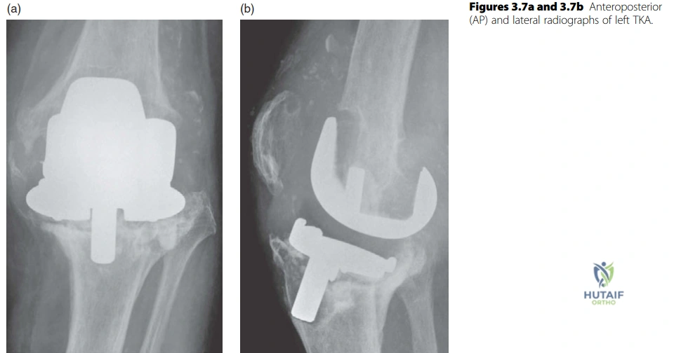
Examine these AP and lateral radiographs of a symptomatic left total knee replacement. Describe your findings and formulate a differential diagnosis.
Candidate: The radiographs show a left TKR with evidence of component loosening. There is significant radiolucency around the femoral and tibial components, suggestive of osteolysis. The tibial baseplate is in varus and extension. There is anterior femoral notching and potential soft tissue calcification. I would suspect aseptic loosening or PJI and require further comparison films.
Jumping immediately to "it's loose" without describing the specific radiographic findings (e.g., varus/extension, notching) or failing to mention the critical differential of PJI. Candidates often forget to ask for comparison films, which is essential to assess the progression of loosening.
Start with a structured description: (1) Alignment and position of components (tibial varus/extension, anterior notching), (2) Bone-implant interface (radiolucency, osteolysis), (3) Soft tissues (calcification/vascularity), and (4) Comparative analysis. Conclude with a clear differential: "Radiographically this represents component failure; clinically, infection must be excluded until proven otherwise."
You have confirmed aseptic loosening and the patient is discussed for revision. Can you describe the AORI classification for bone loss and explain its clinical utility in pre-operative planning?
Candidate: The AORI classification assesses femur (F) and tibia (T) independently. Type 1 has intact metaphyseal bone. Type 2 involves metaphyseal damage (A is one condyle/plateau, B is both). Type 3 involves severe metaphyseal deficiency often requiring stemmed implants or custom solutions.
Failing to emphasize that Type 2 and Type 3 defects often necessitate specialized reconstructive tools like augments, sleeves, or long stems. Candidates often memorize the definitions but fail to translate them into the surgical strategy (reconstruction of the joint line).
Systematically explain Type 1, 2, and 3. Add the "clinical pivot": Explain that the classification dictates the constraint requirement (e.g., AORI Type 3 often requires highly constrained implants or hinged designs) and the method of fixation (e.g., metaphyseal sleeves for Type 2/3 to bypass weak metaphyseal bone).
You mentioned constraint. Please delineate the "Constraint Ladder" in revision TKR and the clinical scenarios that necessitate moving up this ladder.
Candidate: The ladder goes from: 1) Cruciate Retaining (CR) - limited constraint; 2) Posterior Stabilized (PS) - cam-post for A-P stability; 3) Varus-Valgus Constrained (VVC/LCCK) - for collateral insufficiency; 4) Rotating Hinge (RHK) - for global instability or severe bone loss.
Suggesting hinged knees for straightforward cases. Hinged knees have higher stress at the bone-implant interface and are reserved for salvage situations due to potential for aseptic loosening of the stem.
Structure the answer by level of stability required. 1. CR: Needs intact PCL. 2. PS: Substitutes PCL, allows rollback. 3. VVC: Provides coronal stability when ligaments are incompetent. 4. Hinged: For global instability (e.g., MME/LLE loss). Always conclude by saying: "The goal is to use the least amount of constraint necessary to achieve stability to minimize bone-implant stress."
